Abstract
Delphinidin-3-glucoside (Dp-3-g) is one of the predominant bioactive compounds of anthocyanins in many plant foods. Although several anthocyanin compounds have been reported to be protective against cardiovascular diseases (CVDs), the direct effect of anthocyanins on platelets, the key players in atherothrombosis, has not been studied. The roles of Dp-3-g in platelet function are completely unknown. The present study investigated the effects of Dp-3-g on platelet activation and several thrombosis models in vitro and in vivo. We found that Dp-3-g significantly inhibited human and murine platelet aggregation in both platelet-rich plasma and purified platelets. It also markedly reduced thrombus growth in human and murine blood in perfusion chambers at both low and high shear rates. Using intravital microscopy, we observed that Dp-3-g decreased platelet deposition, destabilized thrombi, and prolonged the time required for vessel occlusion. Dp-3-g also significantly inhibited thrombus growth in a carotid artery thrombosis model. To elucidate the mechanisms, we examined platelet activation markers via flow cytometry and found that Dp-3-g significantly inhibited the expression of P-selectin, CD63, CD40L, which reflect platelet α- and δ-granule release, and cytosol protein secretion, respectively. We further demonstrated that Dp-3-g downregulated the expression of active integrin αIIbβ3 on platelets, and attenuated fibrinogen binding to platelets following agonist treatment, without interfering with the direct interaction between fibrinogen and integrin αIIbβ3. We found that Dp-3-g reduced phosphorylation of adenosine monophosphate-activated protein kinase, which may contribute to the observed inhibitory effects on platelet activation. Thus, Dp-3-g significantly inhibits platelet activation and attenuates thrombus growth at both arterial and venous shear stresses, which likely contributes to its protective roles against thrombosis and CVDs.
🔬 Techniques
💻 Software
✨ Fluorophores
🧪 Sample Preparation
🏭 Microscope Brands
🧪 Reagent Suppliers
💻 Software Details
🏛️ Research Organizations (ROR)
Affiliated research institutions:
📋 Methods
Ethics Statement
For research involving human participants, written informed consent was obtained in accordance with the Declaration of Helsinki and studies were approved by the St. Michael's Hospital Research Ethics Board. All animal procedures were approved by the Animal Care Committee at St. Michael's Hospital (protocol numbers 213 and 181). Mice were housed in the research vivarium at St. Michael's Hospital.
Reagents
Dp-3-g was purchased from Polyphenol AS (Norway). Thrombin receptor activating peptide (TRAP; AYPGKF-NH 2 ) was purchased from Peptides International (Louisville, MO, USA). Thrombin and ADP were purchased from Sigma-Aldrich (Oakville, ON, Canada). Type-I collagen fibrils (equine collagen Horm) was purchased from Nycomed (Roskilde, Denmark). DiOC6 dye was purchased from Invitrogen (Burlington, ON, Canada). PE-conjugated mouse anti-human PAC-1 antibody, FITC-conjugated mouse anti-human CD63 antibody, FITC-conjugated mouse anti-human CD40L antibody, FITC-conjugated mouse anti-human CD62P antibody, PE-conjugated mouse anti-mouse CD62P antibody, FITC-conjugated mouse IgG1κ Isotype control, PE-conjugated anti-mouse IgG antibody, FITC-conjugated mouse anti-human IgG1κ Isotype control and PE-conjugated anti-human IgG1κ Isotype control were purchased from BD Biosciences (Mississauga, ON, Canada). PE-conjugated goat anti-mouse JON/A antibody was purchased from Emfret Analytics (Eibelstadt, Germany). Rabbit antibodies against phosphorylated AMPK (pAMPK, Thr172) and AMPK were purchased from Cell Signaling Technology (Danvers, MA, USA). Fg from human plasma, Alexa Fluor 488 conjugate and Calcein AM was purchased from Invitrogen (Burlington, ON, Canada).
Show full methods section
Ethics Statement
For research involving human participants, written informed consent was obtained in accordance with the Declaration of Helsinki and studies were approved by the St. Michael's Hospital Research Ethics Board. All animal procedures were approved by the Animal Care Committee at St. Michael's Hospital (protocol numbers 213 and 181). Mice were housed in the research vivarium at St. Michael's Hospital.
Reagents
Dp-3-g was purchased from Polyphenol AS (Norway). Thrombin receptor activating peptide (TRAP; AYPGKF-NH 2 ) was purchased from Peptides International (Louisville, MO, USA). Thrombin and ADP were purchased from Sigma-Aldrich (Oakville, ON, Canada). Type-I collagen fibrils (equine collagen Horm) was purchased from Nycomed (Roskilde, Denmark). DiOC6 dye was purchased from Invitrogen (Burlington, ON, Canada). PE-conjugated mouse anti-human PAC-1 antibody, FITC-conjugated mouse anti-human CD63 antibody, FITC-conjugated mouse anti-human CD40L antibody, FITC-conjugated mouse anti-human CD62P antibody, PE-conjugated mouse anti-mouse CD62P antibody, FITC-conjugated mouse IgG1κ Isotype control, PE-conjugated anti-mouse IgG antibody, FITC-conjugated mouse anti-human IgG1κ Isotype control and PE-conjugated anti-human IgG1κ Isotype control were purchased from BD Biosciences (Mississauga, ON, Canada). PE-conjugated goat anti-mouse JON/A antibody was purchased from Emfret Analytics (Eibelstadt, Germany). Rabbit antibodies against phosphorylated AMPK (pAMPK, Thr172) and AMPK were purchased from Cell Signaling Technology (Danvers, MA, USA). Fg from human plasma, Alexa Fluor 488 conjugate and Calcein AM was purchased from Invitrogen (Burlington, ON, Canada).
Mice
C57BL/6J mice were purchased from Charles River Laboratories International (Wilmington, MA). Murine platelet and plasma preparation Mice (6–8 weeks old) were anesthetized and bled from the retro-orbital plexus with the use of heparin-coated glass capillary tubes. Blood was collected into tubes containing either 3% ACD (1/9 vol/vol) or 25 U/mL heparin. PRP was obtained by centrifugation at 300 g for 7 min. Platelet-poor plasma (PPP) was prepared by centrifugation at 1500 g for 20 min. The PPP was further centrifuged at 10,000 g for 5 min to remove any remaining cells. Gel-filtered platelets were prepared as previously described [27] , [28] . Briefly, platelets were isolated from citrated PRP using a Sepharose 2B column in PIPES buffer (5 mM PIPES, 1.37 mM NaCl, 4 mM KCl, 0.1% (wt/vol) glucose, pH 7.0).
Human blood preparation
Blood was obtained from healthy human subjects who had not taken any anti-platelet medication in the prior two week period. Blood was obtained by venupuncture into Li-Heparin Vacutainers and was allowed to rest at 37°C for 10 min prior to preparation of PRP. Whole anticoagulated blood was spun at 300 g for 7 min. The PRP was transferred to a fresh tube and stored at 37°C until use. Gel-filtered platelets were prepared using the same method as for preparation of murine gel-filtered platelets. Platelet Aggregation Aggregation of PRP and gel-filtered platelets were performed at 37°C with a sample stir speed of 1000 rpm using a computerized aggregometer (Chrono-Log Corp, Havertown, PA) as we previously described [10] , [11] . PRP and gel-filtered platelets were pre-incubated with the indicated doses of anthocyanin or control buffer for 40 min at 37°C. For PRP, the baseline was adjusted with PPP, and for gel-filtered platelets, equal amounts of platelets were mixed with PIPES (final concentration ∼2.5×10 8 platelets/mL). A total of 250 µL of each sample was added to an aggregation cuvette and incubated for 2 min. Aggregation was induced by 5 µM ADP, 100 µM TRAP, 0.1 U/mL thrombin or 2 µg/mL collagen in the presence of 1 mM Ca 2+ . Aggregation was recorded for 8 min, and data were expressed as a percentage of maximum aggregation of the control without pre-incubation with Dp-3-g.
Perfusion flow assays
The ex vivo perfusion chamber thrombosis model was performed at low (600 s −1 ) and high (1800 s −1 ) shear rates, as we previously described [10] . Briefly, rectangular (0.1×1 mm) glass capillary microslides were coated with 100 µg/mL type-I collagen fibrils overnight at 4°C. Where indicated, whole blood was pre-incubated with different levels of Dp-3-g or control buffer alone for 40 min at 37°C prior to perfusion, followed by washout with phosphate buffered saline (PBS). Platelet adhesion, aggregation, and thrombus formation were recorded in real-time over the course of perfusion under bright field with a Zeiss Axiovert 135 inverted microscope and computer (IBM IntelliStation Z Pro) using the Slidebook program (Intelligent Imaging Innovations). Surface coverage and thrombus size were calculated from the light microscope images using ImageJ software.
Intravital microscopy thrombosis models
The process of thrombus formation in arterioles was monitored in 3–4 week old mice, as we previously described [9] – [11] , [27] , [29] . Briefly, blood was collected into ACD (1∶10) from the experimental mice. PRP was prepared by centrifugation. Platelets were separated from plasma by gel-filtration, labeled with 1 mg/mL of Calcein AM at room temperature for 20 min, and then injected into the experimental mouse (1.25×10 6 platelets/g) via the tail vein. Mice were then anesthetized and the mesentery was externalized. In each mouse, a single mesenteric arteriole of 100–120 µm diameter was chosen and injury was induced by topical application of 30 µL of 250 mM FeCl 3 . Control buffer or Dp-3-g was injected via the tail vein 40 min before FeCl 3 application. Images of thrombus formation and dissolution were visualized with a fluorescence microscope (Zeiss Axiovert 135; Zeiss Oberkochen) and compared between groups based on the time to complete vessel occlusion, as previously described [27] , [29] .
Carotid artery thrombosis model
C57BL/6 mice (6-weeks old) were injected with 50 µM Dp-3-g or control buffer via the tail vein. Following anesthetization, the right carotid artery was dissected and held with a miniature Doppler flow probe (TS420 transit-time perivascular flowmeter, Transonic Systems Inc.). Carotid artery injury was induced with a strip of Whatman filter paper saturated with 10% ferric chloride [30] . The blood flow was monitored until complete vessel occlusion was observed.
Bleeding time assay
C57BL/6 mice (6–8 weeks old) were injected with either control buffer or Dp-3-g intravenously via the tail vein 40 min before the bleeding time was assessed. Mice were anesthetized with 2.5% tribromoethanol (0.015 mL/g) and maintained at 37°C on a heating pad during the experiment. The tip of the tail (5 mm) was cut off with a sharp scalpel, and the tail was immediately placed into warm saline at 37°C. The bleeding time was the period from the moment blood began emerging from the cut until the moment bleeding ceased.
Detection of P-selectin expression on human and mouse platelets
Human and mouse
PRP and gel-filtered platelets were incubated with different concentrations of Dp-3-g or control buffer for 40 min at 37°C. Aliquots of sample (100 µL) were transferred to tubes containing saturating concentrations of the following fluorescently-labeled monoclonal antibodies: PE-conjugated anti-human CD62P antibody, FITC-conjugated anti-mouse CD62P antibody, PE/FITC-conjugated anti-mouse/human IgG1κ (isotope controls), and incubated for 15 min at room temperature. PRP or gel-filtered platelets were activated with either 200 µM ADP, 250 µM TRAP, 10 µg/mL collagen or 1 U/mL thombin, for 5 min at room temperature. PBS (0.5 mL) was added to each sample immediately before acquisition. All samples were analyzed via a calibrated FACSCalibur flow cytometer (BD Biosciences). Ten thousand events per sample were acquired; light scatter and fluorescence channels were set at a logarithmic gain, the platelet population was analyzed for mean fluorescence intensity (MFI). For each experiment, all samples for comparison were acquired at the same settings.
Detection of CD63 and CD40L expression on human platelets
Human gel-filtered platelets were incubated with different concentrations of Dp-3-g or control buffer for 40 min at 37°C. To label CD63 and CD40L, platelets were incubated with FITC-conjugated mouse anti-human CD63 or anti-human CD40L for 15 min at room temperature. Gel-filtered platelets were activated with 1 U/mL thrombin, 250 µM TRAP or 10 µg/mL collagen in the presence of 1 mM Ca 2+ for 5 min. The platelets were fixed with 1% paraformaldehyde before being analyzed via flow cytometry. Detection of activated integrin αIIbβ3 expression on human and mouse platelets The expression of activated integrin αIIbβ3 on human and murine platelets, measured by PAC-1 and JON/A antibody, respectively, were tested as we previously described [31] . PRP or gel-filtered platelets were incubated in polystyrene tubes for 40 min at 37°C with different concentrations of Dp-3-g or control buffer. Aliquots of sample (100 µL) were transferred to tubes containing saturating concentrations of the following fluorescently-labeled monoclonal antibodies: FITC-conjugated anti-human PAC-1 antibody, FITC-conjugated anti-human IgG1κ (isotype control), PE-conjugated anti-mouse JON/A antibody or PE-conjugated mouse IgG1κ (isotype control), and incubated for 15 min at room temperature. Samples in the presence or absence of the agonists 200 µM ADP, 250 µM TRAP, or 10 µg/mL collagen were incubated for 5 min at room temperature. The samples were then fixed in filtered 1% paraformaldehde (pH 7.2) and stored in the dark at 4°C. All samples were analyzed via flow cytometry.
Detection of platelet-bound fibrinogen on human and mouse platelets
Resting gel-filtered platelets were incubated with different concentrations of Dp-3-g or control buffer for 40 min at 37°C, and were then incubated with FITC-conjugated Fg for 15 min at room temperature. Platelet activation was induced by 200 µM ADP, 250 µM TRAP, 10 µg/mL collagen or 1 U/mL thrombin for 5 min at room temperature. The samples were then fixed in filtered 1% paraformaldehyde (pH 7.2) and stored in the dark at 4°C. All samples were analyzed via flow cytometry. Detection of threonine phosphorylation of AMPK in human and mouse platelets Gel-filtered platelets were pre-incubated with the indicated doses of Dp-3-g or control buffer for 40 min at 37°C. Platelets were activated with 25 µg/mL collagen for 90 s at room temperature. Platelets were solubilized in Triton X-100 lysis buffer, and 20 µg of soluble proteins were separated by sodium dodecyl sulfate–polyacrylamide gel electrophoresis (SDS-PAGE). Western blotting was performed as previously described [32] . Membranes were blocked with 5% (w/v) skim milk powder in Tris-buffered saline-Tween (TBS-T; 20 mM Tris, 137 mM NaCl, 0.1% (v/v) Tween 20, pH 7.6). Primary antibody that specifically recognizes phosphorylated AMPK (pAMPK) residue Thr172 and horseradish peroxide (HRP)-conjugated secondary antibody (anti-rabbit IgG-HRP) were diluted 1∶1000 and 1∶2000, respectively, in TBS-T containing 5% (w/v) bovine serum albumin (BSA). Blots were developed with ECL detection reagents (Thermo Scientific) according to the manufacturer's instructions. Following detection, the same membranes were stripped with stripping buffer and re-blotted with a primary antibody that recognizes total AMPK.
Statistical analysis
Data from each treatment or control group were analyzed by one-way ANOVA coupled with the Student-Newman-Keuls multiple comparison test using the SPSS 16.0 statistical package. Differences were considered significant if P
📊 Figures
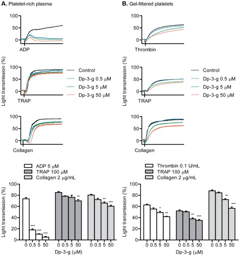
Figure 1
Effects of Dp-3-g on human platelet aggregation.
Human PRP and gel-filtered platelets were pre-incubated with control buffer (black), 0.5 u00b5M Dp-3-g (blue), 5 u00b5M (green) or 50 u00b5M Dp-3-g (red) for 40 min at 37u00b0C. Platelet aggregation w...
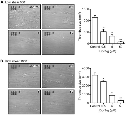
Figure 2
Effects of Dp-3-g on human thrombus formation under flow.
Ex vivo thrombus formation was monitored on type-I collagen at 600 s u22121 or 1800 s u22121 using human whole blood with different concentration of Dp-3-g or control buffer. A) Low shear rate of 600 ...
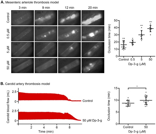
Figure 3
Effects of Dp-3-g on FeCl 3 -induced thrombosis in vivo .
A) Thrombus formation was initiated by topical application of FeCl 3 on mesenteric arterioles in C57BL/6 male mice, which were injected with fluorescently-labeled platelets and different concentration...
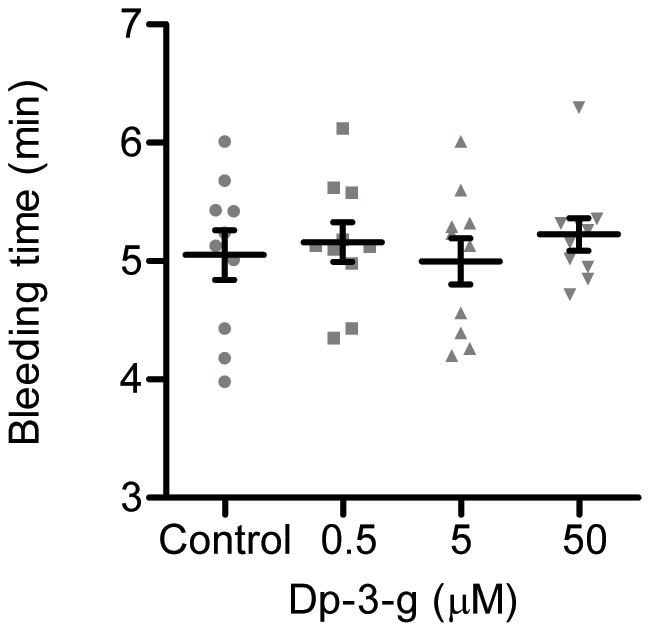
Figure 4
Effects of Dp-3-g on bleeding times in mice.
Tail-vein bleeding times were examined in C57BL/6 mice. Either PBS (control) or different concentrations of Dp-3-g were administered via the tail vein 40 min before the bleeding time was determined. V...
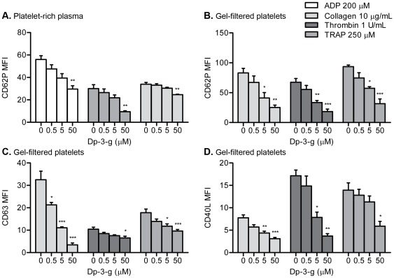
Figure 5
Effects of Dp-3-g on human platelet activation.
Human PRP or gel-filtered platelets were incubated with control buffer, 0.5 u00b5M Dp-3-g, 5 u00b5M Dp-3-g or 50 u00b5M Dp-3-g for 40 min at 37u00b0C. Platelet activation markers were analyzed via flo...
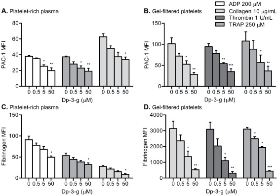
Figure 6
Effects of Dp-3-g on human platelet u03b1IIbu03b23 activation and fibrinogen binding.
Human PRP or gel-filtered platelets were incubated with control buffer, 0.5 u00b5M Dp-3-g, 5 u00b5M Dp-3-g or 50 u00b5M Dp-3-g for 40 min at 37u00b0C. Platelet activation markers were analyzed via flo...
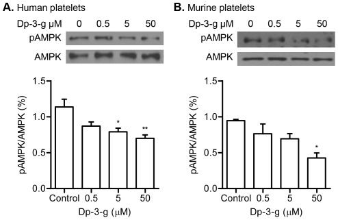
Figure 7
Effects of Dp-3-g on human and murine platelet phosphorylation of threonine residues of AMPK.
Platelets activated with collagen in the presence of Dp-3-g were lysed and proteins were separated by SDSu2013PAGE and immunoblotted to detect phospho-threonine residues. A) Western blot analysis of A...
Figure images are served from the NIH/NLM PubMed Central Open Access Subset or Europe PMC; copyright remains with the publishers and authors.
💬 Discussion
0 commentsNo comments yet. Be the first to start a discussion!
Leave a Comment