Abstract
Platelets play an essential role in hemostasis and atherothrombosis. Owing to their short storage time, there is constant demand for this life-saving blood component. In this study, we report that it is feasible to generate functional megakaryocytes and platelets from human embryonic stem cells (hESCs) on a large scale. Differential-interference contrast and electron microscopy analyses showed that ultrastructural and morphological features of hESC-derived platelets were indistinguishable from those of normal blood platelets. In functional assays, hESC-derived platelets responded to thrombin stimulation, formed microaggregates, and facilitated clot formation/retraction in vitro. Live cell microscopy demonstrated that hESC-platelets formed lamellipodia and filopodia in response to thrombin activation, and tethered to each other as observed in normal blood. Using real-time intravital imaging with high-speed video microscopy, we have also shown that hESC-derived platelets contribute to developing thrombi at sites of laser-induced vascular injury in mice, providing the first evidence for in vivo functionality of hESC-derived platelets. These results represent an important step toward generating an unlimited supply of platelets for transfusion. Since platelets contain no genetic material, they are ideal candidates for early clinical translation involving human pluripotent stem cells.
🔬 Techniques
💻 Software
✨ Fluorophores
🧪 Sample Preparation
🔬 Cell Lines
🏭 Microscope Brands
🧪 Reagent Suppliers
📷 Detectors
🔎 Objectives
💻 Software Details
🏛️ Research Organizations (ROR)
Affiliated research institutions:
📋 Methods
Generation of MKs and platelets from hESCs via hemangioblasts/BCs Hemangioblast/BC generation from hESC lines (HuES-3, MA01 and MA09) was performed as previously reported 16 , 17 . IL-6, IL-9, IL-11, basic FGF, VEGF, TPO, and SCF were obtained from Peprotech, BMP-4 was obtained from Stemgent. BCs from day 6 to 8 cultures were purified and plated (1 to 5 × 10 5 /ml) in “TSI medium” which consisted of Stemline II (Sigma-Aldrich, St Louis, MO, USA) supplemented with 50 ng/ml TPO, 20 ng/ml SCF, and 20 ng/ml IL-11 (all from Peprotech) to induce BC differentiation toward MKs. Half of the MK culture medium was replaced with fresh TSI medium every 2 or 3 days. Platelet generation from MKs was performed under either stroma feeder-free or stroma feeder co-culture conditions, as indicated. For feeder-free platelet generation, MKs from day 4 to 6 cultures were simply maintained in TSI medium for an additional 4-8 days, whereupon platelets were collected intermittently for analysis. In some experiments, GM6001 (100 μM) was added to late stage differentiation cultures. For feeder co-culture experiments, OP9 or C3H 10T1/2 stromal cells were maintained in OP9 culture medium (α-MEM with 15% Hyclone fetal bovine serum). Confluent OP9 or C3H 10T1/2 cells were treated with 100 ng/ml mitomycin-C the day before co-culture, gently washed twice with PBS and allowed to recover overnight in OP9 culture medium. MKs from day 4 to 6 cultures were collected and replated onto mitotically inactivated OP9 or C3H 10T1/2 stromal cells in OP9 culture medium plus 100 ng/ml TPO, 50 ng/mlSCF, and 25 U/ml sodium heparin, as previously described 15 . Platelets were intermittently collected 4 to 8 days later for various analyses. Hematopoietic colony formation assays Megacult-C Kit (Stem Cell Technology, Canada) was used for CFU-MK colony formation assays. After 10 day of plating, CFU-MK cultures were dehydrated, fixed, and stained with anti-CD41 antibody as suggested by the manufacturer. CFU-MK colonies were scored according to the standards provided in the Megacult-C protocol.
Show full methods section
Generation of MKs and platelets from hESCs via hemangioblasts/BCs Hemangioblast/BC generation from hESC lines (HuES-3, MA01 and MA09) was performed as previously reported 16 , 17 . IL-6, IL-9, IL-11, basic FGF, VEGF, TPO, and SCF were obtained from Peprotech, BMP-4 was obtained from Stemgent. BCs from day 6 to 8 cultures were purified and plated (1 to 5 × 10 5 /ml) in “TSI medium” which consisted of Stemline II (Sigma-Aldrich, St Louis, MO, USA) supplemented with 50 ng/ml TPO, 20 ng/ml SCF, and 20 ng/ml IL-11 (all from Peprotech) to induce BC differentiation toward MKs. Half of the MK culture medium was replaced with fresh TSI medium every 2 or 3 days. Platelet generation from MKs was performed under either stroma feeder-free or stroma feeder co-culture conditions, as indicated. For feeder-free platelet generation, MKs from day 4 to 6 cultures were simply maintained in TSI medium for an additional 4-8 days, whereupon platelets were collected intermittently for analysis. In some experiments, GM6001 (100 μM) was added to late stage differentiation cultures. For feeder co-culture experiments, OP9 or C3H 10T1/2 stromal cells were maintained in OP9 culture medium (α-MEM with 15% Hyclone fetal bovine serum). Confluent OP9 or C3H 10T1/2 cells were treated with 100 ng/ml mitomycin-C the day before co-culture, gently washed twice with PBS and allowed to recover overnight in OP9 culture medium. MKs from day 4 to 6 cultures were collected and replated onto mitotically inactivated OP9 or C3H 10T1/2 stromal cells in OP9 culture medium plus 100 ng/ml TPO, 50 ng/mlSCF, and 25 U/ml sodium heparin, as previously described 15 . Platelets were intermittently collected 4 to 8 days later for various analyses. Hematopoietic colony formation assays Megacult-C Kit (Stem Cell Technology, Canada) was used for CFU-MK colony formation assays. After 10 day of plating, CFU-MK cultures were dehydrated, fixed, and stained with anti-CD41 antibody as suggested by the manufacturer. CFU-MK colonies were scored according to the standards provided in the Megacult-C protocol.
Flow cytometry analysis
Cells from blast cultures or MK cultures were monitored routinely by flow cytometry on a FACSCalibur (Becton Dickinson) or Accuri C6 Cytometer (Accuri Cytometers). Fluorochrome-conjugated antibodies for lineage markers, CD41a, CD42a, CD42b, and CD235a (BD Biosciences) were used to characterize MK and erythroid lineages. Antibodies were freshly prepared (1:100 dilution for CD42a and CD42b antibodies; 1:250 dilution for CD41a antibody; 1:2 000 for CD235a antibody) in PBS buffer with 5% FBS. Typically 1 to 2 × 10 5 cells were used for antibody labeling. Cells were stained in a 100 μl antibody cocktail for 1 h on ice, then washed twice with buffer, and resuspended in 250 μl buffer supplemented with 1 μg/ml propidium iodide. To detect the expression level of HLA-ABC, platelets were incubated with a fluorescein isothiocyanate (FITC)-conjugated anti-human HLA-ABC antibody or FITC-conjugated mouse immunoglobulin G (IgG) as a control. The samples were then processed on a FACSCalibur and data was analyzed using Cellquest Pro (BD Biosciences) or Flowjo (Tree Star) software.
DNA content analysis
For polyploidy analysis, cells from day 4-6 MK cultures were fixed in 70% ethanol for 2 h. Cells were then washed once in PBS buffer before staining with 20 μg/ml propidum iodide (Sigma-Aldrich), 20 μg/ml RNase A (Sigma-Aldrich) in PBS buffer overnight at 4 °C. Cellular DNA content was analyzed on an Accuri C6 flow cytometer. Cytospin preparation, Giemsa staining, and immunofluorescence Cells (1 to 2 × 10 4 ) from either blast cultures or MK cultures were cytospun onto polylysine-coated slides (Wessco). Slides were either used for Wright-Giemsa (Sigma-Aldrich) staining or immunofluoroscence. All incubations were performed at room temperature. Cells were blocked with animal-free blocker (Vector Laboratories) for 30 min, incubated with primary antibodies for 1 h, and then washed three times with PBS. For MK identification, primary antibodies include anti-CD41 (1:100; Dako cytomation, Carpinteria, CA, USA) and anti-vWF (1:200, Dako). Cells were incubated with secondary antibodies (1:200 each) for 30 min in the dark, and washed again three times in PBS. DAPI (1 μg/ml) in PBS was used to stain nuclei for 5 min followed by three more PBS washes. Slides were then mounted and examined under Olympus BX51 fluorescence microscope (MVI, Avon, MA, USA). Fluorescent images were captured using a QICAM Fast camera (QImaging, Surrey, BC, Canada) and analyzed with Q Capture Pro version 5.1 software (Media Cybernetics, Bethesda, MD, USA). Phase contrast live cell images were captured using a Nikon microscope, PAXCAM digital camera and PAX-it software.
Preparation of human blood platelets and hESC-derived platelets
Human platelets were isolated as previously described 38 . Briefly, human platelet-rich plasma was prepared by centrifugation of sodium citrate-treated human blood at 200× g for 20 min. The supernatant was collected and centrifuged at 700× g for 10 min in the presence of 0.5 μM PGE1 and 10% sodium citrate buffer. The pellet was resuspended with HEPES-Tyrode buffer (12 mM NaHCO 3 , 138 mM NaCl, 5.5 mM Glucose, 2.9 mM KCl, 0.42 mM NaHPO 4 , 10 mM HEPES, 1 mM CaCl 2 , 1 mM MgCl 2 ) containing 0.15 μM PGE1, and centrifuged at 800× g for 5 min. The pellet was resuspended in RPMI1640 containing 0.1% fatty acid-free bovine serum albumin, 2 mM CaCl 2 , and 1 mM MgCl 2 . Final suspensions of washed platelets were adjusted to 1 × 10 7 platelets/ml. Approval to obtain blood samples was obtained from the University of Illinois-Chicago review board. Informed consent was provided according to the Declaration of Helsinki. In some experiments, human blood samples were also obtained from a commercial source (AllCells, Emeryville, CA, USA). For hESC-PLTs, culture media containing hESC-PLTs were gently collected with apyrase (1 U/ml) and EDTA (5 mM; Sigma-Aldrich) being added to prevent platelet activation. hESC-PLTs were enriched and washed as described above. Washed blood platelets and hESC-PLTs were incubated at 37 °C for 0.5-2 h before functional assays were performed.
Differential interference contrast and electron microscopy analyses
To study cell shape, hESC-PLTs were fixed with 1% glutaraldehyde in platelet buffer for 20 min, moved to a coverslip chamber, and viewed with a 63× differential interference contrast objective. Images were obtained using a cooled CCD camera with Metamorph software (Universal Imaging). Anti-tubulin immunofluorescence microscopy was performed as described previously 39 . Images were analyzed using the Metamorph image analysis software (Molecular Devices, Sunnyvale, CA, USA). Plot profiles were generated using ImageJ (NIH, http://rsb.info.nih.gov/ij/ ). Thin-section electron microscopy was carried out as previously described 40 . Anti-P-selectin and anti-PF-4 immunogold electron microscopy was carried out as described previously 41 . Live cell video microscopy of platelets undergoing activation was performed using previously described methods 42 . Images of spreading platelets were captured every 5 s for 10 min. Platelet spreading on various substrates Spreading and adhesion of blood platelets and hESC-PLTs on immobilized surfaces of fibrinogen, vWF, type I collagen, and BSA were performed as reported 21 , 43 . Chamber slides with microtiter wells (Nalgen Nunc, Rochester, NY, USA) or coverslips were coated with 100 μg/ml fibrinogen or vWF (30 μg/ml; Sigma-Aldrich), acid-soluble fibrillar type I collagen (20 μg/ml, Millipore) or 1% fatty acid-free BSA (Sigma-Aldrich) in 0.1 M NaHCO 3 (pH 8.3) at 4 °C overnight. Washed human blood platelets or hESC-PLTs (1 × 10 7 /ml) were allowed to adhere and spread on protein-coated wells at 37 °C for 90 min. In some experiments, platelets were preincubated with an αIIbβ3 integrin antagonist (RGDS peptide) for 5 min before loading. In other experiments, platelets were mixed with ADP (20 μM) or thrombin (1 U/ml; Sigma-Aldrich), or were pretreated with 20 μg/ml of isotype control mouse IgG1 or a blocking anti-α2β1 antibody (BHA2.1, Millipore) for collagen-coated surfaces, and immediately loaded onto fibrinogen-, vWF-, or collagen-coated wells, as indicated. After washing with PBS buffer, cells were fixed, permeabilized, and stained with Alexa Fluor 568 phalloidin (Molecular Probes, Eugene, OR, USA), FITC conjugated anti-human CD41a antibody (Dako cytomation) and DAPI. Adherent platelets were viewed with an Olympus BX51 fluorescence microscope using a PlanApo lens at 100×/1.40 oil objective, or using a Nikon microscope (E400) equipped with a 100×/1.3 NA oil objective. Images were acquired using a QICAM Fast camera and processed with Q Capture version 5.1 software, or using a CoolSNAP camera (ES2, 1 392 × 1 040 imaging pixels, Photometrics, Tucson, AZ, USA), and NIS Element software (Advanced, v3.1). Formation of platelet microaggregates Washed human blood platelets and hESC-PLTs were resuspended in modified Tyrode buffer and labeled with a PKH67 Green Fluorescent Cell Linker (10 μM, Sigma-Aldrich). Human blood platelets (6 × 10 7 ) were mixed with fluorescence-labeled human blood platelets (3 × 10 5 ) or hESC-PLTs (3 × 10 5 ) in a 450 μl cuvette (Chronolog, Havertown, PA, USA), treated with thrombin (0.5 U/ml) and stirred at 1 200 r.p.m. at 37 °C to trigger platelet aggregation. In control experiments, platelets were preincubated with RGDS peptide at 37 °C for 5 min before the addition of thrombin, and then aggregation assays were performed as above. Platelet microaggregates in 50 μl buffer were spread onto glass slides and visualized under an Olympus BX51 fluorescence microscope.
PAC-1 binding assay
Human blood platelets or hESC-PLTs with or without thrombin stimulation (1 U/ml, incubation at room temperature for 20 min) were stained with APC-conjugated CD41a, PE-conjugated CD42b, and FITC-conjugated PAC-1 antibodies in modified Tyrode's buffer. The samples were then analyzed using a FACSCalibur. Forward vs side scatter gating was determined using human blood platelets as controls. Flow cytometry data was analyzed using Cellquest Pro or Flowjo software. Clot formation and retraction Human blood platelets or hESC-PLTs (approximately 1.5 × 10 7 /ml) were resuspended in 50 μl platelet-depleted plasma in a siliconized glass tubes (Kimble Chase, Vineland, NJ, USA). Thrombin (2 U/ml) was added to the cells to induce clot formation and retraction. The clots were allowed to retract at 37 °C for 1 h and photographed. Clots were embedded in Tissue-Tek OCT compound and Tissue-Tek Cryomolds (Sakura Finetek, Torrance, CA, USA), and then frozen in dry ice. 10 μm clot sections were made using a cryostat microtome Microm HM 560 (Thermo Scientific, Kalamazoo, MI, USA). Slides were fixed with methanol/acetone (1:3) for 30 min, washed with PBS, and permeabilized with 0.1% Triton X-100, 0.1 M Tris, 10 mM EGTA, 0.15 NaCl, 5 mM MgCl 2 , and 1% BSA, pH 7.5. After blocking with 5% BSA and washing with PBS, sections were immunostained with rabbit anti-human integrin αIIb (clone H-160) and mouse anti-human fibrin (clone UC45) antibodies (Santa Cruz Biotech, Santa Cruz, CA, USA). Sections were then incubated for 1 h in a 1:200 dilution of rhodamine-anti-rabbit IgG and FITC-anti-mouse IgM (Jackson ImmunoResearch Laboratory, Bar Harbor, ME, USA). Images were taken as described above.
Intravital microscopy
Male wild-type mice (C57BL/6, 6-8 week old) were purchased from the Jackson ImmunoResearch Laboratory. The University of Illinois Institutional Animal Care and Use Committee approved all animal care and experimental procedures. Wide field, multichannel intravital microscopy of the cremaster muscle microcirculation was performed as previously described 29 , 44 . Male mice were anesthetized by an intraperitoneal injection of ketamine (125 mg/kg, Bedford Laboratories, Bedford, OH, USA) and xylazine (25 mg/kg, Akorn, Decatur, IL, USA). A tracheal tube was inserted and the mouse was placed on a thermo-controlled blanket (37 °C). To maintain anesthesia, 50 μl of the ketamine and xylazine solution was administered every 30 min through a cannulus placed in the jugular vein. An additional cannulus, filled with saline containing 10 U/ml heparin, was placed in the femoral artery. After the scrotum was incised, the cremaster muscle was carefully exteriorized onto an intravital microscopy tray so that it could be monitored without detaching it from the body. The muscle preparation was superfused with thermo-controlled (37 °C) and aerated (95% N 2 , 5% CO 2 ) bicarbonate-buffered saline throughout the experiment. The cremaster muscle arteriolar wall was injured by laser ablation using a Micropoint Laser System (Photonics Instruments, South Windsor, CT, USA) as described previously 29 . The developing mouse platelet thrombus was monitored by infusion of Dylight 649 (red)-labeled anti-mouse CD42 (Emfret Analytics, 0.05 μg/g body weight). One or two pulses ablated an inside vessel wall to stimulate platelet thrombus formation. To determine if they could incorporate into the developing mouse platelet thrombus, human blood platelets or hESC-PLTs, labeled with calcein AM, (green, Invitrogen; 50-100 μl per 5-10 × 10 5 platelets) were infused through a femoral artery cannulus while the mouse platelet thrombus (red) was growing. Multiple thrombi were studied in one mouse, with each new ablation being made upstream of the earlier one to avoid any previous thrombus contribution to the newly forming one. Labeled human platelets, which had been previously infused into a mouse were allowed to completely clear the microcirculation before a new vascular ablation was made. To determine whether their incorporation was dependent on αIIbβ3 integrin signaling, human blood platelets, or hESC-PLTs were pretreated with ReoPro (20 μg for 2 × 10 6 human platelets in 200 μl, Centocor) before infusion. ReoPro is a Fab fragment of a human-murine chimeric monoclonal antibody that specifically binds to αIIbβ3 integrin on human platelets and inhibits its function 30 . In general, two to three ablation-induced thrombi were allowed to form in each mouse to establish human platelet incorporation parameters without ReoPro. Then, another two to three thrombi were generated to examine incorporation of the ReoPro-treated human platelets. Human blood platelets and hESC-PLTs were studied in different mice. Microvessel data were obtained using an Olympus BX61WI microscope with a 60× objective. Digital images were captured with a high-speed digital camera (Hamamatsu C9300) through an intensifier (Video Scope International, Dulles, VA, USA). Fluorescence images were analyzed using Slidebook v5.0 (Intelligent Imaging Innovations, Denvor, CO, USA). Fluorescence images were captured at exposure times of 10-100 milliseconds and bright field images were captured with exposure times of 20 milliseconds. Data were collected for 3 min following vessel wall injury. To simplify the image analysis, the dynamic range of the intensity of each pseudocolor was binarized. The number of human platelets circulating in the microvessel and incorporated into the developing mouse platelet thrombus at the site of vessel injury was counted over 3-4 min after vascular injury. The kinetics of mouse platelet thrombus formation were analyzed by determining median fluorescence intensity values of anti-CD42 antibody over time in approximately five to eight thrombi in three mice 29 . T max and 1/2 T max were defined as the time points at which the mouse platelet thrombus had reached its maximum and 1/2 maximum size, respectively.
Statistical analysis
Data were statistically analyzed by Student's t -test for comparison of two groups using GraphPad Prism (GraphPad Software). Differences were considered significant at P < 0.05.
Supplementary Information Supplementary information, Figure S1 Development of hematopoietic colony forming-unit-megakaryocyte (CFU-MK) from hemangioblasts/blast cells derived from three human embryonic stem cell (hESC) lines. Click here for additional data file. Supplementary information, Figure S2 Effects of different cytokines and growth factors on MK differentiation and expansion from blast cells. Click here for additional data file. Supplementary information, Figure S3 Characterization of MKs generated from hESCs. Click here for additional data file. Supplementary information, Figure S4 Flow cytometry analyses of hESC-PLTs generated under serum- and feeder-free conditions. Click here for additional data file. Supplementary information, Figure S5 hESC-PLTs generated under feeder and serum-free condition spread on fibrinogen or vWF surface. Click here for additional data file. Supplementary information, Figure S6x Characterizations of hESC-PLTs generated under OP9 cell co-culture condition. Click here for additional data file. Supplementary information, Figure S7 hESC-platelets adhere to collagen coated surfaces under static conditions in an α2β1 integrin-dependent manner. Click here for additional data file. Supplementary information, Figure S8 Adhesion of hESC-PLTs on bovine serum albumin (BSA) surface. Click here for additional data file. Supplementary information, Figure S9 Functional characterization of hESC-PLTs generated under OP9 cell co-culture condition. Click here for additional data file. Supplementary information, Figure S10 Kinetics of mouse platelet thrombus formation following laser-induced vascular injury in vivo . Click here for additional data file. Supplementary information, Video S1 Live cell video microscopy of activating platelets was performed using previously described methods (Barkalow KL et al. , 2003, Am J Physiol, 285:C797-C805). Images of spreading platelets were captured every 5 seconds for 12 minutes with Metamorph software. Structural changes that occur following activation of hESC-PLTs were observed using differential interference contrast optics in a light microscope. hESC-PLTs were found to spread, form broad flat lamellipodia and finger-like filopodia. Ruffling activity was observed along the cell edges, and some of them were tethered together. Click here for additional data file. Supplementary information, Video S2 Human blood platelets incorporate into the developing mouse platelet thrombus in an αIIbβ3-dependent manner, at the site of laser-induced arteriolar injury in living mice. Dylight 649-labeled anti-mouse CD42 (0.05 μg/g body weight) was infused to monitor a mouse platelet thrombus. Calcein AM-labeled human blood platelets, 50-100 μl (5-10 × 10 5 platelets), were pretreated without ( Video 2 ) or with ( Video 3 ) ReoPro (20 μg for 2 × 10 6 platelets in 200 μl) and infused through a femoral artery cannulus immediately after laser-induced arteriolar wall injury. Mouse platelets (red) accumulated as fast as 5-20 seconds after vessel injury. Pretreatment with ReoPro reduced the number of human platelets within the growing mouse platelet thrombus. Circulating human blood platelets and human platelets incorporated into the developing mouse platelet thrombus are shown in green and yellow, respectively. Real time is shown in the left upper corner (hours:minutes:seconds:milliseconds). Click here for additional data file. Supplementary information, Video S3 Human blood platelets incorporate into the developing mouse platelet thrombus in an αIIbβ3-dependent manner, at the site of laser-induced arteriolar injury in living mice. Dylight 649-labeled anti-mouse CD42 (0.05 μg/g body weight) was infused to monitor a mouse platelet thrombus. Calcein AM-labeled human blood platelets, 50-100 μl (5-10 × 10 5 platelets), were pretreated without ( Video 2 ) or with ( Video 3 ) ReoPro (20 μg for 2 × 10 6 platelets in 200 μl) and infused through a femoral artery cannulus immediately after laser-induced arteriolar wall injury. Mouse platelets (red) accumulated as fast as 5-20 seconds after vessel injury. Pretreatment with ReoPro reduced the number of human platelets within the growing mouse platelet thrombus. Circulating human blood platelets and human platelets incorporated into the developing mouse platelet thrombus are shown in green and yellow, respectively. Real time is shown in the left upper corner (hours:minutes:seconds:milliseconds). Click here for additional data file. Supplementary information, Video S4 hESC-PLTs incorporate into the developing mouse platelet thrombus in an αIIbβ3-dependent manner, at the site of laser-induced arteriolar injury in living mice. Dylight 649-labeled anti-mouse CD42 (0.05 μg/g body weight) was infused to monitor a mouse platelet thrombus. Calcein AM-labeled hESC-derived platelets, 50-100 μl (5-10 × 10 5 platelets), were pretreated without ( Video 4 ) or with ( Video 5 ) ReoPro (20 μg for 2 × 10 6 platelets in 200 μl) and infused through a femoral artery cannulus immediately after laser-induced arteriolar wall injury. Mouse platelets (red) accumulated as fast as 5-20 seconds after vessel injury. Pretreatment with ReoPro reduced the number of human ESC-derived platelets within the growing mouse platelet thrombus. Circulating hESC-derived platelets and hESC-derived platelets incorporated into the developing mouse platelet thrombus are shown in green and yellow, respectively. Click here for additional data file. Supplementary information, Video S5 hESC-PLTs incorporate into the developing mouse platelet thrombus in an αIIbβ3-dependent manner, at the site of laser-induced arteriolar injury in living mice. Dylight 649-labeled anti-mouse CD42 (0.05 μg/g body weight) was infused to monitor a mouse platelet thrombus. Calcein AM-labeled hESC-derived platelets, 50-100 μl (5-10 × 10 5 platelets), were pretreated without ( Video 4 ) or with ( Video 5 ) ReoPro (20 μg for 2 × 10 6 platelets in 200 μl) and infused through a femoral artery cannulus immediately after laser-induced arteriolar wall injury. Mouse platelets (red) accumulated as fast as 5-20 seconds after vessel injury. Pretreatment with ReoPro reduced the number of human ESC-derived platelets within the growing mouse platelet thrombus. Circulating hESC-derived platelets and hESC-derived platelets incorporated into the developing mouse platelet thrombus are shown in green and yellow, respectively. Click here for additional data file.
📊 Figures
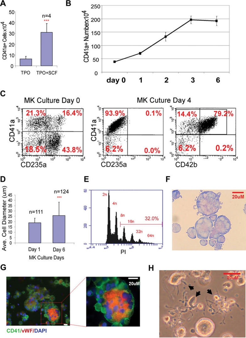
Figure 1
Generation and characterization of megakaryocytes (MKs) derived from human embryonic stem cells (hESCs). (A) Numbers of CD41a+ cells generated from 8 u00d7 10 4 hemangioblasts/blast cells derived from...
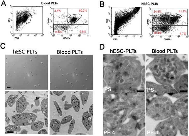
Figure 2
Characterization of platelets generated from human embryonic stem cells (hESC-PLTs). (A) The two panels show flow cytometry profiles of forward scatter (FSC) and side scatter (SSC; left), and CD41a an...
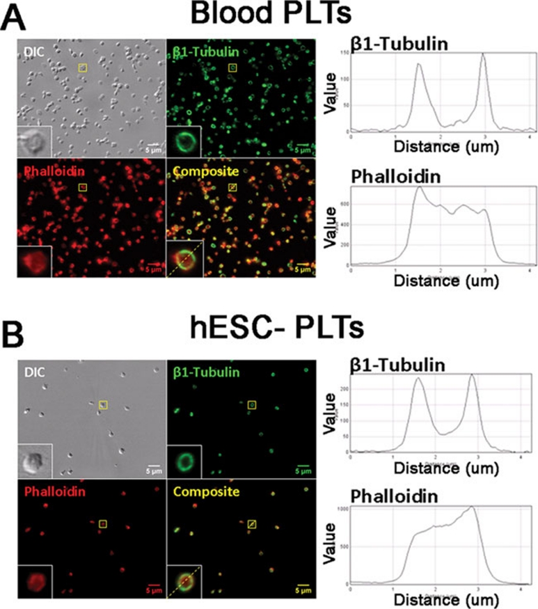
Figure 3
Tubulin and filamentous actin staining in resting blood platelets and hESC-PLTs. Tubulin (green) and filamentous actin staining (red) of resting, blood platelets (A) and hESC-PLTs (B) . Images are pre...
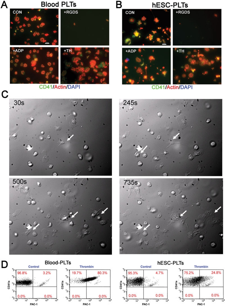
Figure 4
Functional characterization of hESC-PLTs in vitro . (A) Human blood platelets and (B) hESC-PLTs spread on fibrinogen surface: microtiter chamber slides were coated with 100 u03bcg/ml fibrinogen and hu...
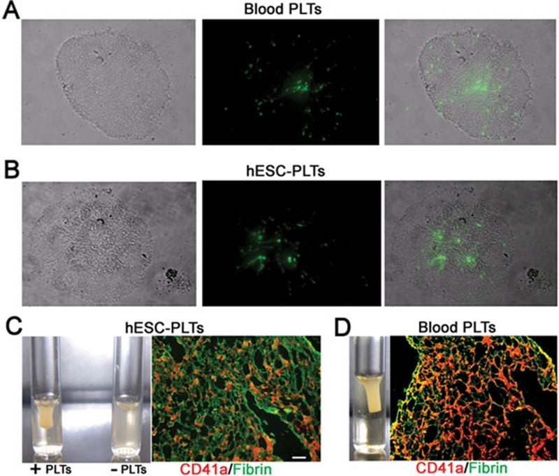
Figure 5
Functional characterization of hESC-PLTs in vitro . ( A and B ) Aggregation assay: 3 u00d7 10 5 PKH67 (green) labeled blood platelets (A) or 3 u00d7 10 5 PKH67 labeled hESC-PLTs (B) were mixed with un...
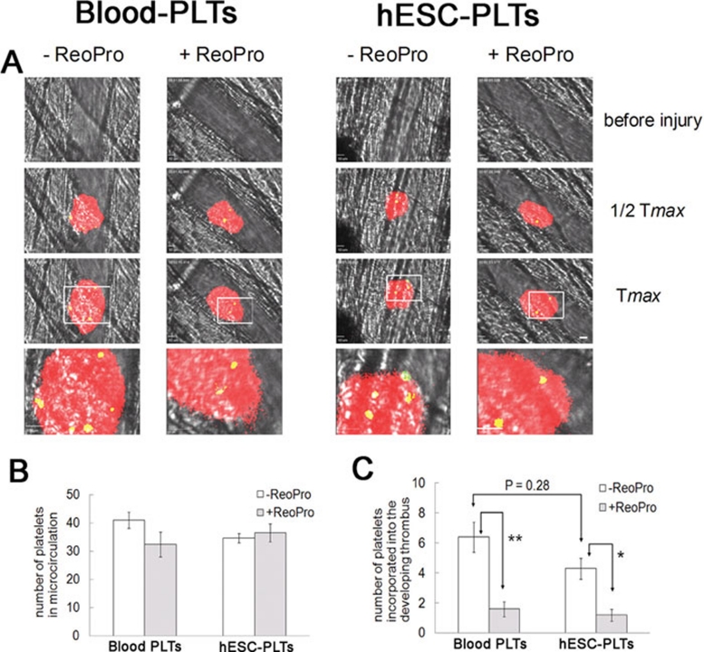
Figure 6
Incorporation of hESC-PLTs into developing mouse platelet thrombi at sites of laser-induced arteriolar injury in living mice. Calcein AM-labeled human blood platelets or hESC-derived platelets, 50-100...
Figure images are served from the NIH/NLM PubMed Central Open Access Subset or Europe PMC; copyright remains with the publishers and authors.
💬 Discussion
0 commentsNo comments yet. Be the first to start a discussion!
Leave a Comment