Abstract
Cell division is characterized by a sequence of events by which a cell gives rise to two daughter cells. Quantitative measurements of cell-cycle dynamics in single cells showed that despite variability in G1-, S-, and G2 phases, duration of mitosis is short and remarkably constant. Surprisingly, there is no correlation between cell-cycle length and mitotic duration, suggesting that mitosis is temporally insulated from variability in earlier cell-cycle phases. By combining live cell imaging and computational modeling, we showed that positive feedback is the molecular mechanism underlying the temporal insulation of mitosis. Perturbing positive feedback gave rise to a sluggish, variable entry and progression through mitosis and uncoupled duration of mitosis from variability in cell cycle length. We show that positive feedback is important to keep mitosis short, constant, and temporally insulated and anticipate it might be a commonly used regulatory strategy to create modularity in other biological systems.
🔬 Techniques
🔭 Microscopes
✨ Fluorophores
🧪 Sample Preparation
🔬 Cell Lines
🏭 Microscope Brands
🧪 Reagent Suppliers
💻 Software Details
🏛️ Research Organizations (ROR)
Affiliated research institutions:
📋 Methods
Cell Lines
All the experiments in this study were performed in human MCF10A, RPE, and HeLa cell lines. Details on growth and maintenance of all the cell lines used can be found in Supplemental Experimental Procedures . Biosensors, shRNAs, and Establishment of Stable Lines cDNAs for Histone H2B fused to Cerulean, Cdt1 (amino acid [aa], 30–120) ( Sakaue-Sawano et al., 2008 ) fused to YFP, PCNA fused with RFP ( Sporbert et al., 2005 ), Cyclin B1 fused with YFP ( Santos et al., 2012 ), Cdk1-AF, Cdk1-wt ( Santos et al., 2012 ), Cdc25C-wt, and Cdc25C-Catalytic dead (C377S) ( Santos et al., 2012 ), NLS (×3) fused to mCherry ( Santos et al., 2012 ) were all cloned into the lentiviral vector CSII-EF-1-MCS-2 by restriction digestion and ligation reactions. The CSII-EF-1-MCS-2 plasmid is a modified CSII-EF-1-MCS backbone vector where a linker TCGAAGCTAGCCCTGCAGGTTAATTAAC has been added to the MCS to increase the number of unique restriction sites. Stable MCF10A, RPE, and HeLa cells lines were made with the following combination of cell-cycle biosensors: Cdt1-YFP, PCNA-mCherry, and H2B-Cerulean or Cyclin B1-YFP and NLS3-mCherry and H2B-CFP. Lentivirus production was carried out in 293T cells transfected with DNA of interest and lentivirus assembly vectors (PAX2 and VSV-G) using with Polyethylenimine (PEI). Cells were infected for 12 hr using polybrene (8 μg). 72 hr post-infection, transduced cells were sorted on a Becton Dickinson FACSAria III influx to obtain pure populations expressing the desired fluorescent reporters. For creation of shMad2 stable lines, a set of two shRNA (GIPZ lentiviral shRNA Pool, Dharmacon, Thermo Scientific) specific to Mad2 in lentiviral constructs were used (clone V3LHS_327851: TGCTGTTGACAGTGAGCGCCTGGTTGTAGTTATCTCAAATTAGTGAAGCCACAGATGTAATTTGAGATAACTACAACCAGTTGCCTACTGCCTCGGA and clone V3LHS_403761:TGCTGTTGACAGTGAGCGCATGGATATTTGTACTGTTTAATAGTGAAGCCACAGATGTATTAAACAGTACAAATATCCATTTGCCTACTGCCTCGGA). Stable lines expressing shEmpty (pGIPZ, Dharmacon, Thermo Scientific) vector and shScramble (GIPZ non-silencing shRNA control. Sequence: TGCTGTTGACAGTGAGCGATCTCGCTTGGGCGAGAGTAAGTAGTGAAGCCACAGATGTACTTACTCTCGCCCAAGCGAGAGTGCCTACTGCCTCGGA) were used as controls for experiments with shMad2. MCF10A, RPE, and HeLa cells were infected with the pool of two shRNAs. Transduced cells were selected with 2 μg/mL of puromycin. Inhibitors The inhibitors used in this study were: Wee1/Myt1 inhibitor, PD 166285, (at 0.5 μM, 1 μM, and 2 μM), SAC inhibitor, Reversine, (at 1 μM), and Leptomycin B (at 100 ng/mL).
Show full methods section
Cell Lines
All the experiments in this study were performed in human MCF10A, RPE, and HeLa cell lines. Details on growth and maintenance of all the cell lines used can be found in Supplemental Experimental Procedures . Biosensors, shRNAs, and Establishment of Stable Lines cDNAs for Histone H2B fused to Cerulean, Cdt1 (amino acid [aa], 30–120) ( Sakaue-Sawano et al., 2008 ) fused to YFP, PCNA fused with RFP ( Sporbert et al., 2005 ), Cyclin B1 fused with YFP ( Santos et al., 2012 ), Cdk1-AF, Cdk1-wt ( Santos et al., 2012 ), Cdc25C-wt, and Cdc25C-Catalytic dead (C377S) ( Santos et al., 2012 ), NLS (×3) fused to mCherry ( Santos et al., 2012 ) were all cloned into the lentiviral vector CSII-EF-1-MCS-2 by restriction digestion and ligation reactions. The CSII-EF-1-MCS-2 plasmid is a modified CSII-EF-1-MCS backbone vector where a linker TCGAAGCTAGCCCTGCAGGTTAATTAAC has been added to the MCS to increase the number of unique restriction sites. Stable MCF10A, RPE, and HeLa cells lines were made with the following combination of cell-cycle biosensors: Cdt1-YFP, PCNA-mCherry, and H2B-Cerulean or Cyclin B1-YFP and NLS3-mCherry and H2B-CFP. Lentivirus production was carried out in 293T cells transfected with DNA of interest and lentivirus assembly vectors (PAX2 and VSV-G) using with Polyethylenimine (PEI). Cells were infected for 12 hr using polybrene (8 μg). 72 hr post-infection, transduced cells were sorted on a Becton Dickinson FACSAria III influx to obtain pure populations expressing the desired fluorescent reporters. For creation of shMad2 stable lines, a set of two shRNA (GIPZ lentiviral shRNA Pool, Dharmacon, Thermo Scientific) specific to Mad2 in lentiviral constructs were used (clone V3LHS_327851: TGCTGTTGACAGTGAGCGCCTGGTTGTAGTTATCTCAAATTAGTGAAGCCACAGATGTAATTTGAGATAACTACAACCAGTTGCCTACTGCCTCGGA and clone V3LHS_403761:TGCTGTTGACAGTGAGCGCATGGATATTTGTACTGTTTAATAGTGAAGCCACAGATGTATTAAACAGTACAAATATCCATTTGCCTACTGCCTCGGA). Stable lines expressing shEmpty (pGIPZ, Dharmacon, Thermo Scientific) vector and shScramble (GIPZ non-silencing shRNA control. Sequence: TGCTGTTGACAGTGAGCGATCTCGCTTGGGCGAGAGTAAGTAGTGAAGCCACAGATGTACTTACTCTCGCCCAAGCGAGAGTGCCTACTGCCTCGGA) were used as controls for experiments with shMad2. MCF10A, RPE, and HeLa cells were infected with the pool of two shRNAs. Transduced cells were selected with 2 μg/mL of puromycin. Inhibitors The inhibitors used in this study were: Wee1/Myt1 inhibitor, PD 166285, (at 0.5 μM, 1 μM, and 2 μM), SAC inhibitor, Reversine, (at 1 μM), and Leptomycin B (at 100 ng/mL).
Microscopy and Data Analysis
Live cell imaging was performed on either ScanR, a fully motorized and automated inverted epifluorescence microscope system IX83 (Olympus) combined with cellVivo (Olympus) or IncuCyte Zoom (Essen BioScience). Both equipped with temperature, humidity, and CO 2 levels control to keep the sample integrity and perfect focus. Details of objectives and lenses used and details on imaging procedures can be found in Supplemental Information . Image analysis was done with scripts written in Matlab (Mathworks) and ImageJ (NIH). Mann-Whitney and Kolmogrov-Smirnov tests were used to estimate p values. Trend lines, R 2 , person correlation coefficient, and mean absolute deviation were calculated using Prism6.
Mathematical Modeling
In brief, the model used consists of three ODEs to simulate the time evolution of the total amount of active Cdk1 ([Cdk1 ∗ ](t)), the synthesis and destruction of the mitotic cyclins, Cyclin B ([cycB](t)), and active APC-cdc20 ([APC](t)). The detailed information on the model construction (equations and parameters used), noise implementation, and the setup of the numerical simulations can be found in the Supplemental Information .
Supplemental Information Document S1. Supplemental Experimental Procedures and Figures S1–S7 Document S2. Article plus Supplemental Information
📊 Figures
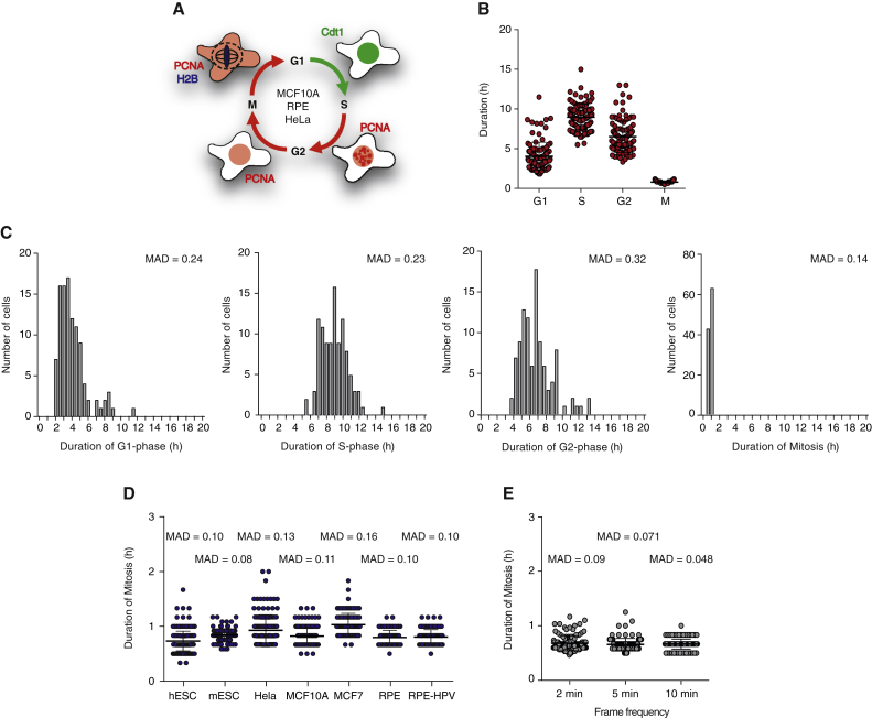
Figureu00a01
Duration of Mitosis Is Short and Constant (A) Schematic of cell lines and biosensors used to measure cell-cycle dynamics in single cells. (B) Duration of G1-, S-, G2-, and M-phases in single MCF10A ce...
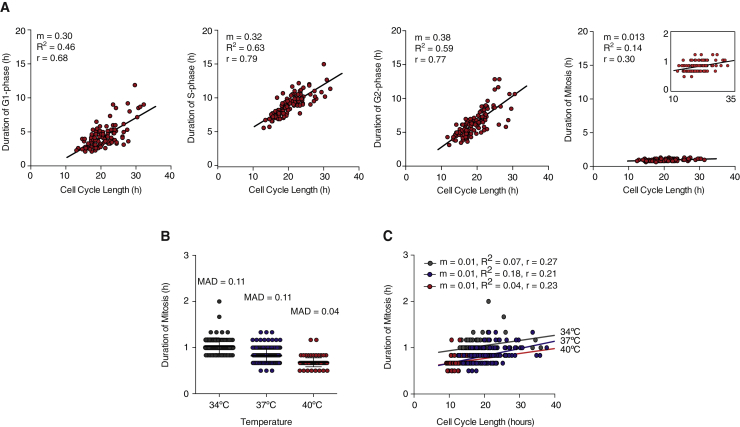
Figureu00a02
Duration of Mitosis Is Independent of Variability in Cell Cycle Length (A) Duration of G1-, S-, G2-, and M-cell-cycle phases in single cells as a function of cell-cycle length measured by single cell ...
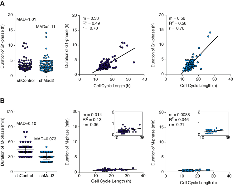
Figureu00a03
Perturbing the Spindle Assembly Does Not Make Mitotic Duration Variable nor Dependent on Cell Cycle Length (A) Left: duration of G1-phase measured in single cells in the presence (shControl) and absen...
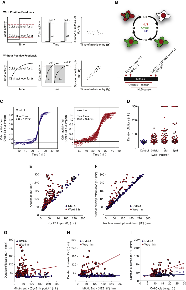
Figureu00a04
Positive Feedback Keeps Mitosis Temporally Insulated from Upstream Cell Cycle Events (A) Schematic of the thought experiment to test importance of positive feedback in keeping duration of mitosis cons...
Figureu00a05
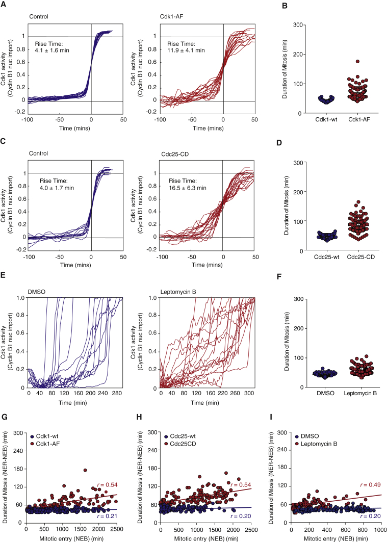
Breaking Cdk1 Activation and Spatial Positive Feedbacks Couples Duration of Mitosis to Upstream Cell-Cycle Events (A) Quantification of Cdk1 activation over time in cells expressing Cdk1-wt (blue) or ...
Figureu00a06
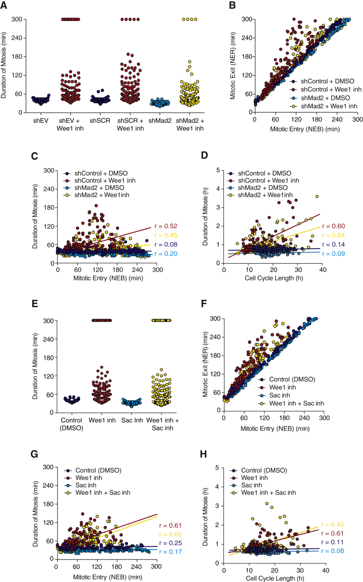
SAC Does Not Contribute to Duration of Mitosis Being Temporally Insulated from Duration of Upstream Cell-Cycle Events (A) Duration of mitosis (measured by the time between NEB and NER) in the presence...
Figureu00a07
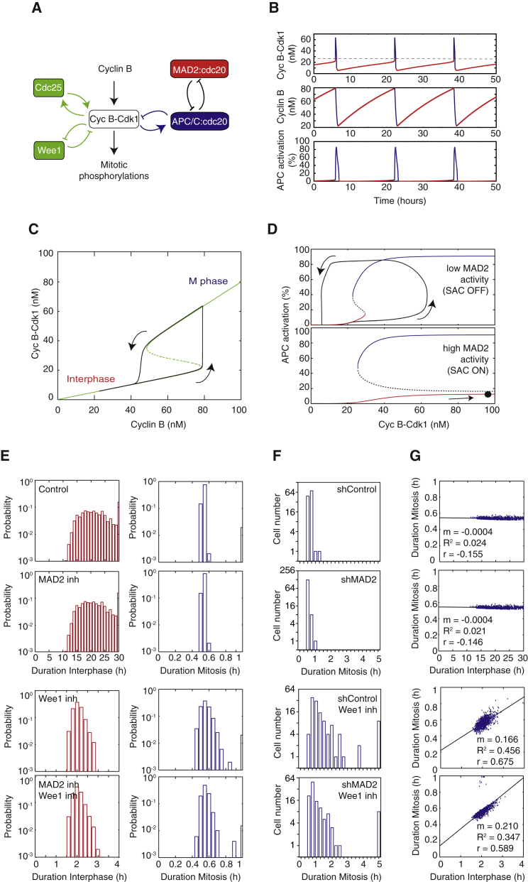
ODE Modeling Predicts that Positive Feedback Promotes Temporal Modularity in Mitosis (A) Wiring diagram showing a simplified Cdk1 regulatory network, including positive and negative feedback loops. (B...
Figure images are served from the NIH/NLM PubMed Central Open Access Subset or Europe PMC; copyright remains with the publishers and authors.
💬 Discussion
0 commentsNo comments yet. Be the first to start a discussion!
Leave a Comment