Abstract
Rabies virus (RABV) is a neurotropic virus that depends on long distance axonal transport in order to reach the central nervous system (CNS). The strategy RABV uses to hijack the cellular transport machinery is still not clear. It is thought that RABV interacts with membrane receptors in order to internalize and exploit the endosomal trafficking pathway, yet this has never been demonstrated directly. The p75 Nerve Growth Factor (NGF) receptor (p75NTR) binds RABV Glycoprotein (RABV-G) with high affinity. However, as p75NTR is not essential for RABV infection, the specific role of this interaction remains in question. Here we used live cell imaging to track RABV entry at nerve terminals and studied its retrograde transport along the axon with and without the p75NTR receptor. First, we found that NGF, an endogenous p75NTR ligand, and RABV, are localized in corresponding domains along nerve tips. RABV and NGF were internalized at similar time frames, suggesting comparable entry machineries. Next, we demonstrated that RABV could internalize together with p75NTR. Characterizing RABV retrograde movement along the axon, we showed the virus is transported in acidic compartments, mostly with p75NTR. Interestingly, RABV is transported faster than NGF, suggesting that RABV not only hijacks the transport machinery but can also manipulate it. Co-transport of RABV and NGF identified two modes of transport, slow and fast, that may represent a differential control of the trafficking machinery by RABV. Finally, we determined that p75NTR-dependent transport of RABV is faster and more directed than p75NTR-independent RABV transport. This fast route to the neuronal cell body is characterized by both an increase in instantaneous velocities and fewer, shorter stops en route. Hence, RABV may employ p75NTR-dependent transport as a fast mechanism to facilitate movement to the CNS.
🔬 Techniques
✨ Fluorophores
🧪 Sample Preparation
🔬 Cell Lines
🏭 Microscope Brands
🧪 Reagent Suppliers
📷 Detectors
💻 Software Details
🏛️ Research Organizations (ROR)
Affiliated research institutions:
📋 Methods
Animals and Dorsal Root Ganglion explants
ICR mice were bred and maintained at the Tel Aviv University animal care facility until the time of sacrifice. Spinal cords were dissected from E12.5–13.5 mice, followed by separation of Dorsal Root Ganglia (DRG) from meninges and additional spinal cord. The Institutional Animal Care Committee at the Tel Aviv University approved all the animal protocols in this work.
Microfluidic chamber preparation
Microfluidic chambers were fabricated using methods previously described in detail [56] . All microfluidic chambers were replica molded using PDMS (#41201841 Dow Corning) from masters that were patterned using the photosensitive epoxy SU-8 (Microchem). All masters consisted of two permanent SU-8 layers on a 3″ silicon wafer and were made in the clean room facility in Tel-Aviv University. The first layer of SU-8 (3 µm depth) contained the microgrooves, which were patterned by photolithography using a high-resolution chromium mask (5 µm minimum feature size; Advance Reproduction Corp.). The second layer of SU-8 (100 µm depth) contained the compartments, which were patterned by photolithography using a 20,000 dpi printed transparency mask (CAD/Art Services, Inc.). Chamber dimensions: channels: length 8.25 mm, width 1.5 mm; grooves: length 400 µm, width 15 µm, height 5 µm ( Fig. 1B ). A single 7 mm well was punctured into the “proximal” or explant channel, into which a “cave” was carved using a scalpel, to prevent explants from floating, 2 additional 1.2 mm wells were punctured into the channel on either side of the “explant” well to allow flow. Two 7 mm wells were punctured into both ends of the “distal” or axons channel to allow control over the distal channel. Microfluidic devices were cleaned of surface particles using adhesive tape and sterilized in 70% high-grade ethanol for 1 h. Devices were allowed to completely air dry under sterile conditions, attached to sterile 50 mm glass bottom dishes (FD5040-100, WPI) using gentle pressure and heated to 70°C for 20′ to improve adhesion to glass. Chambers were coated using 150 µl of 1.5 ng/ml Polyornithine (P-8638, Sigma) in PBS for 24 hours, which was replaced with 150 µl Laminin (L-2020, Sigma) 1∶333 in DDW for 24 hours. Laminin was replaced with culture medium until plating (1–3 days). Assembly and maintenance of DRG explants At the day of plating, media were removed from all wells, and a single DRG was inserted to each explant “cave” using a 20 µl tip. Following 1 hour of incubation at 37°C, 150 µl of medium were added to each well. Basic culture medium consisted of Neurobasal medium (Life Technologies) supplemented with 2% B-27 (Life Technologies), 1% penicillin-streptomycin (Biological Industries, Israel) and 1% Glutamax (Life Technologies). A gradient of murine NGF was created in order to encourage axons to cross the grooves to the distal axon compartment, by adding 100 ng/ml and 62.5 ng/ml murine NGF (Alomone labs) to the distal and proximal wells, respectively. Cultures were maintained at 37°C and 5% CO 2 , and media were refreshed every 2 days. Transport assays were initiated after axons had crossed the grooves and established an axonal network in the distal compartment, 3–5 days from plating ( Fig. 1D ). For TIRF imaging assays, 4–5 DRG were placed on 35 mm glass bottom dishes (FD35-100, WPI) with 20 µl each of DRG culture medium supplemented with 62.5 ng/ml NGF. DRG were incubated in 37°C for 4–5 hours to allow adhesion to glass, then supplemented with 2 ml DRG culture medium supplemented with 62.5 ng/ml NGF. Cultures were maintained at 37°c and 5% CO 2 for 48 hours before imaging. Fluorescent EGFP/mCherry rabies virus production rRABV EGFP-P ΔG/rRABV mCherry-P ΔG are recombinant rabies viruses in which EGFP or mCherry fluorescent reporters were fused to the phosphoprotein P and in which the glycoprotein G gene was deleted. Preparation and amplification was performed as previously described [14] , [57] . In brief, MG-136 cells, expressing the RABV matrix and glycoproteins after induction with doxycycline, were infected with rRABV EGFP-P ΔG. Cells were split, after which media were twice replaced with fresh medium supplemented with 1 mg/ml Doxycycline, to be collected 48 hours later. rRABV EGFP-P ΔG was concentrated from cell culture supernatants using the PEG virus precipitation kit (ab102538, Abcam). EGFP/sh-RNA-p75-EGFP lentivirus production HEK293t cells were transfected using calcium-phosphate precipitation with the viral vector pLL-EGFP and the helper plasmids pVSVG and pGag-PolGpt (kind gift from Eran Bacharach) or mix of 4 shRNA plasmids against murine p75, produced from Mouse GIPZ lentiviral target gene shRNAmir glycerol set (GE healthcare RMM4532-EG18053). Viral particles were collected 48 and 72 hours after transfection, filtered and concentrated using the PEG virus precipitation kit (ab102538, Abcam). DRG cultures, shRNA-p75 and RABV infection DRG were collected from E12.5–13.5 mice, trypsinized with Trypsin-EDTA solution B (Biological industries 03-052-1B) for 5 minutes in 37°C, and washed with complete F12 medium. Cells were dissociated by pipetting, counted and plated on PLO/Laminin coated 96 well plates, in a density of 15K cells per well. Cells were maintained with DRG culture medium supplemented with 62.5 ng/ml NGF. On day of plating, cells were infected with either LV-EGFP or a mix of 4 LV-shRNA-p75-EGFP (MOI≈5 and MOI≈10, respectively, due to high infectivity of LV-EGFP). 3–4 days from plating, cells were infected with ≈120K mCherry RABV particles for 0′, 30′ and 120′ in duplicates after which they were washed ×3 with DRG culture medium. 48 hours later, neurons showing obvious mCherry foci were counted in 6–9 fields per well, and divided by total number of counted neurons. Although cultures varied in axon network, non-neuronal cell populations and LV-infection levels, infection rates were lower in sh-RNA-p75-EGFP groups when compared to GFP control in all experiments. Due to differences in absolute infection rates, these were normalized to the rate obtained at the LV-EGFP control group. Infection of DRG explants in microfluidic chambers were performed on the day of plating, with ≈1×10 6 LV-EGFP particles, or a mix of 4×≈1–2×10 6 LV-sh-RNA-p75-EGFP particles. To estimate knockdown of p75, a DRG culture plated on a glass bottom dish was infected with sh-RNA-p75-EGFP as described above. At 4DIV, culture was treated with 1∶100 anti-p75-550 for 15′ and washed ×3 prior to imaging using spinning disc confocal. Fluorescent beads, markers and antibodies Qdot-NGF was prepared by mixing biotinylated murine-NGF 10 µg/ml (#N-240-B, Alomone Labs) with Quantum-Dot 605 streptavidin conjugate 1 µM ( Q10101 , Molecular Probes) in a molar ratio of 3∶1, respectively. Acidic compartments were delineated using Lysotracker Red DND-99 (Life Technologies) in a final concentration of 50 nM. Mitotracker Deep Red FM (Life Technologies) was used to denote mitochondria, in a final concentration of 100 nM. In order to track the co-transport of RABV with its receptors, the fluorescent extracellular antibodies Anti-p75NTR-ATTO-550, Anti-p75-ATTO-488 and Anti-TrkA-ATTO-633 (ANT-007-AO, ANT-007-AG and ANT-0180-FR, respectively, Alomone Labs) were used in a 1∶100 dilution. Live imaging of axonal transport Live imaging was performed on DRG explants grown in compartmental chambers, 3–5 DIV, upon forming an axonal network at the distal axon compartment. Prior to imaging, media from each well were replaced with 150 µl Neurobasal medium supplemented with 1% penicillin-streptomycin and 1% Glutamax (poor medium). Following two hours of starvation, 30 µl of poor medium were added to the proximal well, to induce compartmental separation. 2 µl of concentrated EGFP-RABV (≈120K particles), Qdot-NGF or both were added to one distal well, with additional 5 µl medium, to encourage flow through the distal axon channel ( Fig. 1B,C ). Chambers were incubated for 1–2 hours in 37°C and 5% CO 2 prior their placement on the microscope stage. Time lapse images of axons in the groove area ( Fig. 1B,C ), were acquired at 37°C and CO 2 controlled environment, using Nikon Eclipse Ti microscope equipped with Yokogawa CSU X-1 spinning disc confocal, controlled via iQ software (Andor). Multi-channel time lapses were captured using 60× lens, NA = 1.4 with 2000 msec intervals, with an approximate lag of 100–400 ms between channels. Digital images were taken with Andor iXon DU-897 EM-CCD camera.
Show full methods section
Animals and Dorsal Root Ganglion explants
ICR mice were bred and maintained at the Tel Aviv University animal care facility until the time of sacrifice. Spinal cords were dissected from E12.5–13.5 mice, followed by separation of Dorsal Root Ganglia (DRG) from meninges and additional spinal cord. The Institutional Animal Care Committee at the Tel Aviv University approved all the animal protocols in this work.
Microfluidic chamber preparation
Microfluidic chambers were fabricated using methods previously described in detail [56] . All microfluidic chambers were replica molded using PDMS (#41201841 Dow Corning) from masters that were patterned using the photosensitive epoxy SU-8 (Microchem). All masters consisted of two permanent SU-8 layers on a 3″ silicon wafer and were made in the clean room facility in Tel-Aviv University. The first layer of SU-8 (3 µm depth) contained the microgrooves, which were patterned by photolithography using a high-resolution chromium mask (5 µm minimum feature size; Advance Reproduction Corp.). The second layer of SU-8 (100 µm depth) contained the compartments, which were patterned by photolithography using a 20,000 dpi printed transparency mask (CAD/Art Services, Inc.). Chamber dimensions: channels: length 8.25 mm, width 1.5 mm; grooves: length 400 µm, width 15 µm, height 5 µm ( Fig. 1B ). A single 7 mm well was punctured into the “proximal” or explant channel, into which a “cave” was carved using a scalpel, to prevent explants from floating, 2 additional 1.2 mm wells were punctured into the channel on either side of the “explant” well to allow flow. Two 7 mm wells were punctured into both ends of the “distal” or axons channel to allow control over the distal channel. Microfluidic devices were cleaned of surface particles using adhesive tape and sterilized in 70% high-grade ethanol for 1 h. Devices were allowed to completely air dry under sterile conditions, attached to sterile 50 mm glass bottom dishes (FD5040-100, WPI) using gentle pressure and heated to 70°C for 20′ to improve adhesion to glass. Chambers were coated using 150 µl of 1.5 ng/ml Polyornithine (P-8638, Sigma) in PBS for 24 hours, which was replaced with 150 µl Laminin (L-2020, Sigma) 1∶333 in DDW for 24 hours. Laminin was replaced with culture medium until plating (1–3 days). Assembly and maintenance of DRG explants At the day of plating, media were removed from all wells, and a single DRG was inserted to each explant “cave” using a 20 µl tip. Following 1 hour of incubation at 37°C, 150 µl of medium were added to each well. Basic culture medium consisted of Neurobasal medium (Life Technologies) supplemented with 2% B-27 (Life Technologies), 1% penicillin-streptomycin (Biological Industries, Israel) and 1% Glutamax (Life Technologies). A gradient of murine NGF was created in order to encourage axons to cross the grooves to the distal axon compartment, by adding 100 ng/ml and 62.5 ng/ml murine NGF (Alomone labs) to the distal and proximal wells, respectively. Cultures were maintained at 37°C and 5% CO 2 , and media were refreshed every 2 days. Transport assays were initiated after axons had crossed the grooves and established an axonal network in the distal compartment, 3–5 days from plating ( Fig. 1D ). For TIRF imaging assays, 4–5 DRG were placed on 35 mm glass bottom dishes (FD35-100, WPI) with 20 µl each of DRG culture medium supplemented with 62.5 ng/ml NGF. DRG were incubated in 37°C for 4–5 hours to allow adhesion to glass, then supplemented with 2 ml DRG culture medium supplemented with 62.5 ng/ml NGF. Cultures were maintained at 37°c and 5% CO 2 for 48 hours before imaging. Fluorescent EGFP/mCherry rabies virus production rRABV EGFP-P ΔG/rRABV mCherry-P ΔG are recombinant rabies viruses in which EGFP or mCherry fluorescent reporters were fused to the phosphoprotein P and in which the glycoprotein G gene was deleted. Preparation and amplification was performed as previously described [14] , [57] . In brief, MG-136 cells, expressing the RABV matrix and glycoproteins after induction with doxycycline, were infected with rRABV EGFP-P ΔG. Cells were split, after which media were twice replaced with fresh medium supplemented with 1 mg/ml Doxycycline, to be collected 48 hours later. rRABV EGFP-P ΔG was concentrated from cell culture supernatants using the PEG virus precipitation kit (ab102538, Abcam). EGFP/sh-RNA-p75-EGFP lentivirus production HEK293t cells were transfected using calcium-phosphate precipitation with the viral vector pLL-EGFP and the helper plasmids pVSVG and pGag-PolGpt (kind gift from Eran Bacharach) or mix of 4 shRNA plasmids against murine p75, produced from Mouse GIPZ lentiviral target gene shRNAmir glycerol set (GE healthcare RMM4532-EG18053). Viral particles were collected 48 and 72 hours after transfection, filtered and concentrated using the PEG virus precipitation kit (ab102538, Abcam). DRG cultures, shRNA-p75 and RABV infection DRG were collected from E12.5–13.5 mice, trypsinized with Trypsin-EDTA solution B (Biological industries 03-052-1B) for 5 minutes in 37°C, and washed with complete F12 medium. Cells were dissociated by pipetting, counted and plated on PLO/Laminin coated 96 well plates, in a density of 15K cells per well. Cells were maintained with DRG culture medium supplemented with 62.5 ng/ml NGF. On day of plating, cells were infected with either LV-EGFP or a mix of 4 LV-shRNA-p75-EGFP (MOI≈5 and MOI≈10, respectively, due to high infectivity of LV-EGFP). 3–4 days from plating, cells were infected with ≈120K mCherry RABV particles for 0′, 30′ and 120′ in duplicates after which they were washed ×3 with DRG culture medium. 48 hours later, neurons showing obvious mCherry foci were counted in 6–9 fields per well, and divided by total number of counted neurons. Although cultures varied in axon network, non-neuronal cell populations and LV-infection levels, infection rates were lower in sh-RNA-p75-EGFP groups when compared to GFP control in all experiments. Due to differences in absolute infection rates, these were normalized to the rate obtained at the LV-EGFP control group. Infection of DRG explants in microfluidic chambers were performed on the day of plating, with ≈1×10 6 LV-EGFP particles, or a mix of 4×≈1–2×10 6 LV-sh-RNA-p75-EGFP particles. To estimate knockdown of p75, a DRG culture plated on a glass bottom dish was infected with sh-RNA-p75-EGFP as described above. At 4DIV, culture was treated with 1∶100 anti-p75-550 for 15′ and washed ×3 prior to imaging using spinning disc confocal. Fluorescent beads, markers and antibodies Qdot-NGF was prepared by mixing biotinylated murine-NGF 10 µg/ml (#N-240-B, Alomone Labs) with Quantum-Dot 605 streptavidin conjugate 1 µM ( Q10101 , Molecular Probes) in a molar ratio of 3∶1, respectively. Acidic compartments were delineated using Lysotracker Red DND-99 (Life Technologies) in a final concentration of 50 nM. Mitotracker Deep Red FM (Life Technologies) was used to denote mitochondria, in a final concentration of 100 nM. In order to track the co-transport of RABV with its receptors, the fluorescent extracellular antibodies Anti-p75NTR-ATTO-550, Anti-p75-ATTO-488 and Anti-TrkA-ATTO-633 (ANT-007-AO, ANT-007-AG and ANT-0180-FR, respectively, Alomone Labs) were used in a 1∶100 dilution. Live imaging of axonal transport Live imaging was performed on DRG explants grown in compartmental chambers, 3–5 DIV, upon forming an axonal network at the distal axon compartment. Prior to imaging, media from each well were replaced with 150 µl Neurobasal medium supplemented with 1% penicillin-streptomycin and 1% Glutamax (poor medium). Following two hours of starvation, 30 µl of poor medium were added to the proximal well, to induce compartmental separation. 2 µl of concentrated EGFP-RABV (≈120K particles), Qdot-NGF or both were added to one distal well, with additional 5 µl medium, to encourage flow through the distal axon channel ( Fig. 1B,C ). Chambers were incubated for 1–2 hours in 37°C and 5% CO 2 prior their placement on the microscope stage. Time lapse images of axons in the groove area ( Fig. 1B,C ), were acquired at 37°C and CO 2 controlled environment, using Nikon Eclipse Ti microscope equipped with Yokogawa CSU X-1 spinning disc confocal, controlled via iQ software (Andor). Multi-channel time lapses were captured using 60× lens, NA = 1.4 with 2000 msec intervals, with an approximate lag of 100–400 ms between channels. Digital images were taken with Andor iXon DU-897 EM-CCD camera.
Axonal transport image analysis
Time-lapse image analysis was carried out using Fiji, following subtraction of average intensity z-projection to exclude completely stationary fluorescent artifacts. XY coordinates of tracks were registered from 9 or more grooves (which contains typically 2–5 axons) in each experiment, using Manual Tracking plugin, while distances, velocities and MSD's were computed using MATLAB implementation for Fiji Trackmate plugin [58] . Tracks with run lengths
📊 Figures
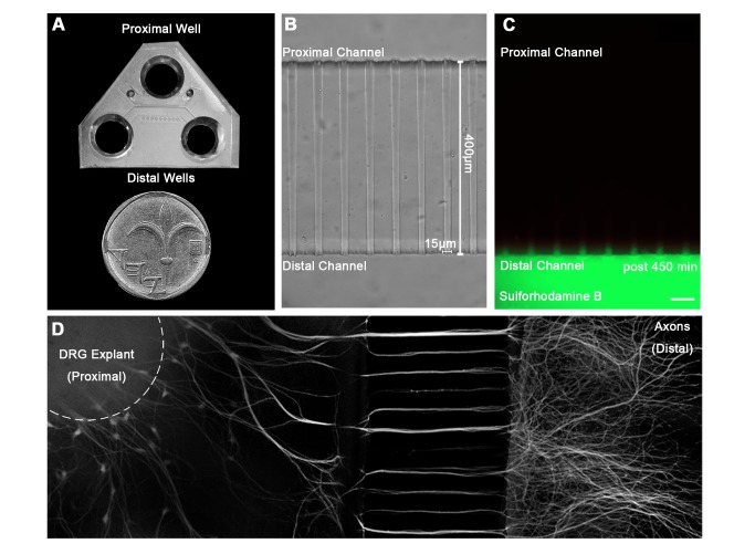
Figure 1
A microfluidic system for tracking retrograde transport in sensory axons.
( A ) A Polydimethylsiloxane (PDMS) microfluidic chamber used for explant culture. ( B,C ) 40 u00b5l interval towards the proximal compartment (top) prevents fluorescent dye from diffusing to the prox...
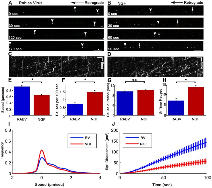
Figure 2
Rabies virus retrograde transport in DRG is faster and more directed than that of NGF.
Retrograde transport of ( A ) EGFP-RABV and ( B ) Quantum-dot conjugated NGF in DRG explants, roughly 2 hours after addition to distal axon compartment. Arrows and arrowheads pointing at transported p...
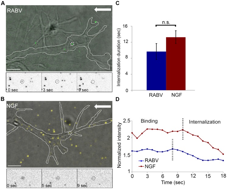
Figure 3
RABV and NGF present similar internalization kinetics at the axon tip.
Live TIRF microscopy was used to track RABV and NGF internalization in DRG neuron tips. ( A ) EGFP-RABV (dashed circles) is detected on the surface of a neuron tip (white line). Lower panels present a...
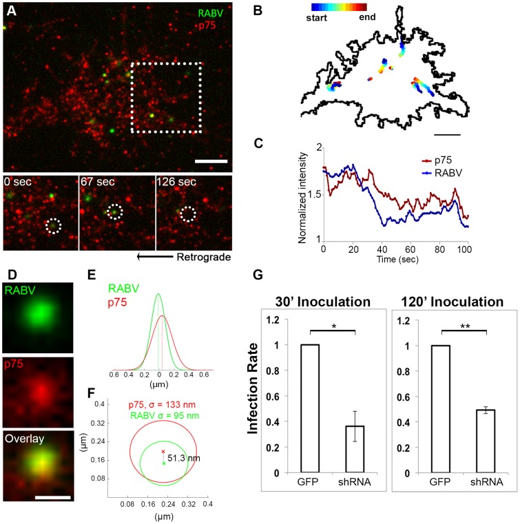
Figure 4
RABV binds and internalizes with p75NTR in DRG neuron tips.
Co-localization of EGFP-RABV with p75NTR is shown by live TIRF imaging and sub-pixel localization algorithms. ( A ) RABV-p75 particles shift from the periphery to the center of the growth cone, where ...
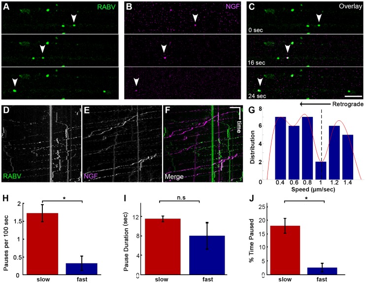
Figure 5
Co-transport of RABV and NGF.
( Au2013C ) EGFP-RABV and Qdot-NGF were simultaneously added to the distal compartment of a DRG explant at 3DIV. Dual-channel live imaging revealed multiple events of RABV co-transported with NGF, ill...
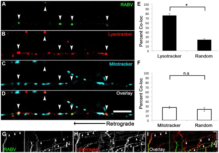
Figure 6
RABV is transported in acidic compartments in sensory axons.
( Au2013D ), Multi-channel live imaging of EGFP-RABV retrograde transport in DRG axons, along with the fluorescent cellular markers Lysotracker Red and Mitotracker Deep Red. Arrowheads: RABV particles...
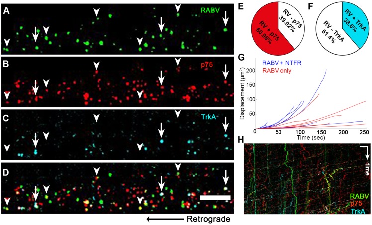
Figure 7
RABV is retrogradely transported with neurotrophin receptors.
( Au2013D ), Retrograde transport of EGFP-RABV, added to the distal axon compartment of DRG explant previously treated with fluorescent antibodies against p75NTR and TrkA. Arrowheads: RABV puncta posi...
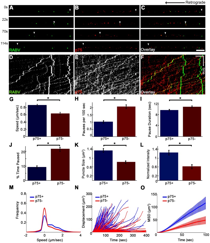
Figure 8
RABV travels faster and is more directed when transported with p75NTR.
( Au2013C ) Multi-channel live imaging of EGFP-RABV 2 hours after addition to distal axon compartment of DRG explant previously treated with a fluorescent antibody against p75NTR. Arrowheads: p75NTR-p...
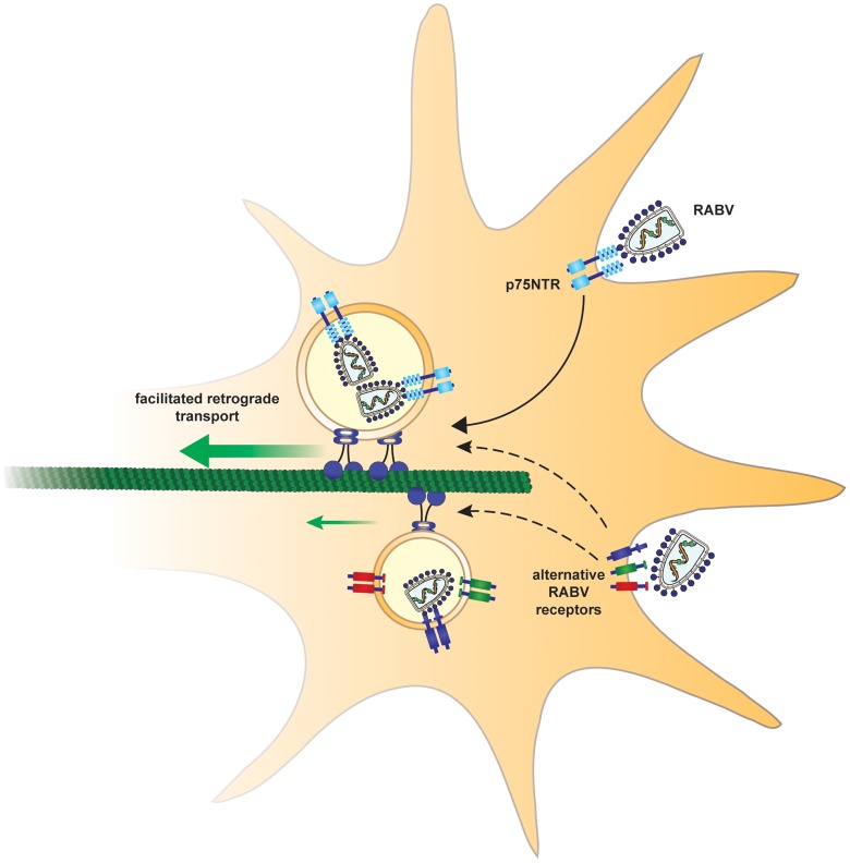
Figure 9
Suggested model.
In order to arrive at the cell body and subsequently the CNS, rabies virus hijacks a fast route using the p75NTR endosomal pathway. In a p75NTR dependent path, RABV manipulates the axonal transport ma...
Figure images are served from the NIH/NLM PubMed Central Open Access Subset or Europe PMC; copyright remains with the publishers and authors.
💬 Discussion
0 commentsNo comments yet. Be the first to start a discussion!
Leave a Comment