Abstract
Abstract Background A central feature of Alzheimer's disease is the cleavage of the amyloid precursor protein (APP) to form beta-amyloid peptide (Aβ) by the β-secretase and γ-secretase enzymes. Although this has been shown to occur after endocytosis of APP from the cell surface, the exact compartments of APP processing are not well defined. We have previously demonstrated that APP and γ-secretase proteins and activity are highly enriched in purified rat liver lysosomes. In order to examine the lysosomal distribution and trafficking of APP in cultured cells, we generated constructs containing APP fused to a C-terminal fluorescent protein tag and N-terminal HA-epitope tag. These were co-transfected with a panel of fluorescent-protein tagged compartment markers. Results Here we demonstrate using laser-scanning confocal microscopy that although APP is present throughout the endosomal/lysosomal system in transfected Cos7 and neuronal SN56 cell lines as well as in immunostained cultured mouse neurons, it is enriched in the lysosome. We also show that the Swedish and London mutations reduce the amount of APP in the lysosome. Surprisingly, in addition to its expected trafficking from the cell surface to the early and then late endosomes, we find that cell-surface labelled APP is transported rapidly and directly from the cell surface to lysosomes in both Cos7 and SN56 cells. This rapid transit to the lysosome is blocked by the presence of either the London or Swedish mutations. Conclusions These results demonstrate the presence of a novel, rapid and specific transport pathway from the cell surface to the lysosomes. This suggests that regulation of lysosomal traffic could regulate APP processing and that the lysosome could play a central role in the pathophysiology of Alzheimer's disease.
🔬 Techniques
🔭 Microscopes
✨ Fluorophores
🧪 Sample Preparation
🔬 Cell Lines
🏭 Microscope Brands
🧪 Reagent Suppliers
🔴 Lasers
💻 Software Details
🏛️ Research Organizations (ROR)
Affiliated research institutions:
📋 Methods
Antibodies and Reagents
Cos7 cells were obtained from Dr. Stephen Ferguson. SN56 cells were obtained from Dr. Jane Rylett [ 30 - 32 ]. Serum for cell culture was purchased from Hyclone and cell culture media and reagents were purchased from Invitrogen. Antibodies used in this study were: rabbit anti-APP-C-terminal (Sigma), monoclonal mouse anti-LAMP1 (A3H4) and rat anti-LAMP1 (1D4B) (Developmental Studies Hybridoma Bank), monoclonal anti-Rab5 (BD Biosciences), monoclonal anti-Rab9 (Affinity Bioreagents), and monoclonal anti-HA antibody (12CA5) (Roche Applied Science). Fluorescently-labeled secondary antibodies, AlexaFluor 488 goat anti-rabbit and AlexaFluor 633 donkey anti-mouse were purchased from Invitrogen.
DNA Constructs
A cDNA encoding APP 750-YFP was a generous gift of Dr. Bradley Hyman. HA-labeled APP constructs were then generated by PCR, first cloning sequence encoding the 17 amino acid signal sequence of APP as well as the L-E residues required for signal peptide cleavage [ 55 ] with a forward primer engineered to add an Nhe1 site and a reverse primer encoding the HA sequence and appending an Mlu1 site. The remainder of the cDNA was cloned using PCR primers to place Mlu1 at the 3-prime end and a Sal1 site at the 5-prime end. These 2 products were then ligated into pEYFP-N1 or pECFP-N1 vectors (Clontech). To reduce the possibility of cleavage of APP by a non-secretase enzyme, we generated a shorter constructs using a forward APP primer which would amplify the sequence coding for the C-terminal 112 amino acids (12 amino acids upstream of the β-cleavage site) and append a short 14 amino acid spacer and an Mlu1 site. This construct is referred to as 'βAPP'. Constructs similar to βAPP have been demonstrated to undergo both beta- and gamma-cleavage [ 56 ]. We engineered London and Swedish mutations into these constructs using PCR (Figure 1 ). LAMP1-YFP was a generous gift from Dr. Walter Mothes and recloned to use mRFP. Cox8-mRFP was a gift from Dr. Mark Huttemann. Plasmids containing mCherry were a gift from Dr. Roger Tsien. Rab9-YFP was obtained from Dr. Susanne Pfeffer and re-cloned to use mCherry.
Show full methods section
Antibodies and Reagents
Cos7 cells were obtained from Dr. Stephen Ferguson. SN56 cells were obtained from Dr. Jane Rylett [ 30 - 32 ]. Serum for cell culture was purchased from Hyclone and cell culture media and reagents were purchased from Invitrogen. Antibodies used in this study were: rabbit anti-APP-C-terminal (Sigma), monoclonal mouse anti-LAMP1 (A3H4) and rat anti-LAMP1 (1D4B) (Developmental Studies Hybridoma Bank), monoclonal anti-Rab5 (BD Biosciences), monoclonal anti-Rab9 (Affinity Bioreagents), and monoclonal anti-HA antibody (12CA5) (Roche Applied Science). Fluorescently-labeled secondary antibodies, AlexaFluor 488 goat anti-rabbit and AlexaFluor 633 donkey anti-mouse were purchased from Invitrogen.
DNA Constructs
A cDNA encoding APP 750-YFP was a generous gift of Dr. Bradley Hyman. HA-labeled APP constructs were then generated by PCR, first cloning sequence encoding the 17 amino acid signal sequence of APP as well as the L-E residues required for signal peptide cleavage [ 55 ] with a forward primer engineered to add an Nhe1 site and a reverse primer encoding the HA sequence and appending an Mlu1 site. The remainder of the cDNA was cloned using PCR primers to place Mlu1 at the 3-prime end and a Sal1 site at the 5-prime end. These 2 products were then ligated into pEYFP-N1 or pECFP-N1 vectors (Clontech). To reduce the possibility of cleavage of APP by a non-secretase enzyme, we generated a shorter constructs using a forward APP primer which would amplify the sequence coding for the C-terminal 112 amino acids (12 amino acids upstream of the β-cleavage site) and append a short 14 amino acid spacer and an Mlu1 site. This construct is referred to as 'βAPP'. Constructs similar to βAPP have been demonstrated to undergo both beta- and gamma-cleavage [ 56 ]. We engineered London and Swedish mutations into these constructs using PCR (Figure 1 ). LAMP1-YFP was a generous gift from Dr. Walter Mothes and recloned to use mRFP. Cox8-mRFP was a gift from Dr. Mark Huttemann. Plasmids containing mCherry were a gift from Dr. Roger Tsien. Rab9-YFP was obtained from Dr. Susanne Pfeffer and re-cloned to use mCherry.
Cell Culture and transfection
SN56 cells and COS7 cells were grown in Dulbecco's minimal Eagle's medium (DMEM), respectively supplemented with 5% (v/v) and 10% (v/v) heat-inactivated fetal bovine serum (Hyclone) respectively, and 100 μg/ml penicillin/streptomycin (Invitrogen). Cells were seeded at a density of 2.5 × 10 6 cells/100-mm dish (Falcon). Cells were transiently transfected using Lipofectamine following manufacturer's instructions (Invitrogen). Following transfection (18 h), the cells were pooled and reseeded into 35-mm glass-bottomed culture dishes (MatTek) for confocal studies. SN56 cells were grown as above, but were differentiated 24 hours before imaging by the addition of 1 mM dibutyryl cyclic AMP (dbcAMP; Sigma) and changed to serum free medium [ 30 - 32 ]. Primary prefrontal cortical neurons were prepared from E18 mouse embryos as described previously [ 57 ], then were seeded in poly-L-ornithine coated plates and maintained in Neurobasal medium supplemented with 1× B27 and 0.8× N2 supplements, 2 mM glutamax and 50 U/ml penicillin/streptomycin (Invitrogen). They were kept at 37°C in a humidified atmosphere containing 5% CO 2 . One third to one half of the volume of neurobasal media was replenished every 3 days. After a growth period of 8-15 days, neurons were processed for immunofluorescence. The University of Western Ontario Animal Care Committee approved all animal protocols.
Immunocytochemistry
Cells were washed twice in Hanks' balanced salt solution (HBSS; 1.2 mM KH 2 PO 4 , 5 mM NaHCO 3 , 20 mM HEPES, 11 mM glucose, 116 mM NaCl, 4.7 mM KCl, 1.2 mM MgSO 4 , 2.5 mM CaCl 2 , pH 7.4) and fixed for 15 min. in fresh methanol-free 4% paraformaldehyde (Electron Microscopy Supply) in PBS. Cells were permeablized with 0.02% Triton in PBS for 10 min and blocked with 3% BSA in PBS for 1 h. Cells were incubated with primary antibody for 1 hour, washed in PBS twice, incubated with secondary antibody for 1 h. After 2 washes with PBS, cells were stained with Hoechst nuclear stain (Sigma) in PBS for 5 min at room temperature and mounted on slides with ImmuMount (Fisher) Confocal Microscopy Imaging was performed on a Zeiss LSM-510 META laser-scanning microscope using a Zeiss 63× 1.4 numerical aperture oil immersion lens. The optical section thickness was typically 1 micron. AlexaFluor 488, EGFP and YFP fluorescence was visualized using a 488 nm excitation laser and a 500-530-nm emission filter set. AlexaFluor 547 and mRFP fluorescence was imaged using a 543 nm excitation laser and BP 565-615 filter set. ECFP fluorescence was imaged using 458 nm laser excitation source and a BP 475-525 filter set. AlexaFluor 647 fluorescence was imaged using 633 nm excitation laser, and a LP 650 filter. Hoechst signal was collected using a Chameleon Multiphoton laser set at 750 nm excitation, and a 390-465 emission filter set.
Cell Surface labeling
Anti-HA antibody was labeled with AlexaFluor 647 using a Zenon labeling kit (Invitrogen) following manufacturer's directions. Live cell imaging was performed in HBSS at 37°C on a BC200 microscope stage warmer with a Bionomic BC100 controller from (20/20 Technologies). Cells normal morphology and strong expression of APP and compartment markers were identified for imaging. In live cell imaging experiments, and images were typically taken at 1-2 frames/minute. For fixed time-course studies, freshly prepared conjugate (10 μg of antibody/ml) was incubated with cells in DMEM on ice for 30 minutes. Conjugate was removed and the cells are washed in cold PBS. Pre-warmed media was then added and cells were incubated at 37°C for the times indicated prior to fixation with 4% paraformaldehyde. Cells were chosen which had strong expression of both the APP and the compartment marker constructs, but had normal morphology and no inclusions. βAPP time courses were performed at least 3 times, with at least 4 representative cells imaged at each time point. For dextran uptake time courses, AlexaFluor 647-labeled 10 kDa Dextran (Invitrogen) was added to media at 100 μg/ml and cells were incubated for the indicated length of time. Media was then aspirated, cells were washed in ice-cold PBS and fixed 4% paraformaldehyde.
Colocalization Analysis
Colocalization analysis was performed on confocal optical sections using Imaris 6.1.5 with Imaris Colocalization module (Biplane) running on an Apple Mac Pro to examine the colocalization of the brightest 2% of pixels in each channel. This allows us to set threshold for colocalization in an unbiased manner using the intrinsic properties of the image, eliminating confounding problems caused by variations in cell-to-cell expression and image brightness/exposure thus allowing direct comparison between experiments. Graphing and statistical analysis was performed using Prism GraphPad 5.0b using one-way ANOVA with Tukey post-test.
Supplementary Material Additional file 1 Image stack demonstrating APP preferentially localized in the lysosome a neuronal SN56 cell . SN56 cells were transiently co-transfected with βAPP-CFP (green) and LAMP1-mRFP (red). Image stack was acquired on a Zeiss LSM510 confocal microscope. The first segment shows βAPP-CFP in green. The second segment of the video shows LAMP1-mrfp overlaid in red. In the third segment, a colocalization channel, showing the colocalization of the brightest 2% of red and green pixels is overlaid in white. QuickTime video was generated using Imaris 6.1.5 software to perform volume rendering and animation. Click here for file Additional file 2 Image stack of an immunostained mouse neuron demonstrating APP in the lysosome . Cultured mouse neurons were fixed and immunolabeled with anti-APP (green) and anti-LAMP1 antibodies (red) and Hoechst nuclear stain (blue). Image stack was acquired on a Zeiss LSM510 confocal microscope. The first segment shows βAPP-CFP in green. The second segment of the video shows LAMP1-mRFP overlaid in red. In the third segment, a channel showing the colocalization of the brightest 2% of red and green pixels is overlaid in white. QuickTime video was generated using Imaris 6.1.5 software to perform volume rendering and animation. Click here for file Additional file 3 Movie demonstrating the internalization of FL-APP to the lysosome in a Cos7 cell . Cos7 cells were transiently transfected with FLAPP-CFP (not shown) and LAMP1-mRFP (red). Fluorescent-labelled anti-HA antibody was added to the media, and a cell was chosen which had good expression of transfected plasmids. Images were acquired using laser-scanning confocal microscopy at approximately 2 frames/min. QuickTime video was generated using Imaris 6.1.5 software. Click here for file Additional file 4 Movie demonstrating the internalization of βAPP to the lysosome in a Cos7 cell . Cos7 cells were transiently transfected with βAPP-CFP (not shown) and LAMP1-mRFP (red). Fluorescent-labelled anti-HA antibody was added to the media, and a cell was chosen which had good expression. Images were acquired using laser-scanning confocal microscopy at approximately 2 frames/min. QuickTime video was generated using Imaris 6.1.5 software. Click here for file
📊 Figures
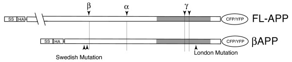
Figure 1
Overview of Constructs . Shaded region denotes the transmembrane domain. Beta-, alpha- and gamma symbols denote secretase cleavage sites. Swedish and London mutations are shown. SS - signal sequence; ...
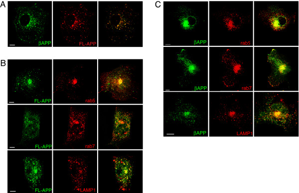
Figure 2
APP is preferential expressed in the lysosome in Cos7 cells . Cos7 cells were transiently co-transfected with the fluorescent -tagged APP and compartment marker proteins and imaged using laser scannin...
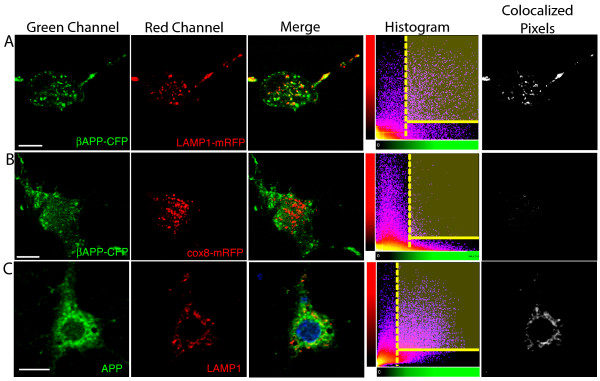
Figure 3
Quantitation of APP enrichment in transfected cells and primary neurons . SN56 cells (A-B) were transfected with transfected with APP-CFP (green channel) and either LAMP1-mRFP or Cox8-mRFP (red channe...
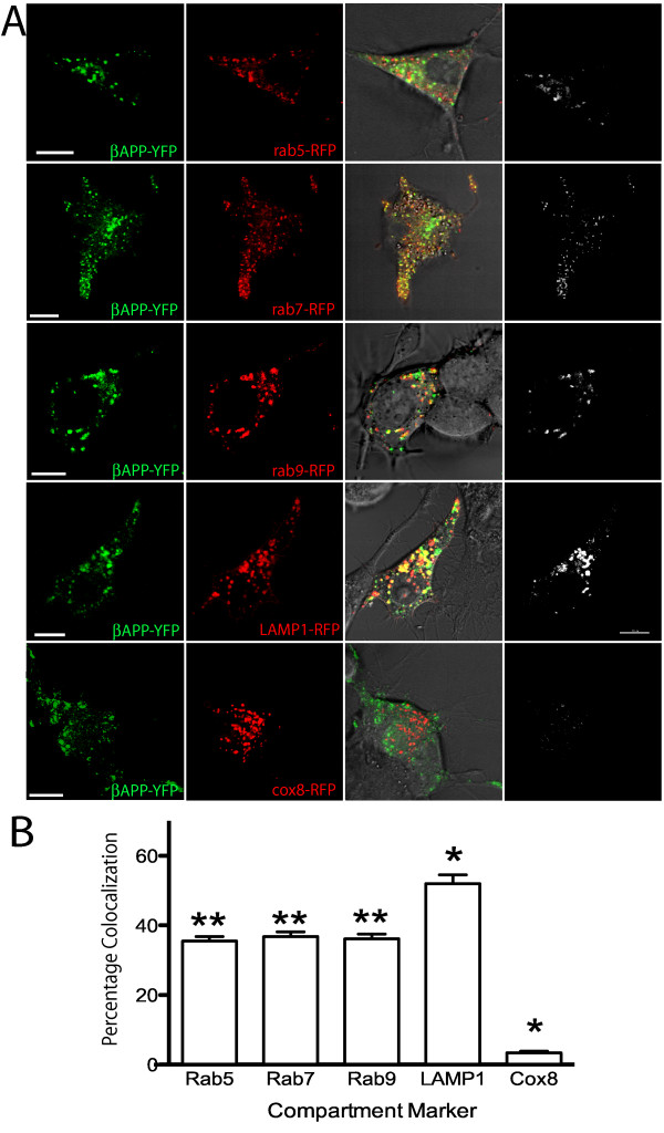
Figure 4
APP is Enriched in the Lysosome in Neuronal SN56 cells . SN56 cells were transiently co-transfected with fluorescent-tagged APP and compartment marker proteins and imaged using laser scanning confocal...
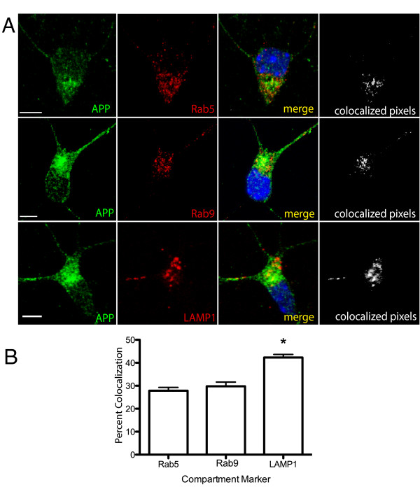
Figure 5
APP is enriched in the lysosome in cultured mouse neurons . A . Cultured mouse neurons were fixed and immunostained with antibodies against APP (green) or compartment markers (red) and nuclei were cou...
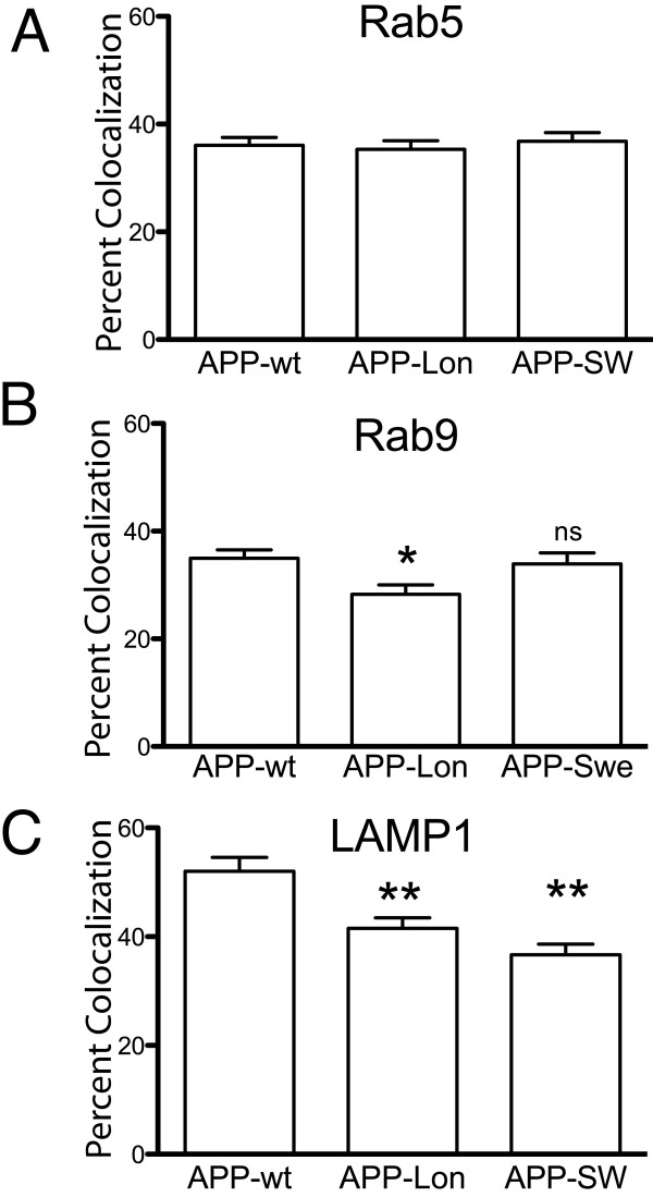
Figure 6
London and Swedish Mutations reduce APP levels in late endosomal and lysosomal compartments . SN56 cells were transiently co-transfected with the u03b2APP-CFP construct with or without the London or t...
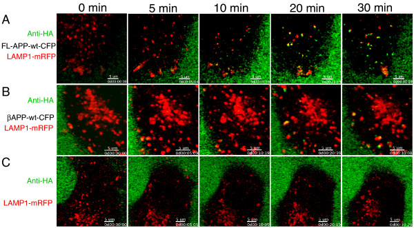
Figure 7
APP is rapidly internalized to the lysosome in live Cos7 cells . Cos7 cells were transiently co-transfected with wild type full length FL-APP-CFP (A) or u03b2APP-CFP (B) constructs along with the LAMP...
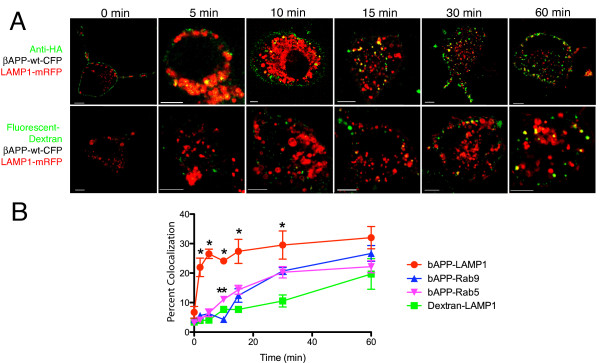
Figure 8
APP internalization into the endosomal/lysosomal system in SN56 cells . A . Representative images of the uptake of anti-HA antibody or fluorescent dextran (green) to lysosomes labeled with LAMP1-mRFP ...
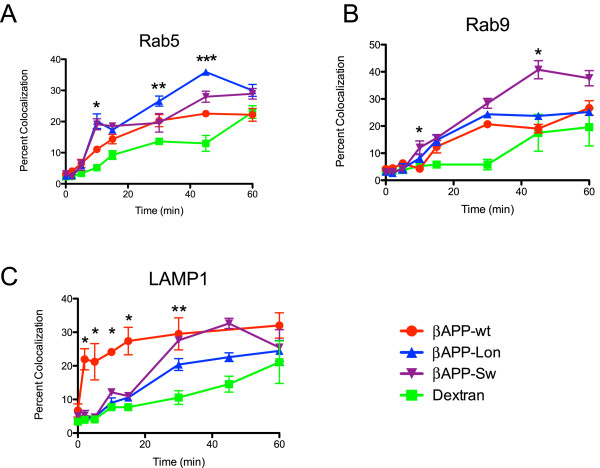
Figure 9
Internalization of APP mutants into the endosomal/lysosomal system in SN56 cells . SN56 cells were cotransfected with the indicated u03b2APP and fluorescent-tagged compartment marker protein construct...
Figure images are served from the NIH/NLM PubMed Central Open Access Subset or Europe PMC; copyright remains with the publishers and authors.
💬 Discussion
0 commentsNo comments yet. Be the first to start a discussion!
Leave a Comment