Abstract
Mounting evidence supports the role of hydrogen peroxide (H2O2) in physiological signaling as well as pathological conditions. However, the subtleties of peroxide-mediated signaling are not well understood, in part because the generation, degradation, and diffusion of H2O2 are highly volatile within different cellular compartments. Therefore, the direct measurement of H2O2 in living specimens is critically important. Fluorescent probes that can detect small changes in H2O2 levels within relevant cellular compartments are important tools to study the spatial dynamics of H2O2. To achieve temporal resolution, the probes must also be photostable enough to allow multiple readings over time without loss of signal. Traditional fluorescent redox sensitive probes that have been commonly used for the detection of H2O2 tend to react with a wide variety of reactive oxygen species (ROS) and often suffer from photostablilty issues. Recently, new classes of H2O2 probes have been designed to detect H2O2 with high selectivity. Advances in H2O2 measurement have enabled biomedical scientists to study H2O2 biology at a level of precision previously unachievable. In addition, new imaging techniques such as two-photon microscopy (TPM) have been employed for H2O2 detection, which permit real-time measurements of H2O2 in vivo. This review focuses on recent advances in H2O2 probe development and optical imaging technologies that have been developed for biomedical applications.
🔬 Techniques
🧬 Organisms
✨ Fluorophores
🔬 Cell Lines
🏛️ Research Organizations (ROR)
Affiliated research institutions:
📊 Figures
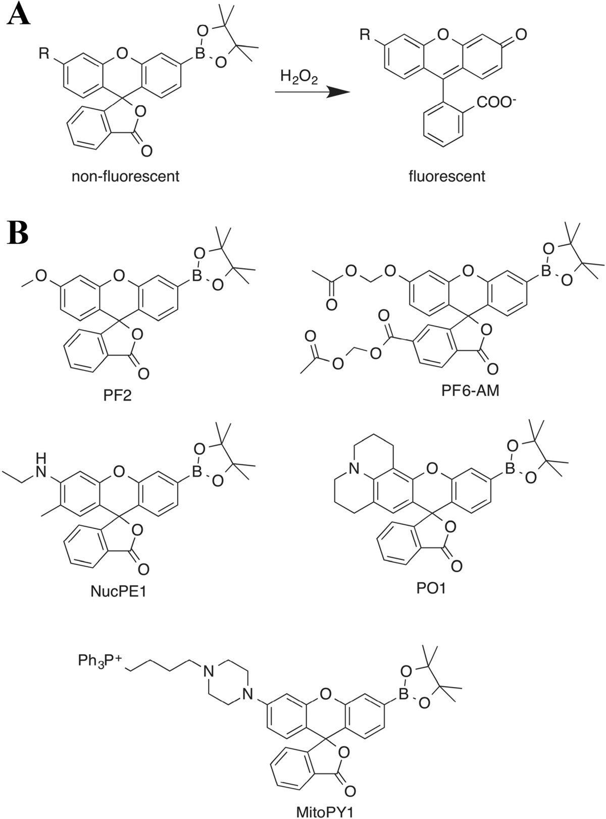
Figure 1
Fluorescent turn-on mechanism and chemical structures of several examples of boronate-based H 2 O 2 fluorescent probes. (A) Lactone-opening mechanism of fluorescence-enhancement for mono-boronate xant...
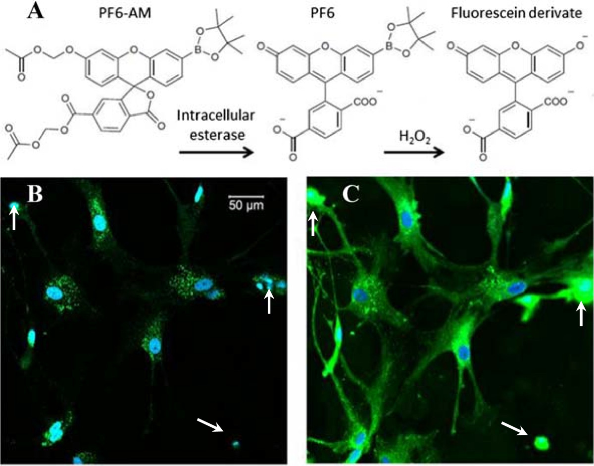
Figure 2
Fluorescence imaging of intracellular H 2 O 2 production using fluorescence probe PF6-AM (green). (A) Mechanism of Chemoselective H 2 O 2 PF6-AM. (B) TPF imaging of H 2 O 2 in astrocytes, fluorescence...
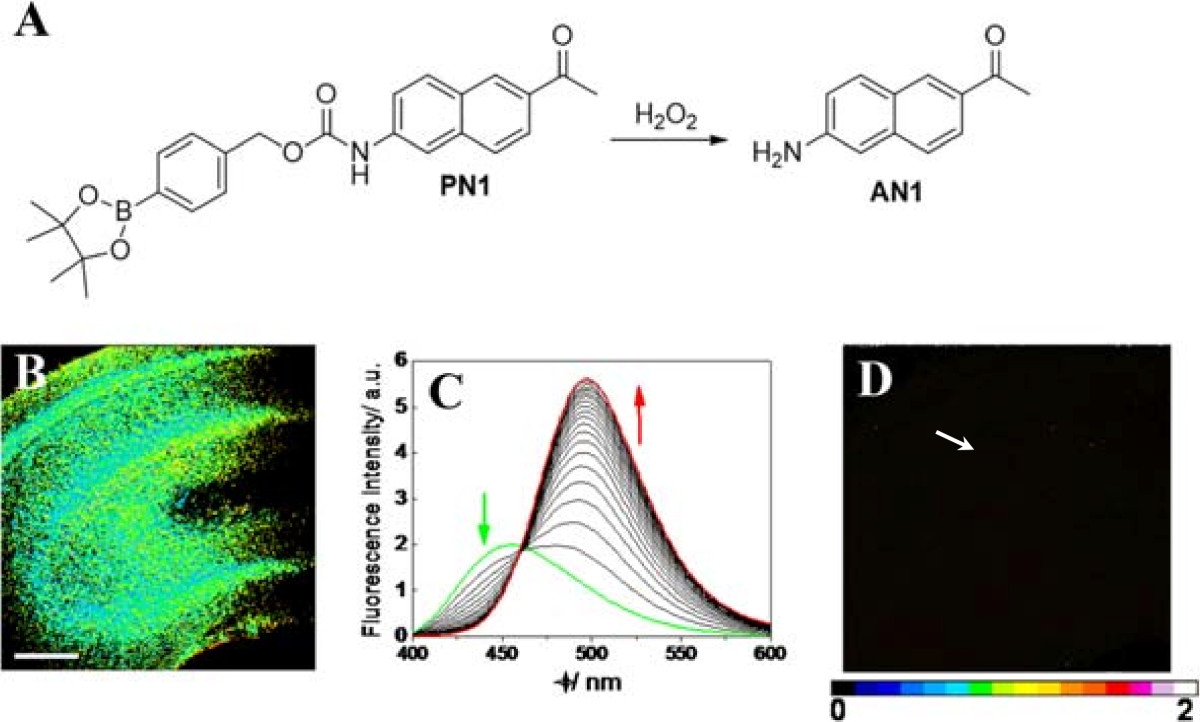
Figure 3
Ratiometric imaging of fresh rat hippocampal slice treated with H 2 O 2 . (A) The reaction between PN1 and H 2 O 2 produced AN1 as the only major fluorescent product. (B) A hippocampal slice labeled w...
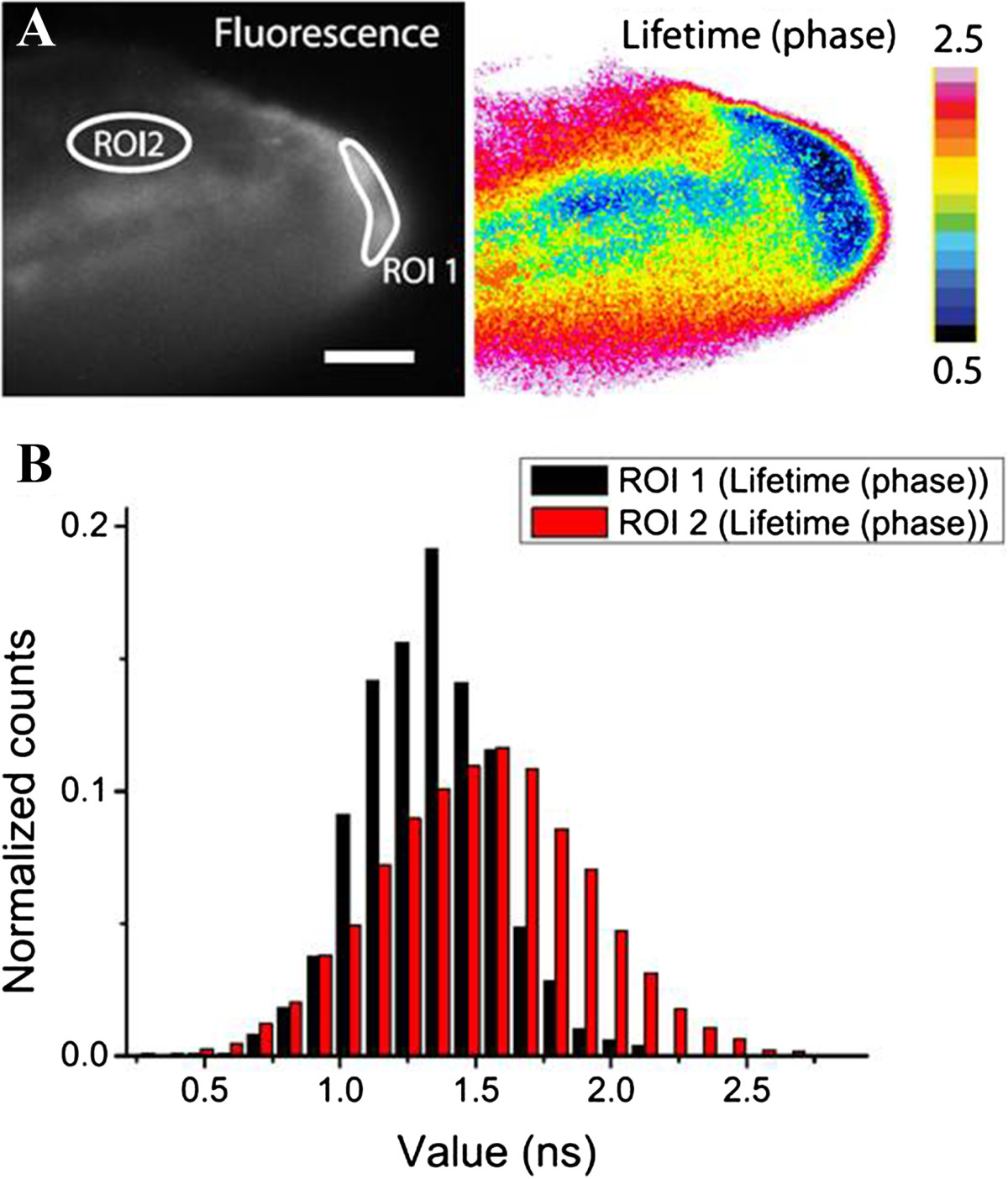
Figure 4
FLIM of HyPer-3 response to H 2 O 2 production induced by inflammation in zebrafish larvae. (A) Left and right panels represent fluorescence intensity and FLIM images, respectively. ROI1 highlights th...
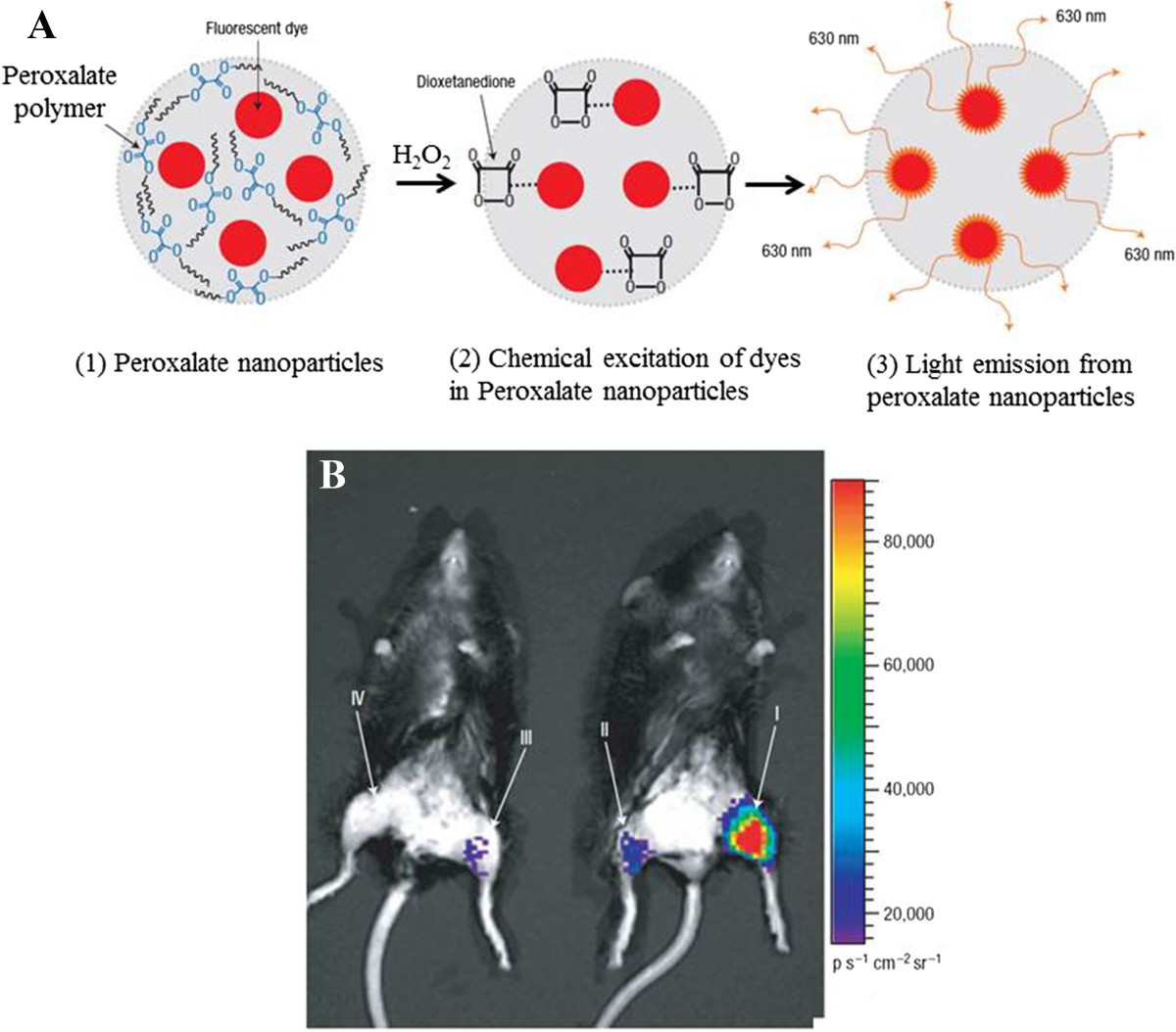
Figure 5
In vivo imaging of H 2 O 2 using peroxalate nanoparticles. (A) Peroxalate nanoparticle. (B) Chemiluminescence imaging. The figures were adapted from ref. [ 29 ] with permission.
Figure images are served from the NIH/NLM PubMed Central Open Access Subset or Europe PMC; copyright remains with the publishers and authors.
💬 Discussion
0 commentsNo comments yet. Be the first to start a discussion!
Leave a Comment