Abstract
Chromatin immunoprecipitation (ChIP) studies provide snapshots of factors on chromatin in cell populations. Here, we use live-cell imaging to examine at high temporal resolution the recruitment and dynamics of transcription factors to the inducible Hsp70 loci in individual Drosophila salivary gland nuclei. Recruitment of the master regulator, HSF, is first detected within 20 s of gene activation; the timing of its recruitment resolves from RNA polymerase II and P-TEFb, and these factors resolve from Spt6 and Topo I. Remarkably, the recruitment of each factor is highly synchronous between different cells. In addition, fluorescence recovery after photobleaching (FRAP) analyses show that the entry and exit of multiple factors are progressively constrained upon gene activation, suggesting the gradual formation of a transcription compartment. Furthermore, we demonstrate that poly(ADP-ribose) (PAR) polymerase activity is required to maintain the transcription compartment. We propose that PAR polymers locally retain factors in a transcription compartment.
🔬 Techniques
🔭 Microscopes
🧬 Organisms
💻 Software
✨ Fluorophores
🧪 Sample Preparation
🏭 Microscope Brands
🧪 Reagent Suppliers
🔴 Lasers
📷 Detectors
🔎 Objectives
💻 Software Details
🏛️ Research Organizations (ROR)
Affiliated research institutions:
📋 Methods
Spinning Disk Microscopy
Drosophila salivary glands, from the crosses described in the Supplemental Experimental Procedures , were dissected from third instar larva as previously described ( Yao et al., 2008 ) and were transferred, immediately with medium to a MatTek glass-bottomed culture dish (P35G-1.0-14-C) and a glass cover-slip was placed on top to reduce evaporation and movement of the glands. A Carl Zeiss Cell Observer SD system with the Yokogawa CSU-X1 spinning disk unit and a Hamamatsu C9100-13 EMCCD was used to obtain confocal 3D stacks with 2-channels and time intervals of 10–30 sec. Two identical Plan-Apochromat 40x/1.3 Oil Iris objectives were used; one was maintained at room temperature and one was heated to 36°C using a Bioptechs Objective Heater. A 40 μm pre-activation z-stack, with 1 μm sections, was taken using the room temperature objective in both channels alternating channels every slice. Then the 36°C objective was moved into position; HS times were started at the moment the objective contacted the slide. A xyzt series with 40 z-sections was obtained after readjusting the focal position. Time intervals were between 10–30 sec and the time series lasted 20 min ( Figure 2A ). Images were taken at a resolution of 512×512 pixels, using 16-bit color depth. Details regarding data analysis can be found in the Supplemental Experimental Procedures .
Laser Scanning Confocal Microscopy and Multiphoton Microscopy
Dissected glands were transferred with medium to a Bioptechs FCS3 Closed Chamber System with a 0.2 mm spacer. We used an upright confocal/multi-photon microscope system (Carl Zeiss LSM510 META). NHS images were obtained with a C-Apochromat 63x, 1.2 NA, water immersion objective. For HS, an identical objective was swapped in for the room temperature objective the same manner as described above.
Show full methods section
Spinning Disk Microscopy
Drosophila salivary glands, from the crosses described in the Supplemental Experimental Procedures , were dissected from third instar larva as previously described ( Yao et al., 2008 ) and were transferred, immediately with medium to a MatTek glass-bottomed culture dish (P35G-1.0-14-C) and a glass cover-slip was placed on top to reduce evaporation and movement of the glands. A Carl Zeiss Cell Observer SD system with the Yokogawa CSU-X1 spinning disk unit and a Hamamatsu C9100-13 EMCCD was used to obtain confocal 3D stacks with 2-channels and time intervals of 10–30 sec. Two identical Plan-Apochromat 40x/1.3 Oil Iris objectives were used; one was maintained at room temperature and one was heated to 36°C using a Bioptechs Objective Heater. A 40 μm pre-activation z-stack, with 1 μm sections, was taken using the room temperature objective in both channels alternating channels every slice. Then the 36°C objective was moved into position; HS times were started at the moment the objective contacted the slide. A xyzt series with 40 z-sections was obtained after readjusting the focal position. Time intervals were between 10–30 sec and the time series lasted 20 min ( Figure 2A ). Images were taken at a resolution of 512×512 pixels, using 16-bit color depth. Details regarding data analysis can be found in the Supplemental Experimental Procedures .
Laser Scanning Confocal Microscopy and Multiphoton Microscopy
Dissected glands were transferred with medium to a Bioptechs FCS3 Closed Chamber System with a 0.2 mm spacer. We used an upright confocal/multi-photon microscope system (Carl Zeiss LSM510 META). NHS images were obtained with a C-Apochromat 63x, 1.2 NA, water immersion objective. For HS, an identical objective was swapped in for the room temperature objective the same manner as described above.
FRAP
Dissected glands were imaged using MPM as described above. Perfusion of 3μM PJ34 or Media alone into the FCS3 chamber occurred as depicted in Figure 6A . We used a circular ROI limited to the dimensions of the Hsp70 loci. eGFP samples were photobleached with a Mai Tai laser (Spectrum Physics) at 910 nm with a power of 15–20 mW (measured after the objective). mRFP photobleaching used the same laser at 800 nm using 40–50 mW. These settings photobleached the samples to 40–60% initial intensity. Images were corrected for acquisition photobleaching by monitoring a small nuclear region. FRAP curves were normalized for pre-bleach images to equal one, and first image after the bleach equal to 0. Recovery times were obtained by fitting the FRAP data to f ( t )= A ×( 1 - C eq × e − koff ×t ) ( Sprague et al., 2004 ).
Supplementary Material 01
📊 Figures

Figure 1
Colocalization of P-TEFb, Spt6 and Topo I with Pol II at Developmental and Hsp70 Loci in Living Polytene Nuclei
LSCM maximum intensity projections of polytene nuclei co-expressing (A, D) mRFP-P-TEFb (left panel), eGFP-Pol II (middle panel), (B, E) eGFP-Spt6 (left panel) and mRFP-Pol II (middle panel) and (C, F)...

Figure 2
Recruitment Timing of TFs
(A) SDCM was used to obtain 3D images of FP-tagged TFs with the complementary FP-tagged Pol II. First, Drosophila salivary glands were imaged using a room temperature objective, then an objective pre-...

Figure 3
Synchrony in the Recruitment of TFs to Hsp70 loci Among Nuclei of the Same Gland or in Different Glands
(Au2013E) Representative SDCM recruitment images and corresponding mean F.I. recruitment plots for (A) mRFP-HSF, (B) Pol II (mRFP and eGFP), (C) mRFP-P-TEFb, (D) eGFP-Spt6 and (E) eGFP-Topo I. The 1 s...
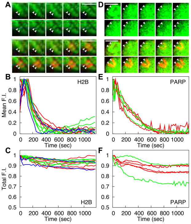
Figure 4
Association of H2B and PARP with Hsp70 Loci after Decondensation
(A and D) Representative time course images illustrating the localization of (A) mRFP-H2B and (D) PARP-eGFP to the Hsp70 loci after HS. Top panel shows the localization of the factor, while the bottom...

Figure 5
FRAP Dynamics of TFs at the Hsp70 Loci Change After Length of HS
(A, C, and E) FRAP of the three TFs after different lengths of HS: (A) mRFP-P-TEFb, (C) eGFP-Spt6 and (E) eGFP-Topo I. Panels show representative FRAP images. The top panel was bleached after 10 min H...

Figure 6
PARP Catalytic Activity is Required for the Maintenance of the Transcription Compartment
(A) Schematic of PJ34 perfusion protocol. Media only or 3u03bcM PJ34 was perfused over the gland for 5 min starting 35 min after HS. FRAP of eGFP-Pol II at the Hsp70 loci was initiated as soon as perf...
Figure images are served from the NIH/NLM PubMed Central Open Access Subset or Europe PMC; copyright remains with the publishers and authors.
💬 Discussion
0 commentsNo comments yet. Be the first to start a discussion!
Leave a Comment