Abstract
Extension of neurites requires the SNARE-dependent fusion of plasmalemmal precursor vesicles with the plasma membrane of growth cones. Here, we show that tomosyn localizes at the palm of growth cones and inhibits the fusion of the vesicles there, thus promoting transport of the vesicles to the plasma membrane of the leading edges of growth cones. Tomosyn localizes because ROCK activated by Rho small G protein phosphorylates syntaxin-1, which increases the affinity of syntaxin-1 for tomosyn and forms a stable complex with tomosyn, resulting in inhibition of the formation of the SNARE complex. In retraction of neurites, tomosyn localizes all over the edges of the neurites and inhibits fusion of the vesicles with the plasma membrane. Thus, tomosyn demarcates the plasma membrane by binding to syntaxin-1 phosphorylated by ROCK, and thereby regulates extension and retraction of neurites.
🔬 Techniques
🧬 Organisms
✨ Fluorophores
🧪 Sample Preparation
🔬 Cell Lines
🧪 Reagent Suppliers
🏛️ Research Organizations (ROR)
Affiliated research institutions:
📋 Methods
Construction of expression vectors
To generate mammalian expression vectors for tomosyn (pCMV-HA-tomosyn and pcDNA3-HA-tomosyn), the full-length of m-tomosyn cDNA was subcloned into the pCMV-HA or pcDNA3-HA vector, respectively. The cDNAs for syntaxin-1A mutants, in which Thr at 5, 21, and 118 aa residues and Ser at 14, 59, and 91 aa residues were replaced to Ala, were generated using a sitedirected mutagenesis kit (Stratagene) and subcloned into pGEX-2T and pCMVHA vectors. The pEF-BOS-myc-C3 was constructed ( Komuro et al., 1996 ). The pCAG-myc-ROCK-Δ3 and pCAG-myc-ROCK-KDIA were provided by Dr. S. Narumiya (Kyoto University Faculty of Medicine, Kyoto, Japan).
Knock-down of tomosyn by the RNA interference method
The mammalian expression vector pSIREN-RetroQ (CLONTECH Laboratories, Inc.), which contains a GFP gene for the selection of transfected cells, was used for expression of small interfering RNA (siRNA) in NG108 cells. Inserts were used as follows: m-tomosyn gene-specific insert was a 21-nt sequence corresponding to nt 2478–2498 (5′-GCGGTACTATATTGAGGTTAA-3′) of m-tomosyn cDNA, which was separated by a 10-nt noncomplementary spacer (5′-TTGATATCCG-3′) from the reverse complement of the same 21-nt sequence. A control (Scramble) insert was a 21-nt sequence (5′-GACGTTATCGGTATGATATAG-3′) with no significant homology to any mammalian gene sequence, which was separated by a 10-nt noncomplementary spacer (5′-TTGATATCCG-3′) from the reverse complement of the same 21-nt sequence.
Quantification of neurites
After the 2-d db-cAMP treatment, NG108 cells were processed ( Martinez-Arca et al., 2000 ). Images were captured using a confocal laser scanning microscope (Radiance 2000; Bio-Rad Laboratories) using a 60× oil immersion objective lens. Collected data were exported as 8-bit TIFF files and were processed using Adobe Photoshop ® software. The assay for neurite outgrowth was performed on 100 cells randomly chosen in each group. The number of primary neurites per cell was defined as the number of thin cell processes with a length >20 μm. The length of individual neurites for each cell was measured by use of the NIH Image software. Neurite length was defined as the traced distance from the neurite tip to the point where neurite emanated from the cell body. The statistical significance of differences between each group was analyzed by the two-tailed t test. Assay for VSV-G transport NG108 cells were transfected with pAR vector encoding either GFP-VSV-G or VSV-G and assayed ( Hirschberg et al., 1998 ). Where indicated, the cells were pulse labeled with [35S]methionine (0.1 mCi/ml) (Tran35S label; ICN Biomedicals) at 37°C for 10 min, chased at 20°C or 37°C for indicated periods of time, and analyzed. To detect surface VSV-G, biotinylation was performed by incubating the cells with 0.5 mg/ml Sulfo-NHS-Biotin (Pierce Chemical Co.) in PBS at 4°C for 30 min and quenched with 50 mM NH4Cl in PBS. The cells were lysed with a RIPA buffer. Total VSV-G was immunoprecipitated with a mouse anti-VSV-G mAb (P5D4), washed, eluted with 0.1 ml of 1% SDS in TBS at 90°C for 3 min, and diluted with 1 ml of 1% Triton X-100 in TBS. Biotinylated VSV-G was recovered with streptavidin-agarose beads (Sigma-Aldrich) and surface VSV-G was eluted with the SDS sample buffer. Assay for the formation of SNARE and tomosyn complexes The lysate of NG108 cells was prepared using a lysis buffer (20 mM Hepes, pH 7.4, 90 mM KOAc, 2 mM Mg(OAc) 2 , 0.5 mM EGTA, 50 mM NaF, 2 mM Na 3 VO 4 , and 1% CHAPS; Fujita et al., 1998). The lysates were incubated with an anti-syntaxin-1 mAb (Synaptic Systems) at 4°C for 2 h. The immune complexes were precipitated with protein G–Sepharose beads (Amersham Biosciences) at 4°C for 1 h, washed with the lysis buffer, resolved in 5–20% gradient SDS-PAGE, and analyzed by immunoblotting with an anti-tomosyn pAb ( Fujita et al., 1998 ), an anti-VAMP-2 pAb (Synaptic systems), an anti-SNAP-25 pAb (Calbiochem), and an anti-syntaxin-1 pAb (Calbiochem). Assay for the binding of syntaxin-1 to tomosyn and VAMP-2 Overlay assay was performed ( Fujita et al., 1998 ). The Sf9 cells expressing tomosyn were collected and boiled with the SDS sample buffer, followed by centrifugation. The GST-VAMP-2 protein was generated in Escherichia coli ( Fujita et al., 1998 ). Tomosyn or VAMP-2 (5 pmol each) was subjected to SDS-PAGE and blotted onto a nitrocellulose membrane sheet. The sheet was incubated with various quantities of syntaxin-1 in 1 ml of TBS containing 5% skim milk for 2 h. The amount of syntaxin-1 bound to the sheet was detected by immunoblotting with the anti-syntaxin-1 mAb. The intensity of the bands was measured with CS-9000 (Shimazu).
Show full methods section
Construction of expression vectors
To generate mammalian expression vectors for tomosyn (pCMV-HA-tomosyn and pcDNA3-HA-tomosyn), the full-length of m-tomosyn cDNA was subcloned into the pCMV-HA or pcDNA3-HA vector, respectively. The cDNAs for syntaxin-1A mutants, in which Thr at 5, 21, and 118 aa residues and Ser at 14, 59, and 91 aa residues were replaced to Ala, were generated using a sitedirected mutagenesis kit (Stratagene) and subcloned into pGEX-2T and pCMVHA vectors. The pEF-BOS-myc-C3 was constructed ( Komuro et al., 1996 ). The pCAG-myc-ROCK-Δ3 and pCAG-myc-ROCK-KDIA were provided by Dr. S. Narumiya (Kyoto University Faculty of Medicine, Kyoto, Japan).
Knock-down of tomosyn by the RNA interference method
The mammalian expression vector pSIREN-RetroQ (CLONTECH Laboratories, Inc.), which contains a GFP gene for the selection of transfected cells, was used for expression of small interfering RNA (siRNA) in NG108 cells. Inserts were used as follows: m-tomosyn gene-specific insert was a 21-nt sequence corresponding to nt 2478–2498 (5′-GCGGTACTATATTGAGGTTAA-3′) of m-tomosyn cDNA, which was separated by a 10-nt noncomplementary spacer (5′-TTGATATCCG-3′) from the reverse complement of the same 21-nt sequence. A control (Scramble) insert was a 21-nt sequence (5′-GACGTTATCGGTATGATATAG-3′) with no significant homology to any mammalian gene sequence, which was separated by a 10-nt noncomplementary spacer (5′-TTGATATCCG-3′) from the reverse complement of the same 21-nt sequence.
Quantification of neurites
After the 2-d db-cAMP treatment, NG108 cells were processed ( Martinez-Arca et al., 2000 ). Images were captured using a confocal laser scanning microscope (Radiance 2000; Bio-Rad Laboratories) using a 60× oil immersion objective lens. Collected data were exported as 8-bit TIFF files and were processed using Adobe Photoshop ® software. The assay for neurite outgrowth was performed on 100 cells randomly chosen in each group. The number of primary neurites per cell was defined as the number of thin cell processes with a length >20 μm. The length of individual neurites for each cell was measured by use of the NIH Image software. Neurite length was defined as the traced distance from the neurite tip to the point where neurite emanated from the cell body. The statistical significance of differences between each group was analyzed by the two-tailed t test. Assay for VSV-G transport NG108 cells were transfected with pAR vector encoding either GFP-VSV-G or VSV-G and assayed ( Hirschberg et al., 1998 ). Where indicated, the cells were pulse labeled with [35S]methionine (0.1 mCi/ml) (Tran35S label; ICN Biomedicals) at 37°C for 10 min, chased at 20°C or 37°C for indicated periods of time, and analyzed. To detect surface VSV-G, biotinylation was performed by incubating the cells with 0.5 mg/ml Sulfo-NHS-Biotin (Pierce Chemical Co.) in PBS at 4°C for 30 min and quenched with 50 mM NH4Cl in PBS. The cells were lysed with a RIPA buffer. Total VSV-G was immunoprecipitated with a mouse anti-VSV-G mAb (P5D4), washed, eluted with 0.1 ml of 1% SDS in TBS at 90°C for 3 min, and diluted with 1 ml of 1% Triton X-100 in TBS. Biotinylated VSV-G was recovered with streptavidin-agarose beads (Sigma-Aldrich) and surface VSV-G was eluted with the SDS sample buffer. Assay for the formation of SNARE and tomosyn complexes The lysate of NG108 cells was prepared using a lysis buffer (20 mM Hepes, pH 7.4, 90 mM KOAc, 2 mM Mg(OAc) 2 , 0.5 mM EGTA, 50 mM NaF, 2 mM Na 3 VO 4 , and 1% CHAPS; Fujita et al., 1998). The lysates were incubated with an anti-syntaxin-1 mAb (Synaptic Systems) at 4°C for 2 h. The immune complexes were precipitated with protein G–Sepharose beads (Amersham Biosciences) at 4°C for 1 h, washed with the lysis buffer, resolved in 5–20% gradient SDS-PAGE, and analyzed by immunoblotting with an anti-tomosyn pAb ( Fujita et al., 1998 ), an anti-VAMP-2 pAb (Synaptic systems), an anti-SNAP-25 pAb (Calbiochem), and an anti-syntaxin-1 pAb (Calbiochem). Assay for the binding of syntaxin-1 to tomosyn and VAMP-2 Overlay assay was performed ( Fujita et al., 1998 ). The Sf9 cells expressing tomosyn were collected and boiled with the SDS sample buffer, followed by centrifugation. The GST-VAMP-2 protein was generated in Escherichia coli ( Fujita et al., 1998 ). Tomosyn or VAMP-2 (5 pmol each) was subjected to SDS-PAGE and blotted onto a nitrocellulose membrane sheet. The sheet was incubated with various quantities of syntaxin-1 in 1 ml of TBS containing 5% skim milk for 2 h. The amount of syntaxin-1 bound to the sheet was detected by immunoblotting with the anti-syntaxin-1 mAb. The intensity of the bands was measured with CS-9000 (Shimazu).
Phosphorylation of syntaxin-1 by ROCK in a cell-free system
Syntaxin-1 was phosphorylated by ROCK in a cell-free system ( Sumi et al., 2001 ). HEK293 cells were transiently transfected with pCAG-myc-ROCK-Δ3 plasmid. Cells were lysed in the lysis buffer and immunoprecipitated with an anti-myc mAb. Myc-ROCK-Δ3 (5 pmol)–bound protein G–Sepharose beads (50 μl slurry) were washed with a kinase buffer (50 mM Tris/HCl, pH 7.5, 7 mM MgCl 2 , 1 mM DTT, 0.06% CHAPS, and 1 mM EDTA) and were incubated at 30°C for indicated periods of time in 100 μl of the kinase buffer containing 50 μM γ[ 32 P]ATP (0.5–1.2 × 10 3 cpm/pmol; Amersham Biosciences) and 30 pmol syntaxin-1 as a substrate. The reaction was stopped by the addition of the SDS sample buffer and boiled for 5 min. Each sample was subjected to SDS-PAGE, followed by protein staining with Coomassie brilliant blue and autoradiography. Relevant gel slices were excised and scintillation counted to determine the radioactivity incorporated into syntaxin-1.
Phosphorylation of syntaxin-1 in intact cells
NG108 cells were transfected with either pCMV-HA-syntaxin-1A or pCMV-HA-S14A-syntaxin-1A, cultured in DME containing 1 mM db-cAMP for 48 h, and allowed to extend neurites. The cells were washed three times with phosphate-free DME and were incubated for 30 min in the phosphate-free DME containing 1 mM db-cAMP and 1× HT. Thereafter, [ 32 P]orthophosphate (phosphorus-32; Amersham Biosciences) was added at a concentration of 0.5 mCi/ml in the phosphate-free DME containing 1 mM db-cAMP and 1× HT. Cells were cultured for 4 h and stimulated by 5 μM LPA for 5 min. Cells were lysed with RIPA buffer and immunoprecipitated with the anti-HA mAb. The immunoprecipitates were subjected to SDS-PAGE, followed by autoradiography. Assay for the phosphorylation of syntaxin-1 at serine 14 in vivo NG108 cells were cultured in DME containing 1 mM db-cAMP for 48 h and were allowed to extend neurites. The cells were preincubated with or without various concentrations of Y27632 for 30 min and were then stimulated by 5 μM LPA for indicated periods of time. Cells were lysed with the SDS-PAGE sample buffer and were subjected to immunoblotting with the anti-phosphorylated syntaxin-1 (S14-phosphospecific) pAb ( Foletti et al., 2000 ).
Knock-down of tomosyn by the RNA interference method
The mammalian expression vector pSIREN-RetroQ (CLONTECH Laboratories, Inc.), which contains a GFP gene for the selection of transfected cells, was used for expression of small interfering RNA (siRNA) in NG108 cells. Inserts were used as follows: m-tomosyn gene-specific insert was a 21-nt sequence corresponding to nt 2478–2498 (5′-GCGGTACTATATTGAGGTTAA-3′) of m-tomosyn cDNA, which was separated by a 10-nt noncomplementary spacer (5′-TTGATATCCG-3′) from the reverse complement of the same 21-nt sequence. A control (Scramble) insert was a 21-nt sequence (5′-GACGTTATCGGTATGATATAG-3′) with no significant homology to any mammalian gene sequence, which was separated by a 10-nt noncomplementary spacer (5′-TTGATATCCG-3′) from the reverse complement of the same 21-nt sequence.
📊 Figures
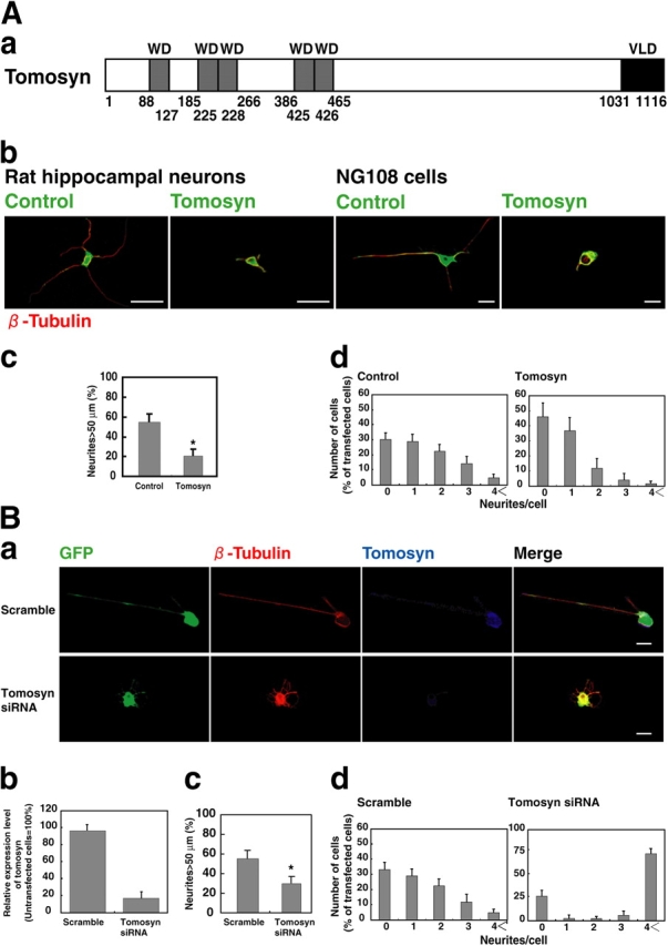
Figure 1.
Inhibition of neurite outgrowth by tomosyn. (A) Effect of overexpression of tomosyn. (Aa) Schematic structure of tomosyn. Gray box (WD), WD40 repeats domain. Black box (VLD), VAMP-like domain. (Ab) Pr...
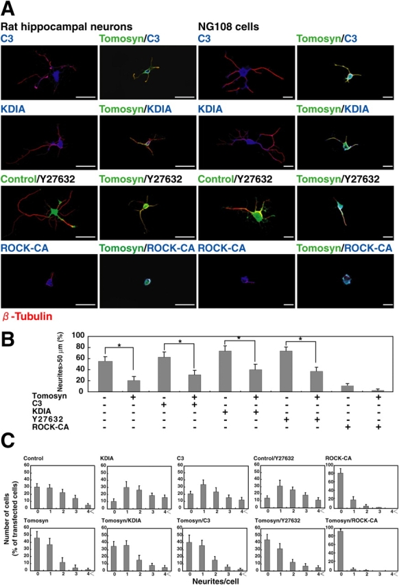
Figure 2.
Involvement of ROCK in the inhibitory effect of tomosyn on neurite outgrowth. (A) Functional interaction between tomosyn and ROCK. Primary cultured rat hippocampal neurons were transfected with myc-C3...
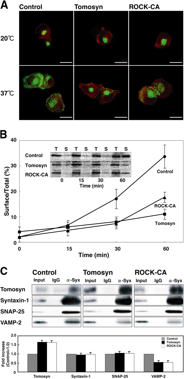
Figure 3.
Inhibition of vesicle transport by tomosyn and ROCK. (A) Transport of VSV-G in NG108 cells. NG108 cells were cotransfected with GFP-VSV-G and either HA-tomosyn, myc-ROCK-u03943 (ROCK-CA), or a null HA...
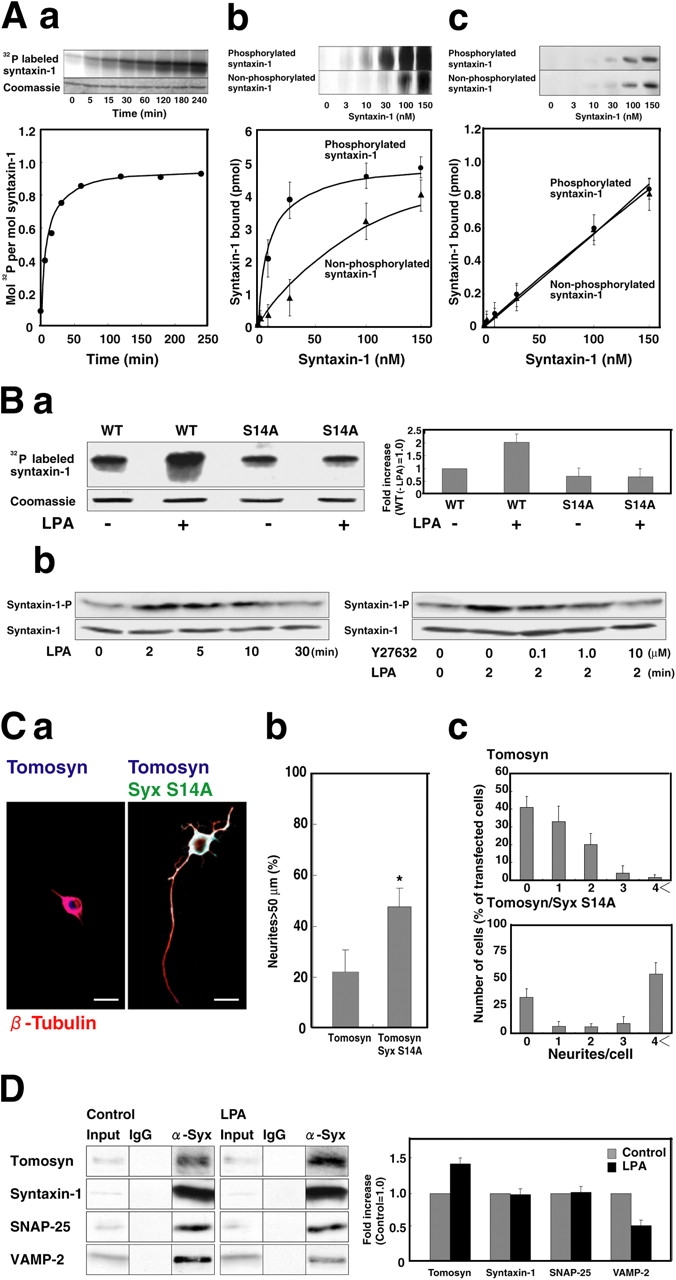
Figure 4.
Molecular link between the tomosyn complex and ROCK in the LPA-induced retraction of neurites. (A) Phosphorylation of syntaxin-1 by ROCK in a cell-free system and increase of the affinity of syntaxin-...
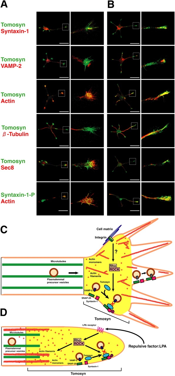
Figure 5.
Localization of tomosyn in extending and retracting neurites and models of a role of tomosyn in extension and retraction of neurites. (A) Localization of tomosyn in extending neurites. Rat hippocampal...
Figure images are served from the NIH/NLM PubMed Central Open Access Subset or Europe PMC; copyright remains with the publishers and authors.
💬 Discussion
0 commentsNo comments yet. Be the first to start a discussion!
Leave a Comment