Abstract
Hypervirulent Klebsiella pneumoniae (HvKP) is hypermucoviscous organism, carrying genes of rmpA and aerobactin, causing serious community-acquired infection and metastatically spread in young healthy hosts. Neutrophils play an important role during innate immune response against bacterial infection by phagocytosis and neutrophil extracellular traps (NETs). Whether neutrophils can effectively defend against HvKP remains unclear. In this study, we observed that the HvKP was significantly more resistant to neutrophil-mediated phagocytosis and intracellular killing than classic Klebsiella pneumoniae (cKP) isolates. Although both HvKP and cKP induced NETs under scanning electron microscopy and confocal microscopy, more cKP than HvKP were trapped in NETs, and the killing by intracellular and extracellular mechanisms of neutrophils was detected only on cKP. Together, our results demonstrated that HvKP resisted to both intracellular and extracellular killing of neutrophils.
🔬 Techniques
🧬 Organisms
✨ Fluorophores
🧪 Sample Preparation
🔬 Cell Lines
🏭 Microscope Brands
🧪 Reagent Suppliers
🏛️ Research Organizations (ROR)
Affiliated research institutions:
📋 Methods
Bacterial strains Fourty-five
Klebsiella pneumoniae strains that were isolated from blood were investigated, including HvKP strains of serotype K1 (n = 16) and serotype K2 (n = 14), non-K1/K2 (cKP) isolates (n = 15). The HvKP-K1 and -K2 isolates were selected based on positive string test, gene amplification of rmpA and aeobactin . While cKP isolates were negative for string test and gene detection for rmpA and aeobactin . All forty-five strains were tested for phagocytosis and bactericidal activity assays. Only 6 strains (including 3 HvKP-K1 strains and 3 cKP strains) were tested for transmission electron microscopic observation and scanning electron microscopic observation. 18 strains (including 9 HvKP-K1 strains and 9 cKP strains) were tested for NETs immunofluorescence staining. Human neutrophils Neutrophils were purified from freshly drawn blood of three healthy human volunteers who signed written consent prior to participation in the study. All experiments were approved by the Ethics Commettee of Chinese General Hospital of People’s Liberation Army. Polymorphprep™ (Axis-Shield) was used according to the manufacturer’s instructions to obtain neutrophil purity of approximately 95~97%. The major contaminant in neutrophil preparation was erythrocytes. Cells were counted in a Neubauer chamber. The purity was determined by the formula that the number of neutrophils divided by the number of total counted cells (at least 200 cells). The neutrophils were resuspended in phosphate-buffered saline (PBS, pH7.4) with concentration adjusted to 1 × 10 7 cell/ml. The viability of the purified neutrophils was greater than 95% by trypan blue exclusion assay. Neutrophils were used immediately after isolation. Fluorescence labeling of bacteria HvKP-K1, HvKP-K2 and cKP isolates incubated on Mueller-Hinton (MH) agar overnight at 37°C were adjusted to a concentration of 0.667 McFarland by spectrophotometer. The bacteria were placed in a 70°C water bath for 60 minutes, then 100 μl bacterial suspension was plated on MH agar to examine whether all bacteria were killed. The killed bacteria were washed with PBS and labeled with fluorescein isothiocyanate [FITC (0.1 mg/ml); Sigma Chemical Co.] in 0.1 M NaHCO 3 , pH 9.0 at 25°C for 1 hour. Unbound FITC was washed off with PBS for three times. FITC-labeled bacteria were resuspended to a concentration of 2 × 10 8 cells/ml in PBS, aliquoted, and stored at -70°C.
Show full methods section
Bacterial strains Fourty-five
Klebsiella pneumoniae strains that were isolated from blood were investigated, including HvKP strains of serotype K1 (n = 16) and serotype K2 (n = 14), non-K1/K2 (cKP) isolates (n = 15). The HvKP-K1 and -K2 isolates were selected based on positive string test, gene amplification of rmpA and aeobactin . While cKP isolates were negative for string test and gene detection for rmpA and aeobactin . All forty-five strains were tested for phagocytosis and bactericidal activity assays. Only 6 strains (including 3 HvKP-K1 strains and 3 cKP strains) were tested for transmission electron microscopic observation and scanning electron microscopic observation. 18 strains (including 9 HvKP-K1 strains and 9 cKP strains) were tested for NETs immunofluorescence staining. Human neutrophils Neutrophils were purified from freshly drawn blood of three healthy human volunteers who signed written consent prior to participation in the study. All experiments were approved by the Ethics Commettee of Chinese General Hospital of People’s Liberation Army. Polymorphprep™ (Axis-Shield) was used according to the manufacturer’s instructions to obtain neutrophil purity of approximately 95~97%. The major contaminant in neutrophil preparation was erythrocytes. Cells were counted in a Neubauer chamber. The purity was determined by the formula that the number of neutrophils divided by the number of total counted cells (at least 200 cells). The neutrophils were resuspended in phosphate-buffered saline (PBS, pH7.4) with concentration adjusted to 1 × 10 7 cell/ml. The viability of the purified neutrophils was greater than 95% by trypan blue exclusion assay. Neutrophils were used immediately after isolation. Fluorescence labeling of bacteria HvKP-K1, HvKP-K2 and cKP isolates incubated on Mueller-Hinton (MH) agar overnight at 37°C were adjusted to a concentration of 0.667 McFarland by spectrophotometer. The bacteria were placed in a 70°C water bath for 60 minutes, then 100 μl bacterial suspension was plated on MH agar to examine whether all bacteria were killed. The killed bacteria were washed with PBS and labeled with fluorescein isothiocyanate [FITC (0.1 mg/ml); Sigma Chemical Co.] in 0.1 M NaHCO 3 , pH 9.0 at 25°C for 1 hour. Unbound FITC was washed off with PBS for three times. FITC-labeled bacteria were resuspended to a concentration of 2 × 10 8 cells/ml in PBS, aliquoted, and stored at -70°C.
FITC-labeled KP strains
(FITC-KP) were analyzed by flow cytometry (FACS Canto II, BD) to ensure the percentage of FITC-KP was greater than 99%. Phagocytosis assay 200 μl of FITC-KP suspension (representing 4 × 10 7 cells) was added to a prewarmed mixture including 100 μl of a neutrophil suspension (representing 1 × 10 6 cells), 100 μl of pooled normal human serum (10% v/v for opsonization), and 600 μl of PBS in a 10 × 75-mm Falcon™ polypropylene tube (BD, Franklin Lakes, NJ). The final volume was 1.0 ml and the multiplicity of infection (MOI, Klebsiella pneumoniae : neutrophils) was 40:1. Each tube was incubated at 37°C in a shaking water bath with continuous agitation except an unincubated tube served as control at 0-min. Tubes were transferred to an ice bath after incubation of 10, 30, 60 min. The superficial fluorescent was quenched by adding 100 μl Trypan Blue (0.04%) to determine the percent of neutrophils with ingested KP. The percentage of neutrophils ingested KP was assessed by flow cytometry (FACS canto II, BD). Neutrophil groups were gated according to forward scatter (FSC) and sideways scatter (SSC) combination. Then FITC positive cell groups were gated based on negative control (neutrophil suspension fixed with 2% paraformaldehyde and incubated with FITC-KP, then quenched), and positive control (neutrophils phagocytosed FITC-KP). A total of 10,000 neutrophils were processed. Phagocytic percentage was determined by the percentage of FITC positive neutrophils. Bactericidal activity assays Neutrophils (1 × 10 6 cells) were cultured to adhere to 24-well flat-bottom plates for 30 min. Then the prepared mixture (100 μl inactivated human normal serum, 200 μl bacterial suspension containing 4 × 10 7 CFU and 600 μl PBS) was added to the 24-well plates with neutrophil adhered which served as an experimental test. A control test absent of neutrophils was set for every isolate. The plates were incubated at 37°C for 60 min. Then neutrophils were lysed with 0.1% TritonX 100 for 15 min on ice. Complete lysis of neutrophils was confirmed by microscopic observation. The suspension was diluted 1000-fold by the addition of PBS. Then 100 μl diluted suspension was plated on MH agar. Colony-forming units (CFUs) were enumerated the following day. Each experiment was repeated twice. The bacterial survival index was calculated with the equation: the number of CFUs in experimental test divided by the number of CFUs in control test. The results of every group indicated as mean ± standard deviations (S.D.). The survival index < 1.0 represented that there were bacteria killed by neutrophils. The survival index ≥ 1.0 represented that bacteria were not killed by neutrophils, or bacteria reproduced. Sample preparation for transmission electron microscopy (TEM) Purified neutrophils were mixed with live HvKP-K1 or cKP, cultured for 30 min and 60 min as described above in phagocytosis assay. To get a visible cell cluster, we enlarged the final volume to 2.0 ml in TEM rather than 1.0 ml for phagocytosis assay, but the proportion of components and MOI were the same as phagocytosis assay. The phagocytosis reaction was stopped by placing the reaction tube to an ice bath. Neutrophils were collected by centrifugation (250 × g , 6 min) and were washed three times with cold PBS. The pellet was fixed in 3% (v/v) glutaraldehyde for 120 min, washed with 0.1 M PB for 10 min, three times in total. The cell pellet was dehydrated in a graded ethanol series and embedded in Eponate-12. Ultra-thin sections were stained with uranyl acetate and lead citrate and examined using transmission electron microscopy (HT7700; Japan). Approximately 100 microscopic fields were observed and 15 images were obtained from each isolate. The number of bacteria ingested by 20 neutrophils at 60 min was counted and the average bacterial number per neutrophil was then calculated for each isolate. Sample preparation for scanning electron microscopy (SEM) Neutrophils (1 × 10 6 cells) were cultured to adhere to coverslips treated with 0.01% polylysine (Sigma-Aldrich) in 24-well flat-bottom plates supplemented with 100 μl pooled normal human serum, treated with PMA (100 ng/ml) (Sigma-Aldrich) or challenged with 200 μl of HvKP-K1 or cKP suspension, containing 4 × 10 7 CFU/ml each. The plates were incubated for 90 min in incubator with carbon dioxide. Next, neutrophils were fixed with 3% glutaraldehyde, then incubated with 1% osmium tetroxide and dehydrated with an ascending ethanol series. After dehydration and critical-point drying, the specimens were coated with gold and analyzed in a Hitachi S4800 scanning electron microscopy. The observation and the images obtained were the same as TEM. NET-bound CFUs were enumerated. Briefly, the number of bacteria trapped in NETs produced by 100 neutrophils was counted. Then the average number of bacteria for each neutrophil related NETs was calculated. Immunostaining Neutrophils were challenged with HvKP-K1 or cKP for 90 min as described above in sample preparation for scanning electron microscopy. The neutrophils were fixed in 4% paraformaldehyde for 30 min at room temperature (RT), then washed with PBS for three times, after which the neutrophils were permeabilized in 0.2% Triton X-100 for 20 min at RT. Blocking was performed with 10% normal goat serum for 30 min at RT. Staining with primary antibodies [mouse anti-Myeloperoxidase antibody [2C7] (Abcam, ab25989) and rabbit anti-Histone H3 antibody (citrulline R2 + R8 + R17) (Abcam, ab5103)] was performed for 1 h at RT at 1:200 in 10% normal goat serum-PBS. Samples were washed with PBS for three times. Secondary antibody staining [goat anti-mouse IgG H&L-Alexa Fluor ® 647 (Abcam, ab150115) and goat anti-rabbit IgG H&L-Alexa Fluor ® 488 (Abcam, ab150077)] was performed for 30 min at RT at 1:500 in 10% normal goat serum-PBS. The unbound secondary antibodies were washed off using PBS for three times. Slides were mounted using mounting medium with DAPI (VECTASHIELD®, H-1200). Specimens were analyzed with a confocal microscopy (OLYMPUS FLUOVIEW1000). Approximately 100 microscopic fields were observed and 20 images obtained from each isolate.
Statistical analysis
The differences of neutrophil phagocytosis and bacterial survival index among three groups were performed using one-way analysis of variance. Differences between groups were assessed by t test. All statistical tests were two sided. The p values less than 0.05 were considered to be statistically significant. Data were presented as mean ± standard deviation (S.D.).
📊 Figures
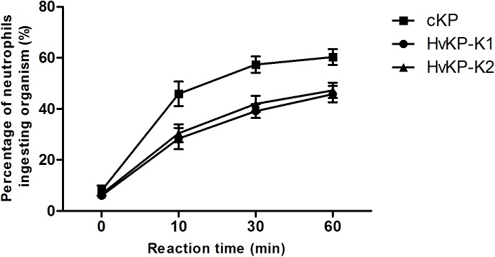
Fig 1
Phagocytosis of neutrophils against HvKP-K1, HvKP-K2 and cKP.
The rate of phagocytosis against cKP (15 isolates) was higher than that against HvKP-K1 (16 isolates) or HvKP-K2 (14 isolates) at 10, 30, 60 min. The mean u00b1 standard deviation (S.D.) of each group...
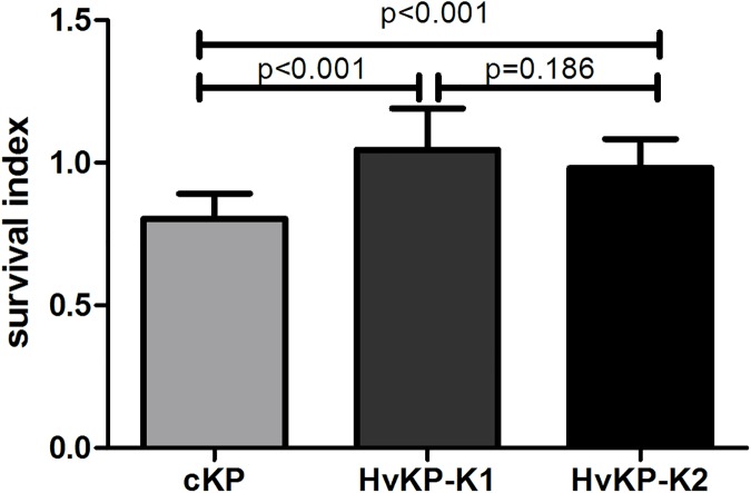
Fig 2
Survival of cKP, HvKP-K1 and HvKP-K2 within human neutrophils.
The survival index was calculated with the equation described in the method. The survival index of cKP (15 isolates) was lower than that of HvKP-K1 (16 isolates) or HvKP-K2 (14 isolates). Each strain ...
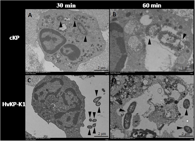
Fig 3
Examination under transmission electron microscopy.
Phagocytosed cKP (3 isolates, A) and HvKP-K1 (3 isolates, C) by neutrophils at 30 min (u00d73K); lysed cKP lack of intact cell wall (3 isolates, B) and binary fission multiplying HvKP-K1 (3 isolates, ...
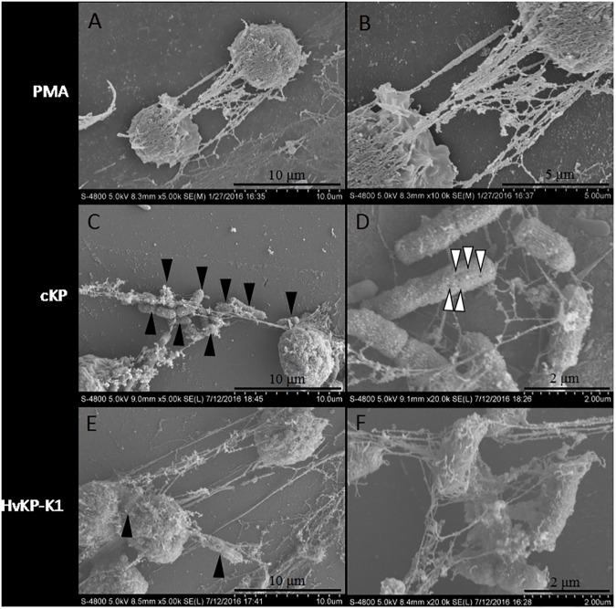
Fig 4
NETs under scanning electron microscopy.
NETs induced by PMA (A and B), cKP (3 isolates, C and D), and HvKP-K1 (3 isolates, E and F). More cKP (C) than HvKP-K1 (E) were trapped in NETs by magnification of 5K. The pores (indicated by white ar...
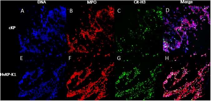
Fig 5
Immunofluorescence staining under confocal microscopy.
Neutrophils challenged by cKP (9 isolates, A-D) and HvKP-K1 (9 isolates, E-H) were stained for DNA (DAPI, blue, A and E), myeloperoxidase (MPO, red, B and F) and citrullinated histone H3 (cit-H3, gree...
Figure images are served from the NIH/NLM PubMed Central Open Access Subset or Europe PMC; copyright remains with the publishers and authors.
💬 Discussion
0 commentsNo comments yet. Be the first to start a discussion!
Leave a Comment