Abstract
Voltage-gated sodium (NaV) channels initiate action potentials in nerve, muscle, and other electrically excitable cells. The structural basis of voltage gating is uncertain because the resting state exists only at deeply negative membrane potentials. To stabilize the resting conformation, we inserted voltage-shifting mutations and introduced a disulfide crosslink in the VS of the ancestral bacterial sodium channel NaVAb. Here, we present a cryo-EM structure of the resting state and a complete voltage-dependent gating mechanism. The S4 segment of the VS is drawn intracellularly, with three gating charges passing through the transmembrane electric field. This movement forms an elbow connecting S4 to the S4-S5 linker, tightens the collar around the S6 activation gate, and prevents its opening. Our structure supports the classical "sliding helix" mechanism of voltage sensing and provides a complete gating mechanism for voltage sensor function, pore opening, and activation-gate closure based on high-resolution structures of a single sodium channel protein.
🔬 Techniques
🔭 Microscopes
🧬 Organisms
💻 Software
✨ Fluorophores
🧪 Sample Preparation
🏭 Microscope Brands
🧪 Reagent Suppliers
💻 Software Details
💾 Data Repositories
🏛️ Research Organizations (ROR)
Affiliated research institutions:
📋 Methods
CONTACT FOR REAGENT AND RESOURCE SHARING
Further information and requests for resources and reagents should be directed to and will be fulfilled by the Lead Contact, William A. Catterall ( wcatt@uw.edu ).
EXPERIMENTAL MODEL AND SUBJECT DETAILS Microbe strains E. coli
GC10 was cultured at 37°C in LB medium supplemented with 100μg/ml of ampicillin for plasmid DNA extraction. E. coli BL21(DE3) was cultured in MagicMedia E. coli expression medium at 37°C for screening of disulfide-crosslinking mutants. E. coli DH10Bac was cultured at 37°C in LB medium supplemented with 50 µg/mL kanamycin sulfate, 7 µg/mL gentamicin and 10 µg/mL tetracycline for bacmid production. Cell lines Sf9 ( Spodoptera frugiperda ) insect cells were maintained in Grace’s Insect Medium and supplemented with 10% FBS and penicillin/streptomycin at 27°C and passaged at 80–95% confluence for electrophysiology. Sf9 and Hi5 ( Trichoplusia ni ) insect cells were maintained and infected in Grace’s Insect Medium supplemented with 7.5% FBS and glutamine/penicillin/streptomycin at 27°C for baculovirus production and protein expression, respectively. METHOD DETAILS Screening of disulfide-crosslinking mutants in E. coli We screened for favorable Cys pairs that could disulfide-lock the resting state of Na V Ab in E. coli because of its ease of expression of large numbers of constructs and its very negative membrane potential (~ −160 mV at early stationary phase) ( Bot and Prodan, 2010 ) that would stabilize the resting state. Double-Cys mutations were designed for disulfide-crosslinking experiments based on a hypothesis that the S4 helix moves vertically across the membrane as a function of membrane potential and exchanges interactions between S4 residues from one subunit with S5 residues from a neighboring subunit ( Shimomura et al., 2011 ). Therefore, favorable interactions could result in inter-subunit disulfide crosslinking that can be visualized by SDS-PAGE and immunoblotting as a measure of formation of higher order oligomers. The gene encoding N-terminal FLAG-tagged Na V Ab was PCR-amplified from pFastBacDual FLAG-Na V Ab vector ( Payandeh et al., 2012 ) and cloned into pET21b vector (Life Technologies). Oligonucleotide primers containing a Cys mutation on S5 helix at positions 143, 146, 147, 150, or 151 were used to generate a set of vectors containing five different S5 Cys mutants by QuickChange Lightening site-directed mutagenesis (Agilent). Oligonucleotide primers containing a Cys mutation on S3-S4 loop to S4 helix at positions S93, G94, F95, E96, I97, L98, R99, V100, R102, L103, R105, or L106 were used to generate a set of double-Cys mutations using the set of vectors containing S5 cysteine mutants as templates. All clones were verified by DNA sequencing to contain correct mutations. The double-Cys vectors (60 in total) were transformed into the BL21(DE3) strain of E. coli , and the cultures were grown in 1 ml of MagicMedia E. coli auto expression medium (Life Technology) for 3 days at 37°C ( Shimomura et al., 2011 ). Cells were harvested by centrifugation at 3000xg for 20 min. Cell pellets were resuspended in a buffer containing 20 mM Tris-HCl and 140 mM NaCl. Ten μl of cell suspension were mixed with 40 μl of 2X LDS sample buffer (Life Technologies) and assayed by SDS-PAGE and immunoblotting. For SDS-PAGE, 10 μl of the LDS samples were loaded on 4–12% Bis-Tris polyacrylamide gel and electrophoresed with 1X MOPS-SDS running buffer (Life Technologies). For immunoblots, protein bands from SDS-PAGE gels were transferred onto nitrocellulose membranes; blocked with 5% non-fat dried milk in Tris-saline buffer with 0.05% Tween 20; probed by mouse anti-FLAG M2 IgG primary antibody (MilliporeSigma) followed by goat anti-mouse IgG-HRP conjugated secondary antibody (Jackson ImmonuResearch Laboratories); detected with AmerSham ECL Prime Western Blotting Detection Reagent (GE Healthcare Life Sciences); and imaged using Bio-Rad ChemiDoc XRS+ Imaging System (Bio-Rad). Disulfide-crosslinking assays in Hi5 ( T. ni ) insect cells Our previous studies showed that the S4 segment of Na V Ab expressed in cultured cells can be disulfide-locked in resting, intermediate, and activated conformations, as assessed by voltage clamp analysis, by insertion of specific pairs of Cys residues and catalysis of disulfide bond formation with Cu-1,10-phenanthroline ( DeCaen et al., 2011 ; DeCaen et al., 2009 ; DeCaen et al., 2008 ). Plasmids containing double-Cys mutations were made using the same procedure as above but pFastBacDual FLAG-Na V Ab was used as a template. Additional mutations including N49K, L109A, M116V, and 28-residue C-terminal truncation with stop codon (Δ28) were introduced by mutagenesis using the same procedure. Baculoviruses containing double-Cys mutants were prepared using the Bac-to-Bac protocol according to the manufacturer with Sf9 insect cells (Life Technologies). Third passage (P3) baculoviruses were used to infect Hi5 cells in single layer-culture dishes, and the cells were incubated at 27°C for 48–72 h. Cells were harvested by centrifugation. For the Control group, cell pellets from half of one dish were resuspended in 10 ml of 20 mM Tris-HCl, pH 7.5 and 140 mM NaCl and incubated at room temperature for 1 h. For the Hyperpolarization-Oxidation group, cell pellets from the other half of a dish were resuspended in 10 ml of 20 mM Tris-HCl (sodium free), pH 7.5, 140 mM choline chloride, and 10 μg/ml Gramicidin (MilliporeSigma) to hyperpolarize cell membrane potential, followed by an addition of 100 μM of copper (II) phenanthroline -- a mild oxidizing agent made freshly by mixing CuSO 4 and 1,10-phenanthroline solutions at 1:3 molar ratio and incubation at room temperature for 1 h. To quench the oxidation reaction, 1 mM EDTA was added to the cell suspension. Cells from both groups were centrifuged and cell pellets were resuspended in 1 mL of 20 mM Tris-HCl and 140 mM NaCl. Five μl of cell suspension were mixed with 45 μl of 2X LDS sample buffer plus 25 mM iodoacetamide (prepared freshly within the hour) and protease inhibitor cocktails (MilliporeSigma). The samples were subsequently assayed by SDS-PAGE and immunoblotting as described above; imaged using Bio-Rad ChemiDoc XRS+ Imaging System; and quantified using Bio-Rad Image Lab Software according to manufacturer (Bio-Rad).
Show full methods section
CONTACT FOR REAGENT AND RESOURCE SHARING
Further information and requests for resources and reagents should be directed to and will be fulfilled by the Lead Contact, William A. Catterall ( wcatt@uw.edu ).
EXPERIMENTAL MODEL AND SUBJECT DETAILS Microbe strains E. coli
GC10 was cultured at 37°C in LB medium supplemented with 100μg/ml of ampicillin for plasmid DNA extraction. E. coli BL21(DE3) was cultured in MagicMedia E. coli expression medium at 37°C for screening of disulfide-crosslinking mutants. E. coli DH10Bac was cultured at 37°C in LB medium supplemented with 50 µg/mL kanamycin sulfate, 7 µg/mL gentamicin and 10 µg/mL tetracycline for bacmid production. Cell lines Sf9 ( Spodoptera frugiperda ) insect cells were maintained in Grace’s Insect Medium and supplemented with 10% FBS and penicillin/streptomycin at 27°C and passaged at 80–95% confluence for electrophysiology. Sf9 and Hi5 ( Trichoplusia ni ) insect cells were maintained and infected in Grace’s Insect Medium supplemented with 7.5% FBS and glutamine/penicillin/streptomycin at 27°C for baculovirus production and protein expression, respectively. METHOD DETAILS Screening of disulfide-crosslinking mutants in E. coli We screened for favorable Cys pairs that could disulfide-lock the resting state of Na V Ab in E. coli because of its ease of expression of large numbers of constructs and its very negative membrane potential (~ −160 mV at early stationary phase) ( Bot and Prodan, 2010 ) that would stabilize the resting state. Double-Cys mutations were designed for disulfide-crosslinking experiments based on a hypothesis that the S4 helix moves vertically across the membrane as a function of membrane potential and exchanges interactions between S4 residues from one subunit with S5 residues from a neighboring subunit ( Shimomura et al., 2011 ). Therefore, favorable interactions could result in inter-subunit disulfide crosslinking that can be visualized by SDS-PAGE and immunoblotting as a measure of formation of higher order oligomers. The gene encoding N-terminal FLAG-tagged Na V Ab was PCR-amplified from pFastBacDual FLAG-Na V Ab vector ( Payandeh et al., 2012 ) and cloned into pET21b vector (Life Technologies). Oligonucleotide primers containing a Cys mutation on S5 helix at positions 143, 146, 147, 150, or 151 were used to generate a set of vectors containing five different S5 Cys mutants by QuickChange Lightening site-directed mutagenesis (Agilent). Oligonucleotide primers containing a Cys mutation on S3-S4 loop to S4 helix at positions S93, G94, F95, E96, I97, L98, R99, V100, R102, L103, R105, or L106 were used to generate a set of double-Cys mutations using the set of vectors containing S5 cysteine mutants as templates. All clones were verified by DNA sequencing to contain correct mutations. The double-Cys vectors (60 in total) were transformed into the BL21(DE3) strain of E. coli , and the cultures were grown in 1 ml of MagicMedia E. coli auto expression medium (Life Technology) for 3 days at 37°C ( Shimomura et al., 2011 ). Cells were harvested by centrifugation at 3000xg for 20 min. Cell pellets were resuspended in a buffer containing 20 mM Tris-HCl and 140 mM NaCl. Ten μl of cell suspension were mixed with 40 μl of 2X LDS sample buffer (Life Technologies) and assayed by SDS-PAGE and immunoblotting. For SDS-PAGE, 10 μl of the LDS samples were loaded on 4–12% Bis-Tris polyacrylamide gel and electrophoresed with 1X MOPS-SDS running buffer (Life Technologies). For immunoblots, protein bands from SDS-PAGE gels were transferred onto nitrocellulose membranes; blocked with 5% non-fat dried milk in Tris-saline buffer with 0.05% Tween 20; probed by mouse anti-FLAG M2 IgG primary antibody (MilliporeSigma) followed by goat anti-mouse IgG-HRP conjugated secondary antibody (Jackson ImmonuResearch Laboratories); detected with AmerSham ECL Prime Western Blotting Detection Reagent (GE Healthcare Life Sciences); and imaged using Bio-Rad ChemiDoc XRS+ Imaging System (Bio-Rad). Disulfide-crosslinking assays in Hi5 ( T. ni ) insect cells Our previous studies showed that the S4 segment of Na V Ab expressed in cultured cells can be disulfide-locked in resting, intermediate, and activated conformations, as assessed by voltage clamp analysis, by insertion of specific pairs of Cys residues and catalysis of disulfide bond formation with Cu-1,10-phenanthroline ( DeCaen et al., 2011 ; DeCaen et al., 2009 ; DeCaen et al., 2008 ). Plasmids containing double-Cys mutations were made using the same procedure as above but pFastBacDual FLAG-Na V Ab was used as a template. Additional mutations including N49K, L109A, M116V, and 28-residue C-terminal truncation with stop codon (Δ28) were introduced by mutagenesis using the same procedure. Baculoviruses containing double-Cys mutants were prepared using the Bac-to-Bac protocol according to the manufacturer with Sf9 insect cells (Life Technologies). Third passage (P3) baculoviruses were used to infect Hi5 cells in single layer-culture dishes, and the cells were incubated at 27°C for 48–72 h. Cells were harvested by centrifugation. For the Control group, cell pellets from half of one dish were resuspended in 10 ml of 20 mM Tris-HCl, pH 7.5 and 140 mM NaCl and incubated at room temperature for 1 h. For the Hyperpolarization-Oxidation group, cell pellets from the other half of a dish were resuspended in 10 ml of 20 mM Tris-HCl (sodium free), pH 7.5, 140 mM choline chloride, and 10 μg/ml Gramicidin (MilliporeSigma) to hyperpolarize cell membrane potential, followed by an addition of 100 μM of copper (II) phenanthroline -- a mild oxidizing agent made freshly by mixing CuSO 4 and 1,10-phenanthroline solutions at 1:3 molar ratio and incubation at room temperature for 1 h. To quench the oxidation reaction, 1 mM EDTA was added to the cell suspension. Cells from both groups were centrifuged and cell pellets were resuspended in 1 mL of 20 mM Tris-HCl and 140 mM NaCl. Five μl of cell suspension were mixed with 45 μl of 2X LDS sample buffer plus 25 mM iodoacetamide (prepared freshly within the hour) and protease inhibitor cocktails (MilliporeSigma). The samples were subsequently assayed by SDS-PAGE and immunoblotting as described above; imaged using Bio-Rad ChemiDoc XRS+ Imaging System; and quantified using Bio-Rad Image Lab Software according to manufacturer (Bio-Rad).
Channel expression for electrophysiology
Sf9 cells below passage 25 were transiently transfected in 24-well plates using a 2:4 ratio of µg DNA: µl PolyJet transfection reagent (SignaGen) in unsupplemented Grace’s insect media with a plasmid encoding GFP-P2A-Na V Ab gene under an OpIE2 promoter modified from pIZT vector (Life Technologies) and purified using an endonuclease-free miniprep kit (Omega). To construct the modified vector, the eGFP was moved from the Zeocin resistance gene to upstream of the FLAG-channel sequence, and a GSG-P2A site (porcine teschovirus-1) was inserted between the two using standard site directed mutagenesis, restriction digestion, and ligation methods. This was done to better identify transfected cells with optimal expression (0.5–3 nA peak current) by utilizing the 1:1 stoichiometric ratio of GFP to the channel conferred by the P2A self-cleaving peptide. Cells were replated onto 10 mm glass coverslips 24 hours post-transfection and used for recording for the following 5–7 days.
Electrophysiological Data Collection 1.5–3
MΩ glass pipettes were used to patch-clamp fluorescent cells, and currents were recorded via whole-cell voltage clamp (EPC10, Pulse, HEKA), sampled at 250 kHz, and filtered at 3 kH. Membrane capacitance ranged from 5–25 pF. Rs was below 10 MΩ, with a 2 µs lag and at least 70% compensation. A P/4 leak subtraction protocol was used. In order to analyze Na V Ab mutants with positively shifted voltage dependence, solutions were designed to give a reversal potential of 0 mV for Na + . Intracellular solution (in mM): NaCl (35) CsF (105) EGTA (10) HEPES (10). Extracellular solution contained: NaCl (35) NMDG-Cl (105) MgCl 2 (2) CaCl 2 (2) HEPES (10). The pH for both solutions was 7.4, adjusted with CsOH and NMDG+, respectively. Osmolarity was adjusted with sucrose to match that of the cells’ media each day. The measured liquid junction potential was −7 mV for all recordings, and the reported voltages were not adjusted. Na V Ab /N49K, which activates at negative voltages, was studied as previously described ( Gamal El-Din et al., 2013 ). Briefly, Hi5 cells were infected with baculovirus and recordings were made 18–24 hours later. The intracellular solution composition was as above, and the extracellular solution was identical except NaCl concentration was 140 mM, no NMDG was used, and pH was adjusted with NaOH. Current families were generated by holding cells at −150 mV (−120 for Na V Ab/N49K) and stimulating with a series of 50-ms pulses at 10 mV intervals. To determine reversal potential of outward-only current, peak current was elicited by pulsing to an appropriate potential for each construct and decreasing the voltage in 10 mV steps until substantial inward current was seen in the tail currents. The time between the start of the initial voltage step and the subsequent steps was determined for each construct as roughly the time to peak (data not shown). An agar bridge consisting of extracellular solution with 3% Agar was placed between the reference electrode and the bath in experiments utilizing perfusion of extracellular solution containing reducing agents.
Expression and protein purification of Na V
Ab activated state disulfide-crosslinked mutant Hi5 cells were infected with P3 virus for Na V Ab V100C/Q150C double-Cys mutant (Δ28 construct ( Gamal El-Din et al., 2018a )) and incubated for 48–72 h at 27°C. Cells were harvested by centrifugation. Cell pellets were subjected to oxidation treatment in Buffer A (50 mM Tris-HCl pH 7.5 and 200 mM NaCl, 20 ml per cells from 1 dish) instead of hyperpolarization buffer using the procedure as described above. Cells were harvested by centrifugation, resuspended in Buffer B (Buffer A supplemented with 1 mM PMSF, 2X SigmaFast protease inhibitor cocktails, benzamidine HCl, and DNAse I), lysed by sonication, and the membranes solubilized as described above. After centrifugation, the supernatant was incubated with FLAG-M2 affinity gel (MilliporeSigma) for 1 hour at 4°C with gentle mixing. The resins were washed with Buffer C (Buffer A supplemented with 0.12% digitonin) and bound protein was eluted with Buffer C supplemented with 200 μM FLAG peptide. Eluted protein was concentrated to 1 ml using Vivaspin20 100 kDa MWCO and further purified with Superdex S200 size-exclusion chromatography with using 10 mM Tris-HCl pH 7.5, 100 mM NaCl, and 0.12% digitonin as a running buffer. Elution fractions were evaluated using SDS-PAGE and peak fractions were combined and concentrated using Vivaspin6 100 kDa MWCO to final concentration ~20 mg/ml. Na V Ab/G94C/Q150C Δ28 mutant was expressed and purified as described above, but without the oxidation treatment. FLAG-eluted protein was treated with 50mM DTT and subsequent purification step contains 5 mM DTT in the buffers. Crystallization of Na V Ab activated state disulfide-crosslinked mutant Na V Ab-bicelle complexes were prepared by mixing Na V Ab double Cys mutant with 10% bicelle (7.5% DMPC and 2.5% CHAPSO) at 1:5 volume ratio and screened for crystallization conditions as described previously. Best crystals appeared under 1.8–2.0 M ammonium sulfate and 0.1 M sodium citrate, pH 4.8–5.0 for Na V Ab/V100C/Q150C Δ28 mutant and pH 5.6 for Na V Ab/G94C/Q150C Δ28 mutant. Crystals were cryo-protected by step-wise transfers to a series of cryo-protectant solutions containing 6–30% glucose with the same concentration of ammonium sulfate and sodium citrate pH 4.8 for Na V Ab/V100C/Q150C Δ28 and pH 6.0 for Na V Ab/G94C/Q150C Δ28. X-ray data collection and structural determination Crystals were tested for diffraction and data were collected at Advanced Light Source (ALS) beamline 8.2.1 and 8.2.2 (HHMI). Diffraction data were processed using HKL2000 software ( Otwinowski and Minor, 1997 ) and structures were determined by molecular replacement using PHASER ( McCoy et al., 2007 ) and refined using REFMAC ( Murshudov et al., 2011 ) in CCP4 program suite ( Winn et al., 2011 ). Manual model building and local real space refinement was carried out in COOT ( Emsley et al., 2010 ), followed by structure refinement in REFMAC ( Table S1 ).
Expression and purification of Na V
Ab/KAV/G94C/Q150C in the resting state We developed a fusion protein strategy to generate MBP-NavAb fusion proteins for structural study by cryo-EM. Several rigid linkers between MBP and Na V Ab were designed and tested by protein purification and cryo-EM imaging using a Tecnai G2 F20 microscope (FEI). The best linker that produced the most homogenous population with good particle image contrast was chosen for the disulfide-locked resting state Na V Ab. The gene encoding MBP from E. coli was N-terminally fused to Na V Ab/KAV/G94C/Q150C with an Asn-Ala linker sequence by PCR-driven overlap extension method ( Heckman and Pease, 2007 ), and the fusion gene was cloned into pFastBacDual vector (Life Technologies). Baculovirus harboring the fusion gene was prepared using the Bac-to-Bac protocol. Hi5 cells were infected with P3 virus and incubated for 72–96 hours at 27°C for maximum expression. Cells were harvested by centrifugation. Cell pellets were subjected to hyperpolarization-oxidation treatment by re-suspending in hyperpolarization buffer (20 mM Tris-HCl (sodium free) pH 7.5, 140 mM choline chloride, 20 ml per cells from 1 dish) plus 100 μM copper (II) phenanthroline prepared as described above and incubated at room temperature for 1 h with gently shaking. The reaction was quenched with 1 mM EDTA, and the cells were pelleted by centrifugation and washed with buffer A followed by centrifugation. Cell pellets were resuspended in Buffer B, lysed by sonication and membranes were solubilized with 1% high purity digitonin (Calbiochem) for 1 h at 4°C with gentle mixing. The mixture was centrifuged at 15,000xg for 30 min at 4°C, and the supernatant was incubated with amylose resin (NEB) for 1 h at 4°C with gentle mixing. The resins were washed with Buffer C and bound protein was eluted with Buffer C supplemented with 10 mM maltose). Eluted protein was concentrated to 1 ml using Vivaspin20 100kDa MWCO and further purified with Superose 6 Increase size-exclusion chromatography using 10 mM Tris HCl pH 7.5, 100 mM NaCl, and 0.12% digitonin as a column running buffer. Elution fractions were evaluated using SDS-PAGE and peak fractions were combined and concentrated using Vivaspin6 100 kDa MWCO to final concentration ~5–8 mg/ml.
Cryo-EM sample preparation and data acquisition
Cryo-EM grids were prepared by applying freshly purified MBP-disulfide locked resting state Na V Ab in digitonin detergent to glow-discharged holey carbon grids (C-Flat Cu R1.2/1.3, EMS). Grids were manually blotted for 4 to 5 s and immediately plunge-frozen in liquid ethane cooled by liquid nitrogen. Cryo-EM data were recorded on a Titan Krios (FEI) operated at 300 kV, equipped with a GIF-quantum energy filter (Gatan) at 20 eV slit width and a K2 Summit direct detector (Gatan). LEGINON ( Suloway et al., 2005 ) was used for automated data collection. Movies were collected at a nominal magnification of 130,000x in super-resolution mode resulting in a pixel size of 0.528 Å, with a small defocus range of –0.5 to –2.5 μm. The dose rate on the camera was set to be ~8 counts per physical pixel per second. The total exposure time was 8.6 s (0.2 s/frame), leading to a total accumulate dose of 60 electrons per Å 2 on the specimen ( Table S2 ).
Cryo-EM Image processing
Movie frames were aligned and 2x binned to a pixel size off 1.056 Å using MotionCor2 ( Zheng et al., 2017 ) and the contrast transfer function parameters for each motion-corrected image were estimated using Gctf ( Zhang, 2016 ). Subsequent image processing steps were performed using RELION ( Scheres, 2012 ) on an NVIDIA GPU-accelerated workstation. Particles were auto-picked using templates derived from 2D class averages that were initially generated from manually picked particles from ~50 images. A total of ~476,000 particles from ~5,000 good images were extracted into 304 × 304-pixel boxes and subjected to several rounds of 2D classification to remove bad particles. 3D classification and refinement were carried out in RELION using the crystal structure of a Na V Ab pore-only model that had been low-pass filtered to 60 Å as an initial template. The resulting 3D reconstructions were routinely better than 5 Å resolution. Subsequently, 333,899 particles were subjected to cycles of global angular search in cisTEM ( Grant et al., 2018 ) with an earlier reconstruction from RELION as a starting reference and a 3D mask that included only the transmembrane region and low-pass filtered the region outside to 50 Å. After global angular search, the particles were further subjected to cycles of local angular search with the resolution limit set to 8 Å for particle alignment and the 3D mask. The resolution was estimated using the gold-standard Fourier shell correlation (FSC) 0.143 criterion ( Henderson et al., 2012 ) to be 4.0 Å ( Table S2 ). Local resolution was evaluated using MonoRes ( Vilas et al., 2018 ). Model building and refinement of the resting state structure The Na V Ab crystal structure (PDB 3RVY) was initially docked into cryo-EM density map using Chimera ( Pettersen et al., 2004 ). The atomic model was then rebuilt using Coot ( Emsley et al., 2010 ) and subsequently refined using the real space refinement feature in Phenix ( Adams et al., 2010 ) ( Table S2 ).
QUANTIFICATION AND STATISTICAL ANALYSIS Analysis of electrophysiological data
Pulse data was analyzed using IGOR Pro (6.37). Peak current at each voltage of the current family was plotted as a function of the stimulus voltage to visualize the current vs voltage (I/V) relationship. As the I/V curves contained only outward current, the reversal potential (V rev ) was determined from the tail currents at peak activation. The instantaneous current of the tail current family was plotted as a function of voltage, fit with a line near 0 mV, and the X intercept was used as V rev to generate the conductance vs voltage (G/V) curve from the I/V curve by calculating G=I/(V m −V rev ). The calculated V rev was near 7 mV for all recordings. Normalized G/V curves were fit with a simple two-state Boltzmann equation 1/(1+exp(V 1/2 −V m )/k) in which V m is the stimulus potential, V 1/2 is the half-activation voltage, and k is a slope factor. I/V plots of inward currents for NaVAb/N49K were fit with (V m −V rev )*(I min /(1+exp((V m −V 1/2 )/k)) to generate the above Boltzmann curve fit to normalized G/V.
Quantification and analysis of immunoblot data
Quantification for bands from immunoblots in Figures S2A , and S2C were performed using Bio-Rad Image Lab Software version 5.1 according to manufacturer. Details of experiments can be found in the figure legends. Data are represented as mean ± standard error of the mean (SEM) from 5 independent experiments (n=5). Significance was determined by Student’s t-test. Estimation Protein concentrations were estimated using an A280 extinction coefficient of 33,920 M −1 cm −1 on a spectrophotometer. Global resolution estimation of cryo-EM density map is based on the 0.143 Fourier Shell Correlation criterion.
DATA AND SOFTWARE AVAILABILITY
The coordinates and structure factors for the reported crystal structures have been deposited in the Protein Data Bank under accession codes 6P6X (NaVAb G94C/Q150C Δ28) and 6P6Y (NaVAb V100C/Q150C Δ28). Cryo-EM map has been deposited in the Electron Microscopy Data Bank (EMDB) under accession code EMD-20265 (MBP-NaVAb KAV/G94C/Q150C). The coordinates of the NaVAb KAV/G94C/Q150C atomic model have been deposited in the PDB under accession code 6P6W.
EXPERIMENTAL MODEL AND SUBJECT DETAILS Microbe strains E. coli
GC10 was cultured at 37°C in LB medium supplemented with 100μg/ml of ampicillin for plasmid DNA extraction. E. coli BL21(DE3) was cultured in MagicMedia E. coli expression medium at 37°C for screening of disulfide-crosslinking mutants. E. coli DH10Bac was cultured at 37°C in LB medium supplemented with 50 µg/mL kanamycin sulfate, 7 µg/mL gentamicin and 10 µg/mL tetracycline for bacmid production. Cell lines Sf9 ( Spodoptera frugiperda ) insect cells were maintained in Grace’s Insect Medium and supplemented with 10% FBS and penicillin/streptomycin at 27°C and passaged at 80–95% confluence for electrophysiology. Sf9 and Hi5 ( Trichoplusia ni ) insect cells were maintained and infected in Grace’s Insect Medium supplemented with 7.5% FBS and glutamine/penicillin/streptomycin at 27°C for baculovirus production and protein expression, respectively.
METHOD DETAILS Screening of disulfide-crosslinking mutants in E. coli We screened for favorable Cys pairs that could disulfide-lock the resting state of Na V Ab in E. coli because of its ease of expression of large numbers of constructs and its very negative membrane potential (~ −160 mV at early stationary phase) ( Bot and Prodan, 2010 ) that would stabilize the resting state. Double-Cys mutations were designed for disulfide-crosslinking experiments based on a hypothesis that the S4 helix moves vertically across the membrane as a function of membrane potential and exchanges interactions between S4 residues from one subunit with S5 residues from a neighboring subunit ( Shimomura et al., 2011 ). Therefore, favorable interactions could result in inter-subunit disulfide crosslinking that can be visualized by SDS-PAGE and immunoblotting as a measure of formation of higher order oligomers. The gene encoding N-terminal FLAG-tagged Na V Ab was PCR-amplified from pFastBacDual FLAG-Na V Ab vector ( Payandeh et al., 2012 ) and cloned into pET21b vector (Life Technologies). Oligonucleotide primers containing a Cys mutation on S5 helix at positions 143, 146, 147, 150, or 151 were used to generate a set of vectors containing five different S5 Cys mutants by QuickChange Lightening site-directed mutagenesis (Agilent). Oligonucleotide primers containing a Cys mutation on S3-S4 loop to S4 helix at positions S93, G94, F95, E96, I97, L98, R99, V100, R102, L103, R105, or L106 were used to generate a set of double-Cys mutations using the set of vectors containing S5 cysteine mutants as templates. All clones were verified by DNA sequencing to contain correct mutations. The double-Cys vectors (60 in total) were transformed into the BL21(DE3) strain of E. coli , and the cultures were grown in 1 ml of MagicMedia E. coli auto expression medium (Life Technology) for 3 days at 37°C ( Shimomura et al., 2011 ). Cells were harvested by centrifugation at 3000xg for 20 min. Cell pellets were resuspended in a buffer containing 20 mM Tris-HCl and 140 mM NaCl. Ten μl of cell suspension were mixed with 40 μl of 2X LDS sample buffer (Life Technologies) and assayed by SDS-PAGE and immunoblotting. For SDS-PAGE, 10 μl of the LDS samples were loaded on 4–12% Bis-Tris polyacrylamide gel and electrophoresed with 1X MOPS-SDS running buffer (Life Technologies). For immunoblots, protein bands from SDS-PAGE gels were transferred onto nitrocellulose membranes; blocked with 5% non-fat dried milk in Tris-saline buffer with 0.05% Tween 20; probed by mouse anti-FLAG M2 IgG primary antibody (MilliporeSigma) followed by goat anti-mouse IgG-HRP conjugated secondary antibody (Jackson ImmonuResearch Laboratories); detected with AmerSham ECL Prime Western Blotting Detection Reagent (GE Healthcare Life Sciences); and imaged using Bio-Rad ChemiDoc XRS+ Imaging System (Bio-Rad). Disulfide-crosslinking assays in Hi5 ( T. ni ) insect cells Our previous studies showed that the S4 segment of Na V Ab expressed in cultured cells can be disulfide-locked in resting, intermediate, and activated conformations, as assessed by voltage clamp analysis, by insertion of specific pairs of Cys residues and catalysis of disulfide bond formation with Cu-1,10-phenanthroline ( DeCaen et al., 2011 ; DeCaen et al., 2009 ; DeCaen et al., 2008 ). Plasmids containing double-Cys mutations were made using the same procedure as above but pFastBacDual FLAG-Na V Ab was used as a template. Additional mutations including N49K, L109A, M116V, and 28-residue C-terminal truncation with stop codon (Δ28) were introduced by mutagenesis using the same procedure. Baculoviruses containing double-Cys mutants were prepared using the Bac-to-Bac protocol according to the manufacturer with Sf9 insect cells (Life Technologies). Third passage (P3) baculoviruses were used to infect Hi5 cells in single layer-culture dishes, and the cells were incubated at 27°C for 48–72 h. Cells were harvested by centrifugation. For the Control group, cell pellets from half of one dish were resuspended in 10 ml of 20 mM Tris-HCl, pH 7.5 and 140 mM NaCl and incubated at room temperature for 1 h. For the Hyperpolarization-Oxidation group, cell pellets from the other half of a dish were resuspended in 10 ml of 20 mM Tris-HCl (sodium free), pH 7.5, 140 mM choline chloride, and 10 μg/ml Gramicidin (MilliporeSigma) to hyperpolarize cell membrane potential, followed by an addition of 100 μM of copper (II) phenanthroline -- a mild oxidizing agent made freshly by mixing CuSO 4 and 1,10-phenanthroline solutions at 1:3 molar ratio and incubation at room temperature for 1 h. To quench the oxidation reaction, 1 mM EDTA was added to the cell suspension. Cells from both groups were centrifuged and cell pellets were resuspended in 1 mL of 20 mM Tris-HCl and 140 mM NaCl. Five μl of cell suspension were mixed with 45 μl of 2X LDS sample buffer plus 25 mM iodoacetamide (prepared freshly within the hour) and protease inhibitor cocktails (MilliporeSigma). The samples were subsequently assayed by SDS-PAGE and immunoblotting as described above; imaged using Bio-Rad ChemiDoc XRS+ Imaging System; and quantified using Bio-Rad Image Lab Software according to manufacturer (Bio-Rad).
Channel expression for electrophysiology
Sf9 cells below passage 25 were transiently transfected in 24-well plates using a 2:4 ratio of µg DNA: µl PolyJet transfection reagent (SignaGen) in unsupplemented Grace’s insect media with a plasmid encoding GFP-P2A-Na V Ab gene under an OpIE2 promoter modified from pIZT vector (Life Technologies) and purified using an endonuclease-free miniprep kit (Omega). To construct the modified vector, the eGFP was moved from the Zeocin resistance gene to upstream of the FLAG-channel sequence, and a GSG-P2A site (porcine teschovirus-1) was inserted between the two using standard site directed mutagenesis, restriction digestion, and ligation methods. This was done to better identify transfected cells with optimal expression (0.5–3 nA peak current) by utilizing the 1:1 stoichiometric ratio of GFP to the channel conferred by the P2A self-cleaving peptide. Cells were replated onto 10 mm glass coverslips 24 hours post-transfection and used for recording for the following 5–7 days.
Electrophysiological Data Collection 1.5–3
MΩ glass pipettes were used to patch-clamp fluorescent cells, and currents were recorded via whole-cell voltage clamp (EPC10, Pulse, HEKA), sampled at 250 kHz, and filtered at 3 kH. Membrane capacitance ranged from 5–25 pF. Rs was below 10 MΩ, with a 2 µs lag and at least 70% compensation. A P/4 leak subtraction protocol was used. In order to analyze Na V Ab mutants with positively shifted voltage dependence, solutions were designed to give a reversal potential of 0 mV for Na + . Intracellular solution (in mM): NaCl (35) CsF (105) EGTA (10) HEPES (10). Extracellular solution contained: NaCl (35) NMDG-Cl (105) MgCl 2 (2) CaCl 2 (2) HEPES (10). The pH for both solutions was 7.4, adjusted with CsOH and NMDG+, respectively. Osmolarity was adjusted with sucrose to match that of the cells’ media each day. The measured liquid junction potential was −7 mV for all recordings, and the reported voltages were not adjusted. Na V Ab /N49K, which activates at negative voltages, was studied as previously described ( Gamal El-Din et al., 2013 ). Briefly, Hi5 cells were infected with baculovirus and recordings were made 18–24 hours later. The intracellular solution composition was as above, and the extracellular solution was identical except NaCl concentration was 140 mM, no NMDG was used, and pH was adjusted with NaOH. Current families were generated by holding cells at −150 mV (−120 for Na V Ab/N49K) and stimulating with a series of 50-ms pulses at 10 mV intervals. To determine reversal potential of outward-only current, peak current was elicited by pulsing to an appropriate potential for each construct and decreasing the voltage in 10 mV steps until substantial inward current was seen in the tail currents. The time between the start of the initial voltage step and the subsequent steps was determined for each construct as roughly the time to peak (data not shown). An agar bridge consisting of extracellular solution with 3% Agar was placed between the reference electrode and the bath in experiments utilizing perfusion of extracellular solution containing reducing agents.
Expression and protein purification of Na V
Ab activated state disulfide-crosslinked mutant Hi5 cells were infected with P3 virus for Na V Ab V100C/Q150C double-Cys mutant (Δ28 construct ( Gamal El-Din et al., 2018a )) and incubated for 48–72 h at 27°C. Cells were harvested by centrifugation. Cell pellets were subjected to oxidation treatment in Buffer A (50 mM Tris-HCl pH 7.5 and 200 mM NaCl, 20 ml per cells from 1 dish) instead of hyperpolarization buffer using the procedure as described above. Cells were harvested by centrifugation, resuspended in Buffer B (Buffer A supplemented with 1 mM PMSF, 2X SigmaFast protease inhibitor cocktails, benzamidine HCl, and DNAse I), lysed by sonication, and the membranes solubilized as described above. After centrifugation, the supernatant was incubated with FLAG-M2 affinity gel (MilliporeSigma) for 1 hour at 4°C with gentle mixing. The resins were washed with Buffer C (Buffer A supplemented with 0.12% digitonin) and bound protein was eluted with Buffer C supplemented with 200 μM FLAG peptide. Eluted protein was concentrated to 1 ml using Vivaspin20 100 kDa MWCO and further purified with Superdex S200 size-exclusion chromatography with using 10 mM Tris-HCl pH 7.5, 100 mM NaCl, and 0.12% digitonin as a running buffer. Elution fractions were evaluated using SDS-PAGE and peak fractions were combined and concentrated using Vivaspin6 100 kDa MWCO to final concentration ~20 mg/ml. Na V Ab/G94C/Q150C Δ28 mutant was expressed and purified as described above, but without the oxidation treatment. FLAG-eluted protein was treated with 50mM DTT and subsequent purification step contains 5 mM DTT in the buffers. Crystallization of Na V Ab activated state disulfide-crosslinked mutant Na V Ab-bicelle complexes were prepared by mixing Na V Ab double Cys mutant with 10% bicelle (7.5% DMPC and 2.5% CHAPSO) at 1:5 volume ratio and screened for crystallization conditions as described previously. Best crystals appeared under 1.8–2.0 M ammonium sulfate and 0.1 M sodium citrate, pH 4.8–5.0 for Na V Ab/V100C/Q150C Δ28 mutant and pH 5.6 for Na V Ab/G94C/Q150C Δ28 mutant. Crystals were cryo-protected by step-wise transfers to a series of cryo-protectant solutions containing 6–30% glucose with the same concentration of ammonium sulfate and sodium citrate pH 4.8 for Na V Ab/V100C/Q150C Δ28 and pH 6.0 for Na V Ab/G94C/Q150C Δ28. X-ray data collection and structural determination Crystals were tested for diffraction and data were collected at Advanced Light Source (ALS) beamline 8.2.1 and 8.2.2 (HHMI). Diffraction data were processed using HKL2000 software ( Otwinowski and Minor, 1997 ) and structures were determined by molecular replacement using PHASER ( McCoy et al., 2007 ) and refined using REFMAC ( Murshudov et al., 2011 ) in CCP4 program suite ( Winn et al., 2011 ). Manual model building and local real space refinement was carried out in COOT ( Emsley et al., 2010 ), followed by structure refinement in REFMAC ( Table S1 ).
Expression and purification of Na V
Ab/KAV/G94C/Q150C in the resting state We developed a fusion protein strategy to generate MBP-NavAb fusion proteins for structural study by cryo-EM. Several rigid linkers between MBP and Na V Ab were designed and tested by protein purification and cryo-EM imaging using a Tecnai G2 F20 microscope (FEI). The best linker that produced the most homogenous population with good particle image contrast was chosen for the disulfide-locked resting state Na V Ab. The gene encoding MBP from E. coli was N-terminally fused to Na V Ab/KAV/G94C/Q150C with an Asn-Ala linker sequence by PCR-driven overlap extension method ( Heckman and Pease, 2007 ), and the fusion gene was cloned into pFastBacDual vector (Life Technologies). Baculovirus harboring the fusion gene was prepared using the Bac-to-Bac protocol. Hi5 cells were infected with P3 virus and incubated for 72–96 hours at 27°C for maximum expression. Cells were harvested by centrifugation. Cell pellets were subjected to hyperpolarization-oxidation treatment by re-suspending in hyperpolarization buffer (20 mM Tris-HCl (sodium free) pH 7.5, 140 mM choline chloride, 20 ml per cells from 1 dish) plus 100 μM copper (II) phenanthroline prepared as described above and incubated at room temperature for 1 h with gently shaking. The reaction was quenched with 1 mM EDTA, and the cells were pelleted by centrifugation and washed with buffer A followed by centrifugation. Cell pellets were resuspended in Buffer B, lysed by sonication and membranes were solubilized with 1% high purity digitonin (Calbiochem) for 1 h at 4°C with gentle mixing. The mixture was centrifuged at 15,000xg for 30 min at 4°C, and the supernatant was incubated with amylose resin (NEB) for 1 h at 4°C with gentle mixing. The resins were washed with Buffer C and bound protein was eluted with Buffer C supplemented with 10 mM maltose). Eluted protein was concentrated to 1 ml using Vivaspin20 100kDa MWCO and further purified with Superose 6 Increase size-exclusion chromatography using 10 mM Tris HCl pH 7.5, 100 mM NaCl, and 0.12% digitonin as a column running buffer. Elution fractions were evaluated using SDS-PAGE and peak fractions were combined and concentrated using Vivaspin6 100 kDa MWCO to final concentration ~5–8 mg/ml.
Cryo-EM sample preparation and data acquisition
Cryo-EM grids were prepared by applying freshly purified MBP-disulfide locked resting state Na V Ab in digitonin detergent to glow-discharged holey carbon grids (C-Flat Cu R1.2/1.3, EMS). Grids were manually blotted for 4 to 5 s and immediately plunge-frozen in liquid ethane cooled by liquid nitrogen. Cryo-EM data were recorded on a Titan Krios (FEI) operated at 300 kV, equipped with a GIF-quantum energy filter (Gatan) at 20 eV slit width and a K2 Summit direct detector (Gatan). LEGINON ( Suloway et al., 2005 ) was used for automated data collection. Movies were collected at a nominal magnification of 130,000x in super-resolution mode resulting in a pixel size of 0.528 Å, with a small defocus range of –0.5 to –2.5 μm. The dose rate on the camera was set to be ~8 counts per physical pixel per second. The total exposure time was 8.6 s (0.2 s/frame), leading to a total accumulate dose of 60 electrons per Å 2 on the specimen ( Table S2 ).
Cryo-EM Image processing
Movie frames were aligned and 2x binned to a pixel size off 1.056 Å using MotionCor2 ( Zheng et al., 2017 ) and the contrast transfer function parameters for each motion-corrected image were estimated using Gctf ( Zhang, 2016 ). Subsequent image processing steps were performed using RELION ( Scheres, 2012 ) on an NVIDIA GPU-accelerated workstation. Particles were auto-picked using templates derived from 2D class averages that were initially generated from manually picked particles from ~50 images. A total of ~476,000 particles from ~5,000 good images were extracted into 304 × 304-pixel boxes and subjected to several rounds of 2D classification to remove bad particles. 3D classification and refinement were carried out in RELION using the crystal structure of a Na V Ab pore-only model that had been low-pass filtered to 60 Å as an initial template. The resulting 3D reconstructions were routinely better than 5 Å resolution. Subsequently, 333,899 particles were subjected to cycles of global angular search in cisTEM ( Grant et al., 2018 ) with an earlier reconstruction from RELION as a starting reference and a 3D mask that included only the transmembrane region and low-pass filtered the region outside to 50 Å. After global angular search, the particles were further subjected to cycles of local angular search with the resolution limit set to 8 Å for particle alignment and the 3D mask. The resolution was estimated using the gold-standard Fourier shell correlation (FSC) 0.143 criterion ( Henderson et al., 2012 ) to be 4.0 Å ( Table S2 ). Local resolution was evaluated using MonoRes ( Vilas et al., 2018 ). Model building and refinement of the resting state structure The Na V Ab crystal structure (PDB 3RVY) was initially docked into cryo-EM density map using Chimera ( Pettersen et al., 2004 ). The atomic model was then rebuilt using Coot ( Emsley et al., 2010 ) and subsequently refined using the real space refinement feature in Phenix ( Adams et al., 2010 ) ( Table S2 ).
Supplementary Material 1 Figure S1. Double-cysteine Substitution Screening in E. coli , Related to Figure 1 (A) Matrix of cysteine substitutions used in this study. Twelve residues on S4 were screened against 5 residues on S5 that yielded a total of 60 double-cysteine variants. Key residues are highlighted in red for resting, yellow for intermediate, and green for activated state crosslinking. (B) Immunoblot result for double-cysteine substitutions in E. coli for Na V Ab/Q150C and S4 cysteine mutants. R99C and V100C (green) show large crosslinking yields while E96C (yellow) shows a small yield with Q150C. (C) Immunoblot result for double-cysteine substitution in E. coli for Na V Ab/N49K/Q150C and S4 cysteine mutants. R99C and V100C (green) shows large crosslinking yields while E96C and I97C (yellow) and G94C (red) shows moderate yield with Q150C. 9 Video S1. Conformational Change of Na V Ab from the Resting Closed State to the Activated Open State from Side (Transmembrane) View, Related to Figure 6 Morph movie between the resting/closed and activated/open states were calculated using Pymol. The VS from S0 to S3 segments are shown in orange, S4 in magenta, the S4-S5 linker in blue and the PM in yellow. Only one VS, and two S4-S5 linkers and PM from two opposing subunits are shown for clarity. Side chains of key residues are rendered as sticks and colored as in Figures 5B and 6D . 10 Video S2. Conformational Change of Na V Ab from the Resting Closed State to the Activated Open State from Bottom (intracellular) View, Related to Figure 6 Morph movie between the resting/closed and activated/open states were calculated using Pymol. The VS from S0 to S3 segments are shown in orange, S4 in magenta, the S4-S5 linker in blue and the PM in yellow. Only one VS is shown for clarity. Side chains of key residues are rendered as sticks and the structure is colored as in Video S1 . I217 at the S6 activation gate is shown in yellow as sticks overlaid with transparent atomic spheres. 2 Figure S2. Disulfide-crosslinking Assays in Insect Cells, Related to Figure 1 (A) Immunoblot result from disulfide-crosslinking assay for Na V Ab double-cysteine substitutions in Hi5 insect cells. Lanes with (−) denotes cells from control experiments; (+) from hyperpolarization/oxidation experiments with gramicidin/Cu-1,10-phenathroline. (B) Quantification of percent crosslink in disulfide-crosslinking assays for Na V Ab double-cysteine substitutions with Q150C. Percent crosslink denotes the extent of crosslinking quantified from intensity of a tetramer band divided by the total intensity from immunoblots. Control (dark red) vs. hyperpolarization/oxidation (gray). (C) Immunoblot result from disulfide-crosslinking assays for Na V Ab KAV double cysteine substitutions in Hi5 insect cells. Lanes with (−) denotes cells from control experiments; (+) from hyperpolarization/oxidation experiments with gramicidin/Cu-1,10-phenathroline. (D) Quantification of percent crosslink in the disulfide-crosslinking assays for Na V Ab double-cysteine substitutions with Q150C. Error bars represent SEM with n = 5. 3 Figure S3.
Electrophysiology Recordings of Na V
Ab/KAV and Na V Ab/KAV Cysteine Mutants, Related to Figure 2 (A) Normalized conductance-voltage (G/V) relationships and Boltzmann fits for full-length (FL) compared to C-terminal truncated (Δ28) channels. Due to decreased current density in cells expressing channels containing G94C, the C-terminal truncation mutation Δ28 was added to all constructs for electrophysiological recordings to increase total signal, but the truncation did not change the voltage-dependent properties of the channels. The G/V relationship was not altered for Na V Ab/KAV (black, circles; V 1/2 =61.3 ± 2.1 mV, k= 7.2 ± 0.4 mV, n = 3) compared to Na V Ab/KAV Δ28 (gray, triangles; V 1/2 =59.1 ± 0.8 mV, k=7.7 ± 0.8 mV, n = 3) nor was it changed in Na V Ab/KAV/Q150C (green, circles; V 1/2 =77.8 ±0.8 mV, k=9.9 ± 0.3 mV, n = 3) compared to Na V Ab/KAV/Q150C Δ28 (blue, triangles; V 1/2 =76.8 ± 3.1 mV, k=10.0 ± 0.9 mV, n = 3). Dashed (FL) and solid (Δ28) curves = 1/(1+ê(( V 1/2 −V m )/k)). Half activation (V 1/2 ) and slope (k) values are averages of individual fits. Data points within 15 mV of V rev were omitted to reduce noise. Markers and error bars represent average G/G max ± SEM. (B) Representative current families of Na V Ab/KAV (black) and Na V Ab/KAV/Q150C (green) full length. Transiently transfected Sf9 cells were held at −150 mV and stimulated for 50 ms to depolarized voltages in 10 mV increments. Scale bars represent 10 msec x 1 nAmp. 4 Figure S4. Structures of Na V Ab G94C/Q150C activated state and Disulfide Crosslinked Na V Ab V100C/Q150C Activated State, Related to Figure 3 (A) Size-exclusion chromatography profile of protein purification on a Superdex 200 column for NavAb/G94C/Q150C Δ28 in the presence of DTT. The protein was expressed and purified as reported previously ( Payandeh et al., 2011 ) except for pre-treatment of FLAG-eluted protein with 50 mM DTT and for inclusion of 5 mM DTT in the running buffer for size-exclusion chromatography. The peak fractions used for protein crystallization are highlighted in light blue. (Inset) SDS-PAGE of the peak fractions of NavAb/G94C/Q150C Δ28 in the presence of DTT. The Na V Ab bands migrated at the molecular weight of a monomer indicating complete reduction of disulfide crosslink. (B) Size-exclusion chromatography profile of protein purification on a Superdex 200 column for disulfide-crosslinked Na V Ab/V100C/Q150C Δ28. Hi5 cells were treated with Cu-1,10-phenathroline prior to cell lysis and protein purification. The peak fractions used for protein crystallization are highlighted in light blue. (inset) SDS-PAGE of the peak fractions of disulfide-crosslinked NavAb/V100C/Q150C Δ28. The Na V Ab bands migrated at the molecular weight of a tetramer indicating complete disulfide crosslinking. (C) Superposition of Na V Ab/V100CC/Q150C Δ28 structure with Na V Ab/WT structure (PDB 4EKW). Cα backbone structures are shown with Na V Ab/WT colored in gray with Cα r.m.s.d. of ~0.9 Å. 5 Figure S5. Cryo-EM of Disulfide Crosslinked Na V Ab/KAV/G94C/Q150C Resting State, Related to Figure 4 (A) Size-exclusion chromatography profile of protein purification on a Superose 6 column for disulfide-crosslinked MBP-Na V Ab/KAV/G94C/Q150C. Hi5 cells were resuspended in a sodium-free choline chloride buffer and treated with Cu-1,10-phenathroline prior to cell lysis and protein purification. The peak fractions used for cryo-EM are highlighted in light blue. (B) SDS-PAGE of purified disulfide crosslinked MBP-Na V Ab/KAV/G94C/Q150C. The protein bands migrated as a molecular weight of a tetramer indicating a complete disulfide crosslink. (C) Representative electron micrograph (left) and contrast transfer function (CTF) estimation (right). (D) Reference-free 2D classification of particle images. The box size is 321 × 321 Å (304 × 304 pixels). The particle largest dimension is ~150 Å. The 2D averages indicate particles of c4 symmetry and contain features of intracellular MBP, Na V Ab transmembrane helices and its intracellular C-terminal tail. 6 Figure S6.
Single Particle Electron Microscopy
Analyses of Disulfide Crosslinked Na V Ab KAV/G94C/Q150C Resting State, Related to Figure 4 (A) Angular distribution of particle orientations in 3D reconstruction. (B) Resolution assessment for 3D reconstruction of the cryo-EM map using Gold Standard Fourier Shell Correlation (FSC) plot from two half maps. Refinement of particle alignment was carried out using resolution limit of 8 Å. The density map has an overall resolution of 4.0 Å using the FSC criterion of 0.143. 7 Figure S7. Cryo-EM Map and Model of Disulfide Crosslinked Na V Ab KAV/G94C/Q150C Resting State, Related to Figure 4 (A) FSC plot of the refined model vs. summed map. The model vs. map correlation is 4.1 Å and 4.5 Å using FSC criterion of 0.143 and 0.5, respectively. (B) Cryo-EM map and model for VS S0 and S1, S2, S3 and S4 segments, S4-S5 linker, and PM S5, P-loop, and S6 segments. Na V Ab stick model is shown superimposed with cryo-EM density in black mesh. High-resolution features for side chains are apparent for the VS S4 segment, the S4-S5 linker, and the PM S5 to S6 segments. 8
📊 Figures
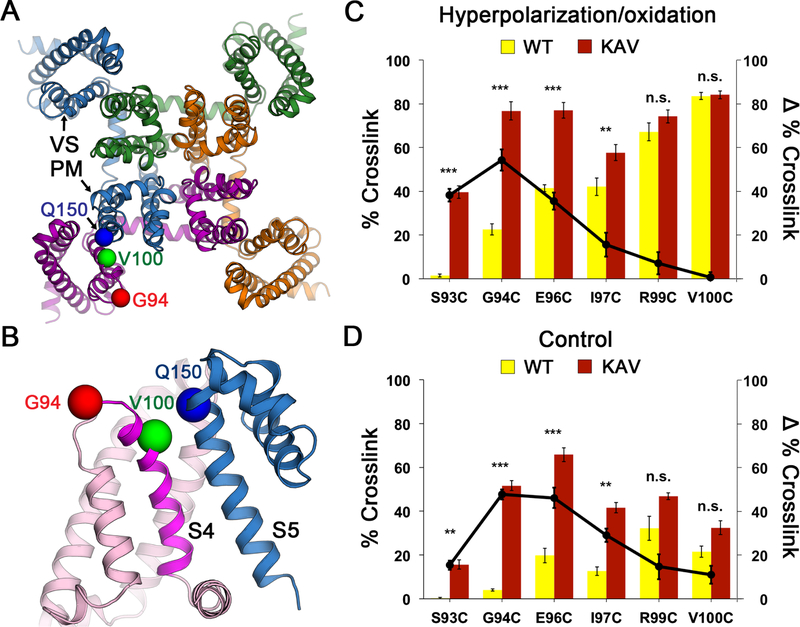
Figure 1
Na V Ab Double Cysteine Substitutions Reveal State-dependent Disulfide Crosslinking
(A) Design of double-cysteine substitution experiment for disulfide crosslinking. The crystal structure of Na V Ab in the activated state ( Payandeh et al., 2011 ) was used as a starting template. The...
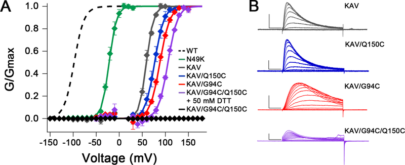
Figure 2
Electrophysiological Recordings of Na V Ab/WT, Na V Ab/N49K, Na V Ab/KAV and Na V Ab/KAV Cysteine Mutants
(A) Normalized conductance-voltage (G/V) relationships and Boltzmann fits for WT and each mutant. Sf9 cells expressing Na V Ab KAV mutants (all C-terminal u039428 truncation constructs) were stimulate...
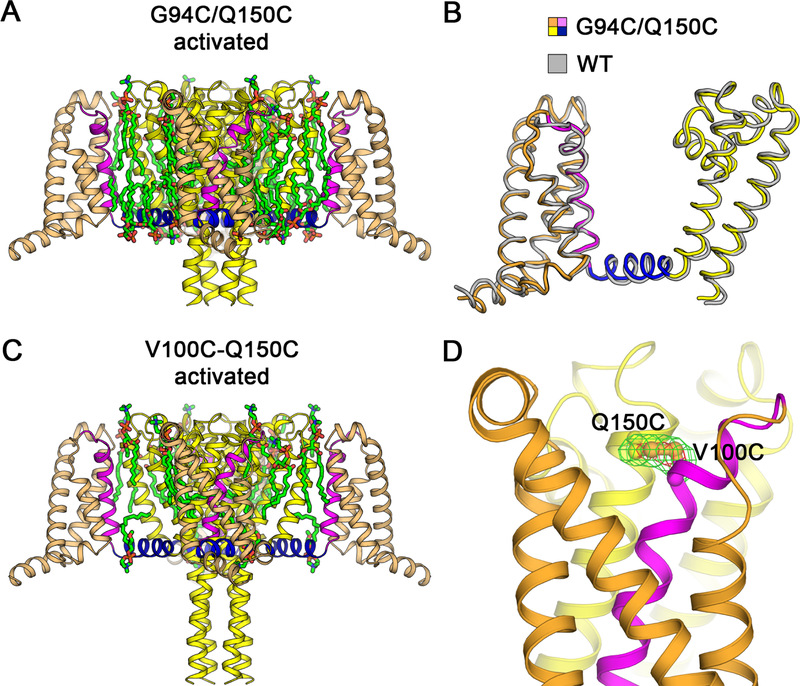
Figure 3
Structures of Na V Ab G94C/Q150C and Disulfide-crosslinked Na V Ab V100C/Q150C in the Activated State
(A) Overall structure of Na V Ab/G94C/Q150C u039428 activated state. The VS S0 to S3 segments are colored in orange and S4 in magenta. The S4-S5 linker is shown in blue and the pore module S5 to S6 in...
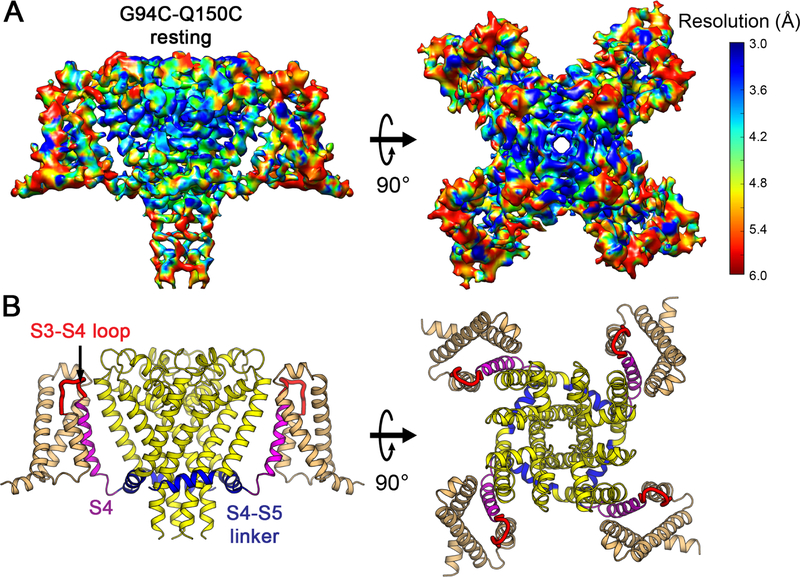
Figure 4
Cryo-EM Structure of Na V Ab Resting State at 4.0 u00c5 Resolution
(A) Cryo-EM density map of MBP-Na V Ab/KAV/G94C/Q150C disulfide-crosslinked resting state in digitonin detergent. Side view (left) and top (extracellular) view (right) are shown. The density map is co...
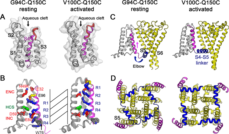
Figure 5
Comparison of Na V Ab Disulfide-crosslinked Structures in the Resting State and the Activated State
(A) Structures of the VS are shown as backbone cartoon superimposed with the solvent accessibility surface. S0 to S3 are shown in gray, S3-S4 loop in red, and S4 in magenta. A wider and shallower aque...
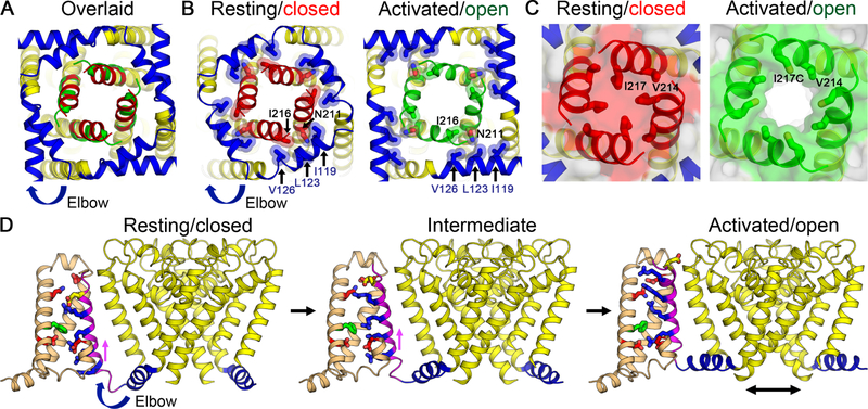
Figure 6
Gating Mechanism for Voltage-gated Sodium Channels
(A) Superposition of structures of Na V Ab in the resting/closed state and the activated/open state (PDB 5VB8) viewed from the intracellular side. Major structural changes are observed in the S4-S5 li...
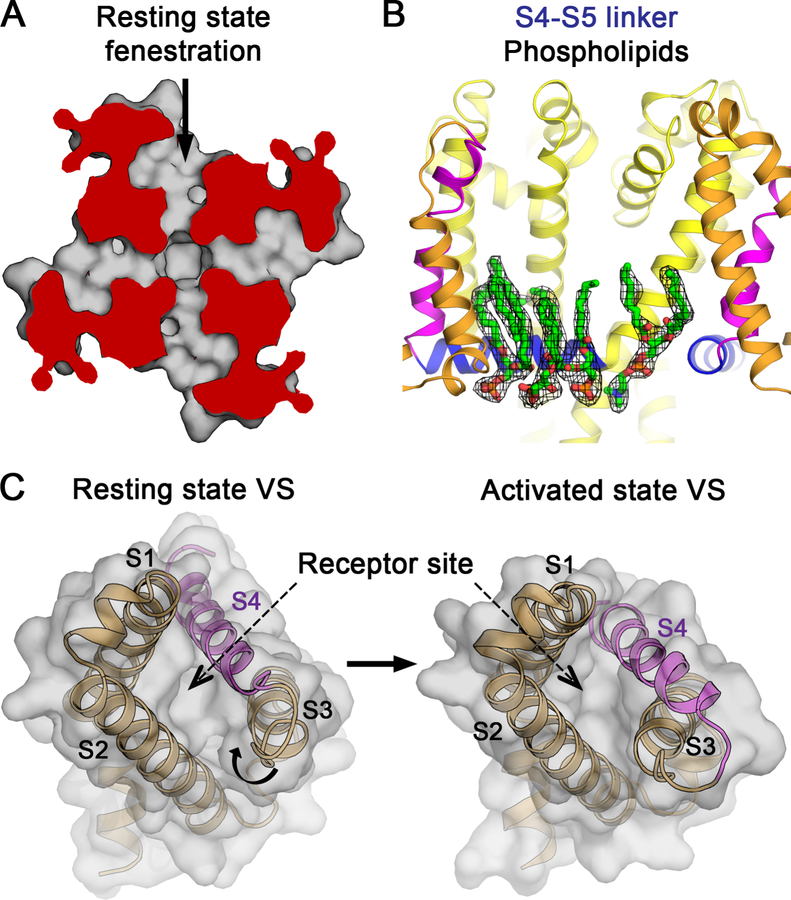
Figure 7
Implications of the Resting State Structure for Sodium Channel Pharmacology.
(A) Drug access to the pore of Na V Ab via the fenestrations in the resting state. Top view cutaway section below the selectivity filter of PM shows fenestration and hydrophobic access to central cavi...
Figure images are served from the NIH/NLM PubMed Central Open Access Subset or Europe PMC; copyright remains with the publishers and authors.
💬 Discussion
0 commentsNo comments yet. Be the first to start a discussion!
Leave a Comment