Abstract
BACKGROUND: Our previous study demonstrated that nitric oxide (NO) contributes to long-term potentiation (LTP) of C-fiber-evoked field potentials by tetanic stimulation of the sciatic nerve in the spinal cord in vivo. Ryanodine receptor (RyR) is a downstream target for NO. The present study further explored the role of RyR in synaptic plasticity of the spinal pain pathway. RESULTS: By means of field potential recordings in the adult male rat in vivo, we showed that RyR antagonist reduced LTP of C-fiber-evoked responses in the spinal dorsal horn by tetanic stimulation of the sciatic nerve. Using spinal cord slice preparations and field potential recordings from superficial dorsal horn, high frequency stimulation of Lissauer's tract (LT) stably induced LTP of field excitatory postsynaptic potentials (fEPSPs). Perfusion of RyR antagonists blocked the induction of LT stimulation-evoked spinal LTP, while Ins(1,4,5)P3 receptor (IP(3)R) antagonist had no significant effect on LTP induction. Moreover, activation of RyRs by caffeine without high frequency stimulation induced a long-term potentiation in the presence of bicuculline methiodide and strychnine. Further, in patch-clamp recordings from superficial dorsal horn neurons, activation of RyRs resulted in a large increase in the frequency of miniature EPSCs (mEPSCs). Immunohistochemical study showed that RyRs were expressed in the dorsal root ganglion (DRG) neurons. Likewise, calcium imaging in small DRG neurons illustrated that activation of RyRs elevated [Ca(2+)]i in small DRG neurons. CONCLUSIONS: These data indicate that activation of presynaptic RyRs play a crucial role in the induction of LTP in the spinal pain pathway, probably through enhancement of transmitter release.
🔬 Techniques
✨ Fluorophores
🧪 Sample Preparation
🏭 Microscope Brands
🧪 Reagent Suppliers
📷 Detectors
🏛️ Research Organizations (ROR)
Affiliated research institutions:
📋 Methods
Sprague-Dawley rats were from Experimental Animal Center, Shanghai Medical College of Fudan University, China. All procedures of the experiments were approved by the Committee of Animal Use for Research and Education of Fudan University, and all efforts were made to minimize the number of animals used and their suffering, in accordance with the ethical guidelines for animal research [ 32 ].
Preparation of spinal cord slices Young
Sprague-Dawley rats (postnatal days 14-21) were deeply anesthetized with diethyl ether, and for LTP recording, about 1 ml lidocaine (5 ml:0.1 g) was injected to both sides of lumbar vertebrae (L 4-5 ). Laminectomy was performed from mid-thoracic to low lumbar levels and the cord was quickly removed to cold modified artificial cerebrospinal fluid (ACSF): (in mM) NaCl, 80; KCl, 2.5; NaH 2 PO4, 1.25; CaCl 2 , 0.5; MgCl 2 , 3.5; NaHCO 3 , 25; sucrose, 75; ascorbate, 1.3; sodium pyruvate, 3.0; oxygenated with 95% O 2 and 5% CO 2 ; pH 7.4; measured osmolarity 310.5 mOsm. Transverse 500 μm slices, with attached dorsal roots, were obtained. Slices were then incubated for at least 1 h at 35°C in a solution that consisted of: (in mM) NaCl, 125; KCl, 2.5; CaCl 2 , 2; MgCl 2 , 1, NaH 2 PO 4 , 1.25; NaHCO 3 , 26; D-glucose, 25; ascorbate, 1.3; sodium pyruvate, 3.0; oxygenated with 95% O 2 and 5% CO 2 , pH 7.4; measured osmolarity 324.5 mOsm. The slice was then transferred into a recording chamber and perfused with oxygenated recording solution at a rate of 5 ml min -1 prior to electrophysiological recordings at room temperature. Field potential and whole-cell patch clamp recordings from spinal cord slices Field potential recordings from the superficial spinal dorsal horn were performed with glass microelectrodes (impedance 2-5 MΩ) filled with (in mM) NaCl 135, KCl 5.4, CaCl 2 1.8, MgCl 2 1, HEPES 5 (pH adjusted to 7.2 with NaOH). A bipolar tungsten electrode was used to stimulate LT. Low-pass filter was set to 1 kHz, amplification 500× (Axopatch 200B, Axon Instruments). To minimize current spread to the dorsal roots and the recording site, the electrode was placed at the most ventrolateral border of LT [ 33 ]. For LTP associated experiments, recordings of fEPSPs were made in the presence of bicuculline (10 μM) and strychnine (1 μM) to block tonic inhibitory action of GABA A and glycine receptors and data are normalized (to mean values of 10 min before HFS or caffeine exposure) mean peak fEPSPs amplitudes. In some of the experiments (caffeine associated), a suction electrode (A-M systems) was used for electrical stimulation of the attached dorsal root. Neurons were identified by infrared differential interference contrast (IR-DIC) video microscopy with an upright microscope (Leica DMLFSA, Germany) equipped with a 40×, 0.80 NA water-immersion objective and a CCD camera (IR-1000E, USA). Patch pipettes (5-10 MΩ) from borosilicate glass were made on a horizontal micropipette puller (P-97, Sutter Instruments, Novato, CA, USA) and were filled with (in mM) potassium gluconate 120, KCl 20, MgCl 2 2, Na 2 ATP 2, NaGTP 0.5, HEPES 20, EGTA 0.5, pH 7.28 with KOH, measured osmolarity 300 mM. To measure EPSCs from neurons in the inner part of lamina II, the dorsal root was stimulated via a suction electrode. Test pulses of 0.1 msec (0.7-1 mA) were given at 30 sec intervals. Data were acquired using an Axopatch 200B patch-clamp amplifier. Responses were low-pass filtered on-line at 2 kHz, digitized at 5 kHz, and analyzed off-line using Clampfit 8.1 software. Membrane potential was held at -70 mV and all recordings were performed at room temperature. Field potential recording from hippocampal slices Young Sprague-Dawley rats (postnatal days 14-21) were deeply anesthetized with diethyl ether. The brain was quickly removed and immersed in ice-cold ACSF bubbled with a gas mixture of 95% O 2 and 5% CO 2 . 400 μM transverse slices were prepared. The slices were then incubated in a chamber maintained at 35°C for at least 1.5 h before recording, and a single slice was then transferred into a recording chamber and perfused with oxygenated recording solution at 3 ml min -1 prior to electrophysiological recordings at room temperature. The composition of the ACSF was (in mM): 124 NaCl, 4.4 KCl, 2.5 CaCl 2 , 1.3 MgSO 4 , 1 NaH 2 PO 4 , 26 NaHCO 3 , and 10 glucose. Field potential recordings were performed as previously described [ 15 ]. Briefly, fEPSPs were recorded with a glass micropipette containing 1 M NaCl in the stratum radiatum of the CA1 region, stimulated via a bipolar tungsten electrode placed along the Schaffer collateral fibers. The stimulation intensity was adjusted to produce a half-maximal field potential amplitude at the beginning of each experiment. Test pulses of 0.1 msec were given at 100 sec intervals. To induce LTP, HFS (100 Hz for 1 sec, 3 times at 10-sec intervals) was delivered, similar to the induction protocol for spinal LTP in vitro . Recording of C-fiber-evoked field potentials in vivo As we described previously [ 14 ], experiments were performed on adult male Sprague-Dawley rats weighing 250-300 g. Pairs of rats were housed in plastic cages and maintained on a 12:12 h light-dark cycle and constant room temperature of 21°C with free access to food and water. Urethane (1.5 g/kg, intraperitoneally) was used to induce and maintain anesthesia. Surgical level of anesthesia was verified by the absence of corneal reflexes and foot withdrawal to pinch. The trachea was cannulated to allow mechanical ventilation with room air, if necessary. One carotid artery was cannulated to monitor mean arterial blood pressure, which ranged from 80 to 100 mmHg. A laminectomy was performed at vertebrae T 13 -L 1 to expose the lumbar enlargement, and the left sciatic nerve was dissected free for bipolar electrical stimulation with a pair of stainless-steel hook electrodes. The exposed nervous tissues were covered with warm paraffin oil. An intrathecal catheter (PE-10 tube) was inserted through the gap between the L 4 and L 5 vertebrae and extended to the subarachniod space of the lumbar enlargement (L 4 and L 5 segments). Colorectal temperature was kept constant at 37-38°C by a feedback-controlled heating blanket. The electrocardiogram was monitored continuously. Following electrical stimulation of the sciatic nerve, the field potentials were recorded in the ipsilateral L 4-5 segments, 300-800 μm from the surface of the cord with glass microelectrodes (impedance 3-6 MΩ) which were driven by an electronically controlled microstepping motor (Narishige Scientific Instrument Co.). The low-pass filter was adjusted to 100 Hz. An A/D converter card (SUMP-PC, Shanghai Medical College, Fudan University, China) was used to digitize and store data at 10 kHz. A single rectangular pulse (0.5 ms, 15-20 V), sufficient to excite C afferent fibers, was applied to sciatic nerve at 1 min intervals as test stimulus. It was easy to distinguish the C-fiber-evoked field potentials from A-fiber-evoked potentials by the latencies of the A and C responses. After recording stable responses for more than 40 min, conditioning tetanic stimuli (0.5 m, 100 Hz, 30-40 V, 10 trains of 2 sec duration at 10 sec intervals) were delivered to the sciatic nerve for inducing LTP of C-fiber-evoked field potentials. The distance from the stimulation site to the recording site was approximately 10 cm. At the end of experiments, rats were killed by an overdose of urethane. The area of C-fiber-evoked field potentials was determined off-line by Average Soft provided by the Department of Physiology, Shanghai Medical College, Fudan University. In each experiment, responses to six consecutive test stimuli were averaged. Data are normalized to mean values of the first 30 min areas of C-fiber evoked field potentials.
Show full methods section
Sprague-Dawley rats were from Experimental Animal Center, Shanghai Medical College of Fudan University, China. All procedures of the experiments were approved by the Committee of Animal Use for Research and Education of Fudan University, and all efforts were made to minimize the number of animals used and their suffering, in accordance with the ethical guidelines for animal research [ 32 ].
Preparation of spinal cord slices Young
Sprague-Dawley rats (postnatal days 14-21) were deeply anesthetized with diethyl ether, and for LTP recording, about 1 ml lidocaine (5 ml:0.1 g) was injected to both sides of lumbar vertebrae (L 4-5 ). Laminectomy was performed from mid-thoracic to low lumbar levels and the cord was quickly removed to cold modified artificial cerebrospinal fluid (ACSF): (in mM) NaCl, 80; KCl, 2.5; NaH 2 PO4, 1.25; CaCl 2 , 0.5; MgCl 2 , 3.5; NaHCO 3 , 25; sucrose, 75; ascorbate, 1.3; sodium pyruvate, 3.0; oxygenated with 95% O 2 and 5% CO 2 ; pH 7.4; measured osmolarity 310.5 mOsm. Transverse 500 μm slices, with attached dorsal roots, were obtained. Slices were then incubated for at least 1 h at 35°C in a solution that consisted of: (in mM) NaCl, 125; KCl, 2.5; CaCl 2 , 2; MgCl 2 , 1, NaH 2 PO 4 , 1.25; NaHCO 3 , 26; D-glucose, 25; ascorbate, 1.3; sodium pyruvate, 3.0; oxygenated with 95% O 2 and 5% CO 2 , pH 7.4; measured osmolarity 324.5 mOsm. The slice was then transferred into a recording chamber and perfused with oxygenated recording solution at a rate of 5 ml min -1 prior to electrophysiological recordings at room temperature. Field potential and whole-cell patch clamp recordings from spinal cord slices Field potential recordings from the superficial spinal dorsal horn were performed with glass microelectrodes (impedance 2-5 MΩ) filled with (in mM) NaCl 135, KCl 5.4, CaCl 2 1.8, MgCl 2 1, HEPES 5 (pH adjusted to 7.2 with NaOH). A bipolar tungsten electrode was used to stimulate LT. Low-pass filter was set to 1 kHz, amplification 500× (Axopatch 200B, Axon Instruments). To minimize current spread to the dorsal roots and the recording site, the electrode was placed at the most ventrolateral border of LT [ 33 ]. For LTP associated experiments, recordings of fEPSPs were made in the presence of bicuculline (10 μM) and strychnine (1 μM) to block tonic inhibitory action of GABA A and glycine receptors and data are normalized (to mean values of 10 min before HFS or caffeine exposure) mean peak fEPSPs amplitudes. In some of the experiments (caffeine associated), a suction electrode (A-M systems) was used for electrical stimulation of the attached dorsal root. Neurons were identified by infrared differential interference contrast (IR-DIC) video microscopy with an upright microscope (Leica DMLFSA, Germany) equipped with a 40×, 0.80 NA water-immersion objective and a CCD camera (IR-1000E, USA). Patch pipettes (5-10 MΩ) from borosilicate glass were made on a horizontal micropipette puller (P-97, Sutter Instruments, Novato, CA, USA) and were filled with (in mM) potassium gluconate 120, KCl 20, MgCl 2 2, Na 2 ATP 2, NaGTP 0.5, HEPES 20, EGTA 0.5, pH 7.28 with KOH, measured osmolarity 300 mM. To measure EPSCs from neurons in the inner part of lamina II, the dorsal root was stimulated via a suction electrode. Test pulses of 0.1 msec (0.7-1 mA) were given at 30 sec intervals. Data were acquired using an Axopatch 200B patch-clamp amplifier. Responses were low-pass filtered on-line at 2 kHz, digitized at 5 kHz, and analyzed off-line using Clampfit 8.1 software. Membrane potential was held at -70 mV and all recordings were performed at room temperature. Field potential recording from hippocampal slices Young Sprague-Dawley rats (postnatal days 14-21) were deeply anesthetized with diethyl ether. The brain was quickly removed and immersed in ice-cold ACSF bubbled with a gas mixture of 95% O 2 and 5% CO 2 . 400 μM transverse slices were prepared. The slices were then incubated in a chamber maintained at 35°C for at least 1.5 h before recording, and a single slice was then transferred into a recording chamber and perfused with oxygenated recording solution at 3 ml min -1 prior to electrophysiological recordings at room temperature. The composition of the ACSF was (in mM): 124 NaCl, 4.4 KCl, 2.5 CaCl 2 , 1.3 MgSO 4 , 1 NaH 2 PO 4 , 26 NaHCO 3 , and 10 glucose. Field potential recordings were performed as previously described [ 15 ]. Briefly, fEPSPs were recorded with a glass micropipette containing 1 M NaCl in the stratum radiatum of the CA1 region, stimulated via a bipolar tungsten electrode placed along the Schaffer collateral fibers. The stimulation intensity was adjusted to produce a half-maximal field potential amplitude at the beginning of each experiment. Test pulses of 0.1 msec were given at 100 sec intervals. To induce LTP, HFS (100 Hz for 1 sec, 3 times at 10-sec intervals) was delivered, similar to the induction protocol for spinal LTP in vitro . Recording of C-fiber-evoked field potentials in vivo As we described previously [ 14 ], experiments were performed on adult male Sprague-Dawley rats weighing 250-300 g. Pairs of rats were housed in plastic cages and maintained on a 12:12 h light-dark cycle and constant room temperature of 21°C with free access to food and water. Urethane (1.5 g/kg, intraperitoneally) was used to induce and maintain anesthesia. Surgical level of anesthesia was verified by the absence of corneal reflexes and foot withdrawal to pinch. The trachea was cannulated to allow mechanical ventilation with room air, if necessary. One carotid artery was cannulated to monitor mean arterial blood pressure, which ranged from 80 to 100 mmHg. A laminectomy was performed at vertebrae T 13 -L 1 to expose the lumbar enlargement, and the left sciatic nerve was dissected free for bipolar electrical stimulation with a pair of stainless-steel hook electrodes. The exposed nervous tissues were covered with warm paraffin oil. An intrathecal catheter (PE-10 tube) was inserted through the gap between the L 4 and L 5 vertebrae and extended to the subarachniod space of the lumbar enlargement (L 4 and L 5 segments). Colorectal temperature was kept constant at 37-38°C by a feedback-controlled heating blanket. The electrocardiogram was monitored continuously. Following electrical stimulation of the sciatic nerve, the field potentials were recorded in the ipsilateral L 4-5 segments, 300-800 μm from the surface of the cord with glass microelectrodes (impedance 3-6 MΩ) which were driven by an electronically controlled microstepping motor (Narishige Scientific Instrument Co.). The low-pass filter was adjusted to 100 Hz. An A/D converter card (SUMP-PC, Shanghai Medical College, Fudan University, China) was used to digitize and store data at 10 kHz. A single rectangular pulse (0.5 ms, 15-20 V), sufficient to excite C afferent fibers, was applied to sciatic nerve at 1 min intervals as test stimulus. It was easy to distinguish the C-fiber-evoked field potentials from A-fiber-evoked potentials by the latencies of the A and C responses. After recording stable responses for more than 40 min, conditioning tetanic stimuli (0.5 m, 100 Hz, 30-40 V, 10 trains of 2 sec duration at 10 sec intervals) were delivered to the sciatic nerve for inducing LTP of C-fiber-evoked field potentials. The distance from the stimulation site to the recording site was approximately 10 cm. At the end of experiments, rats were killed by an overdose of urethane. The area of C-fiber-evoked field potentials was determined off-line by Average Soft provided by the Department of Physiology, Shanghai Medical College, Fudan University. In each experiment, responses to six consecutive test stimuli were averaged. Data are normalized to mean values of the first 30 min areas of C-fiber evoked field potentials.
Calcium imaging
As we previously described [ 34 ], acutely isolated DRG neurons were loaded with 1 μM Fura-2 acetoxymethyl ester (Fura-2/AM; DoJinDo Laboratories, Kumamoto, Japan). The neurons were observed on an inverted microscope (Olympus IX51) with a 40× UV flour oil-immersion objective lens. The fluorescence in individual neurons was recorded by a cooled CCD camera (Hamamatsu, Hamamatsu City, Japan), with a 1 Hz alternating wavelength time scanning with excitation wavelengths of 340 and 380 nm and an emission wavelength of 510 nm (monochromators; Till Polychrome IV, Munich, Germany). Images were captured every 3 sec. Digitized images were acquired and analyzed in a personal computer controlled by SimplePCI (Compix, Lake Oswego, OR). The ratio of the fluorescence at the two excitation wavelengths was used to estimate changes of [Ca 2+ ] i .
Immunohistochemistry
Normal adult rats were given an overdose of urethane (2 g/kg, i.p.) and perfused intracardially with 0.9% sterile saline followed by 4% paraformaldehyde in 0.1 M phosphate buffer (PB, pH 7.4). The L 4-6 DRG were then removed, post-fixed in the same fixative for 2 h at 4°C, and immersed in 20% sucrose in PB for 24 h at 4°C for cryoprotection. Transverse DRG sections (14 μm) were cut in a cryostat and processed for immunofluorescence. All sections were blocked with 10% donkey serum in 0.01 M PBS, pH 7.4 with 0.3% Triton-X-100 for 2 h at room temperature (RT) and incubated 24 h at 4°C with rabbit anti-RyR 1 and 3 antibody (1: 1000, Chemicon, Temecula, CA) or a mixture of rabbit anti-RyR 1 and 3 antibody (1: 1000, Chemicon, Temecula, CA) and goat anti-CGRP antibody (1:500, Santa Cruze) in PBS with 1% normal donkey serum and 0.3% Triton-X-100. Following six 10 min rinses in 0.01 M PBS, the sections were incubated in a mixture of FITC-conjugated Isolectin B4 (IB4) (1:1000, Sigma) and rhodamine red-X-conjugated donkey anti-rabbit IgG (1:100, Jackson ImmunoResearch, West Grove, PA) for 2 h at 37°C, or FITC-conjugated donkey anti-goat IgG (1:100, Jackson ImmunoResearch, West Grove, PA) and rhodamine red-X-conjugated donkey anti-rabbit IgG (1:100, Jackson ImmunoResearch, West Grove, PA) for 120 min at RT, and then washed in PBS. The specificity of immunostaining was verified by omitting the primary antibodies, and immunostaining signal disappeared after omitting primary antibodies. All sections were coverslipped with a mixture of 50% glycerin in 0.01 M PBS, and then observed with a Leica fluorescence microscope, and images were captured with a CCD spot camera. Drugs In vivo experiments: Ryanodine was initially dissolved in dimethylsulfoxide (DMSO) and then diluted in physiological saline to their final concentration immediately before administration. The final DMSO concentration in the diluted working solution was 0.25%. To evaluate the effects of drugs on spinal LTP induction and maintenance, drugs or vehicles were intrathecally injected over a period of 2 min at a volume of 10 μl followed by 5 μl 0.9% sterile saline solution for flushing at 30 min before or 60 min after tetanic stimuli. In vitro experiments: Most of the drugs were purchased from sigma, including bicuculline methiodide (10 μM), strychnine (1 μM), AP5 (50 μM), DNQX (10 μM), dantrolene (10 μM), 2-APB (75 μM), cADPR (5 μM), L-NAME (50 μM). The other drugs were TTX (0.5 μM), ryanodine (2 μM, 20 μM, 20 μM, Calbiochem) and caffeine (10 mM, Lancaster). All drugs were prepared as stock solutions and diluted to the required concentration (1:1000) with ACSF.
Statistics
Data were expressed as mean ± s.e.m. Effects of drugs on LTP were compared with two-way repeated measures (RM) ANOVA (treatment and time) followed by the Holm-Sidak tests. Pared t -test or t -test was used for other electrophysiology experiments. P < 0.05 was considered statistically significant.
📊 Figures
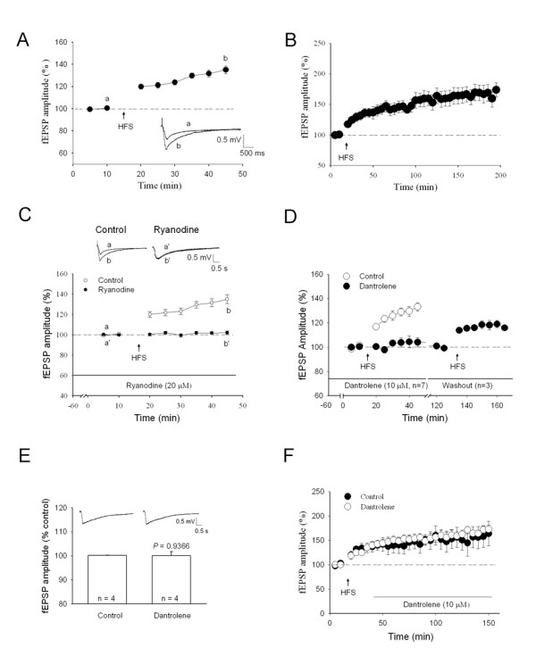
Figure 1
Involvement of RyR in the induction of LTP of fEPSPs in the spinal dorsal horn in vitro . A , fEPSPs were recorded in the presence of 10 u03bcM bicuculline methiodide and 1 u03bcM strychnine. fEPSPs r...

Figure 2
Involvement of RyR in the induction of LTP of fEPSPs in the spinal dorsal horn in vivo . The spinal LTP was blocked by high concentration of ryanodine (1 mM, 10 u03bcl) intrathecally applied 30 min be...

Figure 3
Reversal of L-NAMA-induced inhibition of LTP of fEPSP by cADPR, an endogenous agonist of the RyR . Perfusion with 50 u03bcM L-NAME for 1 h before HFS of LT inhibited the induction of LTP ( P < 0.00...

Figure 4
No effect of IP3R antagonist on induction of spinal LTP in vitro . A , IP 3 R antagonist 2-APB (75 u03bcM) applied for 1 h before HFS of LT had no significant effect either on the induction of LTP of ...

Figure 5
Caffeine-induced LTP of fEPSPs in spinal cord slice . A , After perfusion with 10 mM caffeine for 2 min instantly following washed out, LTP of fEPSPs occurred without conditioning stimulation in the p...

Figure 6
Expression of ryanodine receptors 1 or 3 (RyR1, RyR3) and their co- localization with IB4 or CGRP in DRG neurons in normal rats . A , Co-localization of RyR1 (red) with IB4 (green) in DRG neurons. B ,...
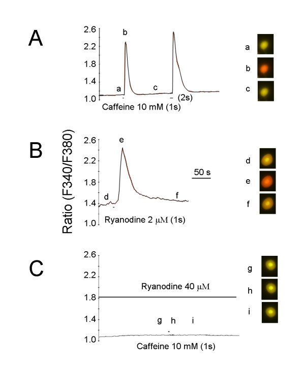
Figure 7
Increase in [Ca 2+ ] i by caffeine in isolated DRG neurons . A , Brief puff of 10 mM caffeine for 1 or 2 s increased [Ca 2+ ] i transients in DRG neurons (n = 9). B , Puff of 2 u03bcM ryanodine increa...

Figure 8
Increase in mEPSCs frequency by caffeine in superficial dorsal horn neurons . A , Sample traces of mESPCs recorded from a lamina II neuron in the presence of 0.5 u03bcM TTX before (left) and after bat...
Figure images are served from the NIH/NLM PubMed Central Open Access Subset or Europe PMC; copyright remains with the publishers and authors.
💬 Discussion
0 commentsNo comments yet. Be the first to start a discussion!
Leave a Comment