Abstract
Following invasion of non‐phagocytic host cells, Salmonella enterica survives and replicates within a phagosome‐like compartment known as the Salmonella ‐containing vacuole (SCV). It is now well established that SCV biogenesis, like phagosome biogenesis, involves sequential interactions with the endocytic pathway. However, Salmonella is believed to limit these interactions and, in particular, to avoid fusion of terminal lysosomes with the SCV. In this study, we reassessed this process using a high‐resolution live‐cell imaging approach and found an unanticipated level of interaction between the SCV and the endocytic pathway. Direct interactions, in which late endosomal/lysosomal content was transferred to SCVs, were detected within 30 min of invasion and continued for several hours. Mechanistically, these interactions were very similar to phagosome–lysosome fusion because they were accompanied by rapid acidification of the SCV, could be blocked by chemical perturbation of microtubules or vacuolar acidification and involved the small GTPase Rab7. In comparison with vacuoles containing internalized Escherichia coli or heat‐killed Salmonella , SCVs did show some delay of fusion and acidification, although, this appeared to be independent of either type III secretion system. These results provide compelling evidence that inhibition of SCV–lysosome fusion is not the major determinant in establishment of the Salmonella replicative niche in epithelial cells.
🔬 Techniques
🔭 Microscopes
🧬 Organisms
💻 Software
✨ Fluorophores
🧪 Sample Preparation
🔬 Cell Lines
🏭 Microscope Brands
🧪 Reagent Suppliers
🎨 Filters
💻 Software Details
💾 Data Repositories
🏛️ Research Organizations (ROR)
Affiliated research institutions:
📋 Methods
Cell culture, bacterial strains and reagents HeLa (human adenocarcinoma cervix epithelial, CCL-2) and C2BBe1 (human polarized adenocarcinoma colon epithelial cells, CRL-2102) were obtained from the American Type Culture Collection (ATCC, Manassas, VA, USA) and grown in a humidified 37°C, 5% CO 2 tissue culture incubator. Cell culture reagents were from Invitrogen (Carlsbad, CA, USA) unless otherwise stated. HeLa cells were maintained in Eagle’s Minimal Essential Media (MEM) containing 2 mM l -glutamine, 1 mM sodium pyruvate and 10% heat-inactivated fetal bovine serum (FBS). C2BBe1 cells were maintained in Dulbecco’s modification of MEM (DMEM) containing 4 mM l -glutamine, 0.01 mg/mL human transferrin and 10% FBS. Low passage number (
📊 Figures
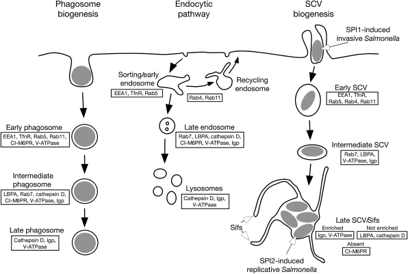
Figure 1:
Markers of the endocytic pathway and their distribution during SCV maturation
According to the prevailing model, the SCV transiently acquires markers of early endosomes and late endosomes during biogenesis, but delays delivery of the lysosomal hydrolase cathepsin D and lysobisp...
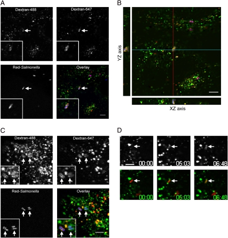
Figure 2:
SCVs acquire content from endosomes and lysosomes
To load lysosomes HeLa (A, B and D), or C2BBe1 (C), cells were preincubated with dextran-647 (red) o/n, then chased in dextran-free media for 3 h before infection with Red- Salmonella (blue). After in...
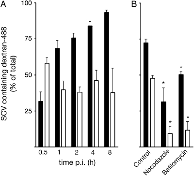
Figure 3:
SCV acquisition of fluid-phase content from endosomes and lysosomes is dependent on V-ATPase and microtubules
To load lysosomes, HeLa cells were preincubated with dextran-488 o/n, then chased in dextran-free media for 3 h before infection with Red- Salmonella . To follow endocytic access to the SCV dextran-48...
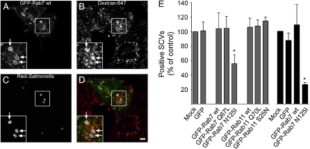
Figure 4:
SCV acquisition of fluid-phase markers is Rab7 dependent
HeLa cells expressing GFP alone or GFP-Rab protein fusions (green), were infected with Red- Salmonella (blue). Dextran-647 (red) was added 2 h p.i. to load the endocytic pathway. Live-cell imaging was...
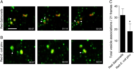
Figure 5:
Increased lysosomal vesicle association with the SCV
LE/Lys were loaded by incubating HeLa cells with dextran-488 o/n, followed by a chase in dextran-free media for 3 h before infection with Red- Salmonella or Red- E. coli pInv. Live-cell imaging was in...
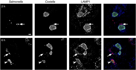
Figure 6:
SCVs fuse with preformed C. burnetii parasitophorous vacuoles in co-infected cells
HeLa cells were infected with C. burnetii for 48 h before infection with wt Salmonella . At 2 or 6 h p.i., cells were fixed and processed for immunofluorescence microscopy using u03b1- Salmonella LPS ...
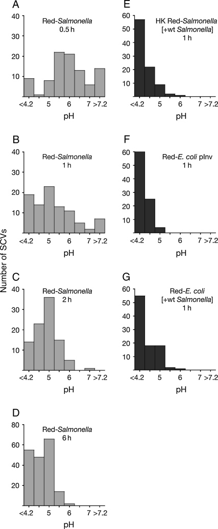
Figure 7:
SCV acidification is delayed in epithelial cells
The endolysomal system and vacuoles containing bacteria were loaded with dextrans by incubating HeLa cells o/n and throughout infection with a mixture of dextran-FITC and dextran-647. Au2013D) pH SCV ...
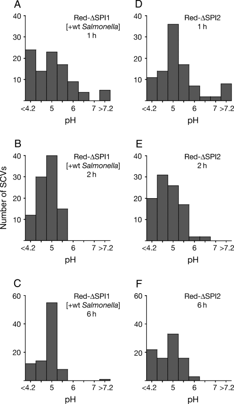
Figure 8:
SCVs containing u0394SPI1 or u0394SPI2 mutants do not acidify more rapidly
HeLa cells were loaded with dextran-FITC and dextran-647 as in Figure 7 . Au2013C) Time course of the pH SCV of u0394SPI1 Red- Salmonella taken up by co-internalization with wt Salmonella . Du2013F) T...
Figure images are served from the NIH/NLM PubMed Central Open Access Subset or Europe PMC; copyright remains with the publishers and authors.
💬 Discussion
0 commentsNo comments yet. Be the first to start a discussion!
Leave a Comment