Abstract
The axon initial segment (AIS) serves as the site of action potential initiation in most neurons, but difficulties in isolating the effects of voltage-gated ion channels in the AIS from those of the soma and dendrites have hampered understanding how AIS properties influence neural coding. Here we have combined confocal microscopy, patch-clamp recordings and light-sensitive channel blockers ('photoswitches') in binaural auditory gerbil neurons to show that hyperpolarization and cyclic-nucleotide-gated (HCN) channels are expressed in the AIS and decrease spike probability, in a manner distinct from that of HCN channels in the soma and dendrites. Furthermore, the control of spike threshold by HCN channels in the AIS can be altered through serotonergic modulation of 5-hydroxytryptamine 1A (5-HT1A) receptors, which hyperpolarizes the activation range of HCN channels. As release of serotonin signals changes in motivation and attention states, axonal HCN channels provide a mechanism to translate these signals into changes in the threshold for sensory stimuli.
🔬 Techniques
🔭 Microscopes
💻 Software
✨ Fluorophores
🧪 Sample Preparation
🏭 Microscope Brands
🧪 Reagent Suppliers
💻 Software Details
🏛️ Research Organizations (ROR)
Affiliated research institutions:
📋 Methods
All procedures were conducted in accordance with The University of Texas at Austin Institutional Animal Care and Use Committee, following guidelines of the National Institutes of Health.
Immunostaining Gerbil brains
(P21) were acutely dissected and drop-fixed in ice cold 4% paraformaldehyde, pH 7.2, for 1 hour. Brains were then transferred to 20% sucrose in 0.1M PB overnight, and then transferred to 30% sucrose in 0.1M PB until sectioning. 40 μm thick coronal sections of brainstem were then cut on a cryostat and mounted on coverslips. Sections were then immunostained as described 51 using mouse antibodies against HCN1 (clone N70/28 NeuroMab), PanNav 52 , or Caspr (clone K65/35 NeuroMab), and rabbit antibodies against βIV spectrin 53 or Nav1.6 54 . AIS lengths were measured using Zeiss Zen software (Zeiss, Thornwood, NY) or Image J (NIH). For AIS lengths, the start and end of the AIS was defined as the point at which the immunoreactivity fell below 10% of the maximum fluorescence intensity along the AIS. Statistics were performed using GraphPad Prism.
Brainstem slice preparation
Mongolian gerbils ( Meriones unguiculatus ) of both sexes were obtained from Charles River Laboratories or bred at the Animal Resource Center of the University of Texas at Austin. Litters were group housed and kept on a 12/12 hour light/dark cycle. Gerbils (P18-P23) were anesthetized with isoflurane, decapitated and the brain rapidly removed in artificial cerebrospinal fluid (ACSF) at 32°C. ACSF was bubbled with 95% O 2 / 5% CO 2 and contained (in mM) 125 NaCl, 25 glucose, 25 NaHCO 3 , 2.5 KCl, 1.25 NaH 2 PO 4 , 1.5 CaCl 2 , 1.5 MgSO 4 , pH 7.45. Horizontal or coronal sections containing the superior olivary complex were cut at a thickness of 200 μM with an oscillating tissue slicer (VT1200S; Leica), incubated at 35°C for at least 30 min, and then held at room temperature until recording. Selected brain slices were biased towards the middle of the dorso-ventral (tonotopic) axis. MSO neurons were identified by their location in the slice, morphology, and distinct electrophysiological characteristics as in previous studies 55 , 56 . Photoswitch experiments Brainstem slices were pre-incubated at room temperature in the dark for 20-30 minutes with either 300 μM AAQ diluted in ACSF for current clamp recordings or 200 μM DENAQ in ACSF for voltage clamp recordings. Thereafter, photoswitch-free ACSF was used for recordings. AAQ and DENAQ were custom synthesized 7 from Jublilant Chemsys., Ltd, Uttar Pradesh, India. Recordings were performed within 1 hour of photoswitch incubation to avoid time-dependent changes in the concentration of photoswitches in the membrane. To visualize the axon, soma and dendrites of MSO neurons, 40 μM Alexa Fluor 568 hydrazide (Invitrogen, Carlsbad, CA) was included in patch pipette solutions, and allowed to dialyze into neurons for at least 5 minutes after establishing a whole-cell recording. Excitation of Alexa 568 was achieved using the 543 nm laser line from a 1 mW HeNe laser through the 40x objective of a fixed stage upright confocal microscope (Leica TCS SP5 II). Excitation at 568 nm did not affect the conformation of AAQ or the intrinsic membrane properties of MSO neurons. Scan speed was 400 Hz (pixel dwell time 4.9 μs), and ROIs generally occupied ~250 and ~600 pixels for the AIS and soma, respectively. Current-clamp electrophysiology and imaging with AAQ All current-clamp recordings were performed at 35°C in ACSF unless otherwise noted. Recordings were made using heat-polished borosilicate patch pipettes (1.65 mm outer diameter; World Precision Instruments, Sarasota, FL), and had resistances of 3-5 ΜΩ in ACSF. The internal solution in patch pipettes contained the following (in mM): 115 potassium gluconate, 20 KCl, 10 sodium phosphocreatine, 10 HEPES, 0.5 EGTA, 4 MgATP, 0.3 NaGTP, and the pH adjusted to 7.3 with KOH. Recordings were made with a Dagan (Minneapolis, MN) BVC-700A amplifier in current-clamp mode. To focally block voltage-gated ion channels in restricted sub-regions of MSO neurons, we combined whole-cell recordings, confocal microscopy, and the use of AAQ in gerbil brainstem slices. After identifying the intact AIS using 568 nm scans under the confocal microscope, the cell was continuously illuminated with 380 nm light through the epifluorescence port of the microscope, which maintains AAQ in the cis (non-blocking) conformation. The AIS, soma, or whole cell was then scanned with 488 nm light driving AAQ in to the trans (blocking) conformation, while simultaneously discontinuing the 380 nm illumination and initiating data acquisition of electrophysiological responses to real or simulated synaptic stimuli. Since all of the effects of this “photoblockade” were rapidly reversible upon reintroduction of 380 nm light, electrophysiological responses under 380 nm and 488 nm illumination were interleaved to control both for time dependent concentration changes in AAQ in cell membranes and AAQ molecules entering a non-reversible configuration. Data acquisition was controlled by custom macros programmed in Igor Pro (WaveMetrics, Lake Oswego, OR). The effective spatial resolution of photoswitches was assessed with AAQ. Using a 30 μm ROI over the AIS, we measured how the change in resting potential at 488 nm was affected by moving the ROI perpendicular to the long axis of the axon in 0.5 μm steps ( Supplementary Fig. 7 ). The change in membrane potential decreased exponentially with a distance constant of 0.37 μm, close to the theoretical point spread function of 0.31 μm at 488 nm, demonstrating that the compartment-specific effects of AAQ blockade are minimally affected by light scattering in the slice. Recordings were only included if the series resistance was
Show full methods section
All procedures were conducted in accordance with The University of Texas at Austin Institutional Animal Care and Use Committee, following guidelines of the National Institutes of Health.
Immunostaining Gerbil brains
(P21) were acutely dissected and drop-fixed in ice cold 4% paraformaldehyde, pH 7.2, for 1 hour. Brains were then transferred to 20% sucrose in 0.1M PB overnight, and then transferred to 30% sucrose in 0.1M PB until sectioning. 40 μm thick coronal sections of brainstem were then cut on a cryostat and mounted on coverslips. Sections were then immunostained as described 51 using mouse antibodies against HCN1 (clone N70/28 NeuroMab), PanNav 52 , or Caspr (clone K65/35 NeuroMab), and rabbit antibodies against βIV spectrin 53 or Nav1.6 54 . AIS lengths were measured using Zeiss Zen software (Zeiss, Thornwood, NY) or Image J (NIH). For AIS lengths, the start and end of the AIS was defined as the point at which the immunoreactivity fell below 10% of the maximum fluorescence intensity along the AIS. Statistics were performed using GraphPad Prism.
Brainstem slice preparation
Mongolian gerbils ( Meriones unguiculatus ) of both sexes were obtained from Charles River Laboratories or bred at the Animal Resource Center of the University of Texas at Austin. Litters were group housed and kept on a 12/12 hour light/dark cycle. Gerbils (P18-P23) were anesthetized with isoflurane, decapitated and the brain rapidly removed in artificial cerebrospinal fluid (ACSF) at 32°C. ACSF was bubbled with 95% O 2 / 5% CO 2 and contained (in mM) 125 NaCl, 25 glucose, 25 NaHCO 3 , 2.5 KCl, 1.25 NaH 2 PO 4 , 1.5 CaCl 2 , 1.5 MgSO 4 , pH 7.45. Horizontal or coronal sections containing the superior olivary complex were cut at a thickness of 200 μM with an oscillating tissue slicer (VT1200S; Leica), incubated at 35°C for at least 30 min, and then held at room temperature until recording. Selected brain slices were biased towards the middle of the dorso-ventral (tonotopic) axis. MSO neurons were identified by their location in the slice, morphology, and distinct electrophysiological characteristics as in previous studies 55 , 56 . Photoswitch experiments Brainstem slices were pre-incubated at room temperature in the dark for 20-30 minutes with either 300 μM AAQ diluted in ACSF for current clamp recordings or 200 μM DENAQ in ACSF for voltage clamp recordings. Thereafter, photoswitch-free ACSF was used for recordings. AAQ and DENAQ were custom synthesized 7 from Jublilant Chemsys., Ltd, Uttar Pradesh, India. Recordings were performed within 1 hour of photoswitch incubation to avoid time-dependent changes in the concentration of photoswitches in the membrane. To visualize the axon, soma and dendrites of MSO neurons, 40 μM Alexa Fluor 568 hydrazide (Invitrogen, Carlsbad, CA) was included in patch pipette solutions, and allowed to dialyze into neurons for at least 5 minutes after establishing a whole-cell recording. Excitation of Alexa 568 was achieved using the 543 nm laser line from a 1 mW HeNe laser through the 40x objective of a fixed stage upright confocal microscope (Leica TCS SP5 II). Excitation at 568 nm did not affect the conformation of AAQ or the intrinsic membrane properties of MSO neurons. Scan speed was 400 Hz (pixel dwell time 4.9 μs), and ROIs generally occupied ~250 and ~600 pixels for the AIS and soma, respectively. Current-clamp electrophysiology and imaging with AAQ All current-clamp recordings were performed at 35°C in ACSF unless otherwise noted. Recordings were made using heat-polished borosilicate patch pipettes (1.65 mm outer diameter; World Precision Instruments, Sarasota, FL), and had resistances of 3-5 ΜΩ in ACSF. The internal solution in patch pipettes contained the following (in mM): 115 potassium gluconate, 20 KCl, 10 sodium phosphocreatine, 10 HEPES, 0.5 EGTA, 4 MgATP, 0.3 NaGTP, and the pH adjusted to 7.3 with KOH. Recordings were made with a Dagan (Minneapolis, MN) BVC-700A amplifier in current-clamp mode. To focally block voltage-gated ion channels in restricted sub-regions of MSO neurons, we combined whole-cell recordings, confocal microscopy, and the use of AAQ in gerbil brainstem slices. After identifying the intact AIS using 568 nm scans under the confocal microscope, the cell was continuously illuminated with 380 nm light through the epifluorescence port of the microscope, which maintains AAQ in the cis (non-blocking) conformation. The AIS, soma, or whole cell was then scanned with 488 nm light driving AAQ in to the trans (blocking) conformation, while simultaneously discontinuing the 380 nm illumination and initiating data acquisition of electrophysiological responses to real or simulated synaptic stimuli. Since all of the effects of this “photoblockade” were rapidly reversible upon reintroduction of 380 nm light, electrophysiological responses under 380 nm and 488 nm illumination were interleaved to control both for time dependent concentration changes in AAQ in cell membranes and AAQ molecules entering a non-reversible configuration. Data acquisition was controlled by custom macros programmed in Igor Pro (WaveMetrics, Lake Oswego, OR). The effective spatial resolution of photoswitches was assessed with AAQ. Using a 30 μm ROI over the AIS, we measured how the change in resting potential at 488 nm was affected by moving the ROI perpendicular to the long axis of the axon in 0.5 μm steps ( Supplementary Fig. 7 ). The change in membrane potential decreased exponentially with a distance constant of 0.37 μm, close to the theoretical point spread function of 0.31 μm at 488 nm, demonstrating that the compartment-specific effects of AAQ blockade are minimally affected by light scattering in the slice. Recordings were only included if the series resistance was
📊 Figures
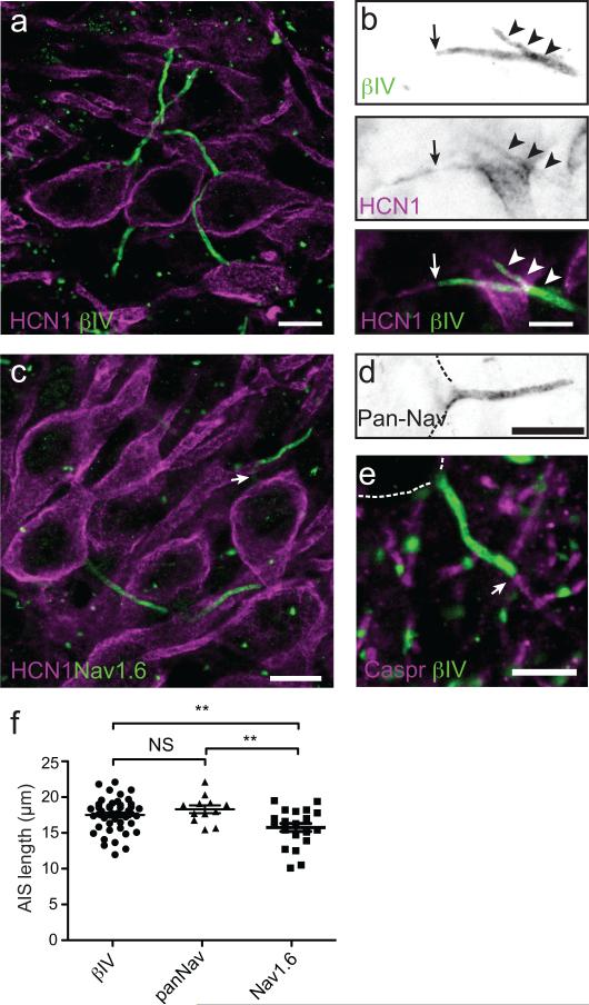
Figure 1
Expression of HCN1 subunits in the AIS of MSO principal neurons
(a ) Immunostaining of MSO neurons using antibodies against HCN1 (red) and u03b2IV spectrin (green). Scale = 10 um. ( b ) Immunostaining of MSO neuron AIS using antibodies against HCN1(red) and u03b2I...
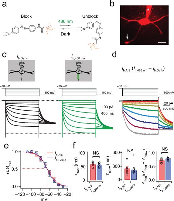
Figure 2
Activation properties of axonal I h , as revealed by laser scanning of the photoswitch DENAQ
(a ) The cis , unblocking conformation of DENAQ is maintained at 488 nm light (green) while the trans , blocking conformation is maintained in the dark (black). (b ) Confocal image of an MSO principal...
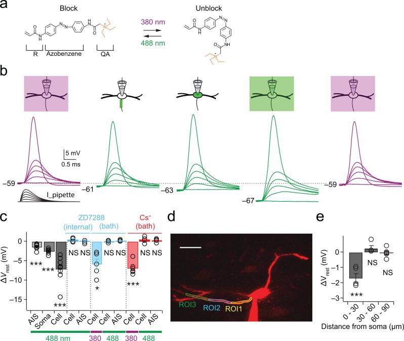
Figure 3
Compartment-specific block of resting conductances by laser scanning of AAQ
(a ) The unblocking and blocking conformations of AAQ are maintained at 380 and 488 nm light, respectively. R, acrylamide group; QA, quaternary ammonium group. (b ) Scanning block of different cellula...
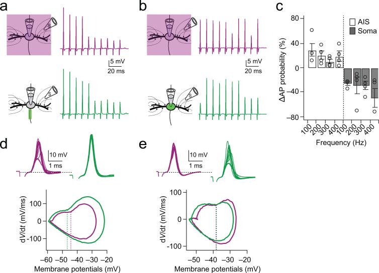
Figure 4
HCN channels in the AIS but not the soma and dendrites decrease spike probability by raising threshold
(a ) Responses to synaptic stimulation during a whole-cell current clamp recording with AAQ maintained in the unblocked conformation with 380 nm field illumination (purple traces) and during AIS block...
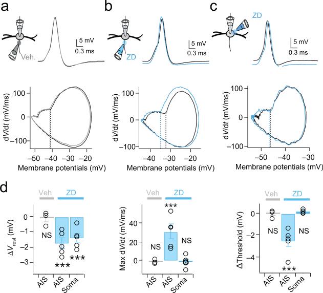
Figure 5
Local pharmacological blockade of HCN channels in the AIS mimics the effects of photoswitches
( a ) Top: First suprathreshold spike in response to a train of somatic simulated EPSCs (100 Hz; 0-6 nA, 0.4 pA steps) in a whole-cell current clamp recording in control (black) and following brief fo...
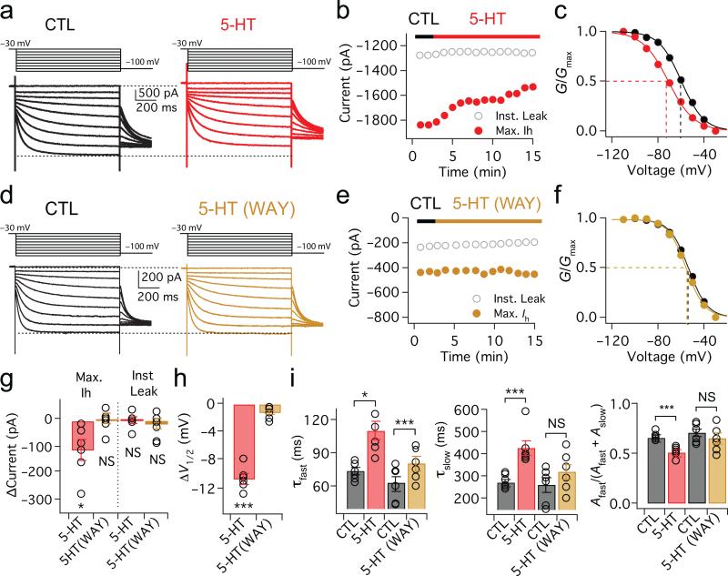
Figure 6
Serotonin modulates the activation range of HCN channels through 5-HT 1A receptors
( a ) Voltage dependence of I h activation in whole-cell voltage-clamp recordings (voltage steps: u221230 mV to u2212110 mV in u221210 mV steps, 1 s duration). Red traces: responses after application ...
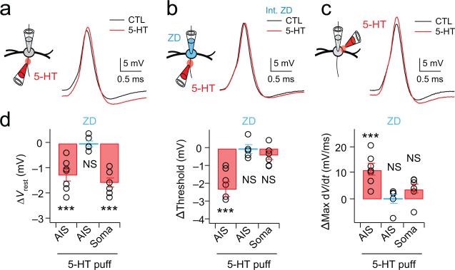
Figure 7
Spike threshold can be controlled by serotonergic modulation of HCN channels in the AIS
( a ) Focal application of 300 u03bcM 5-HT onto the AIS (red traces) hyperpolarizes the resting membrane potential as well as action potential threshold as compared to the ACSF control (black traces)....
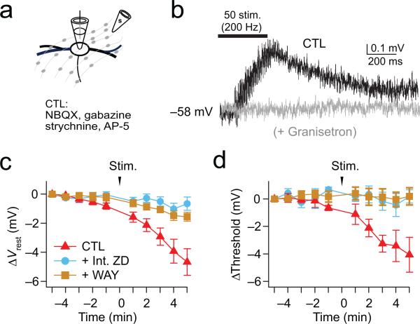
Figure 8
Axonally stimulated release of serotonin modulates the resting potential and spike threshold of MSO neurons
( a ) Axonal stimulation (200Hz, 50 pulses) of serotonergic fibers. Excitatory and inhibitory inputs are blocked with 10 u03bcM NBQX, 50 u03bcM AP-5, 5 u03bcM gabazine, 1 u03bcM strychnine. ( b ) Resp...
Figure images are served from the NIH/NLM PubMed Central Open Access Subset or Europe PMC; copyright remains with the publishers and authors.
💬 Discussion
0 commentsNo comments yet. Be the first to start a discussion!
Leave a Comment