Abstract
Silica nanoparticle supported cationic lipids can effectively bind plasmid DNAs and transfect mammalian cells with an efficiency that depends on both the particle size and lipid composition; here the gene delivery and expression process has been confirmed by confocal fluorescence microscopy.
🔬 Techniques
✨ Fluorophores
🧪 Sample Preparation
🔬 Cell Lines
🏛️ Research Organizations (ROR)
Affiliated research institutions:
📊 Figures
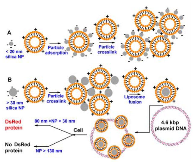
Fig. 1
(A) Small silica NPs are adsorbed onto positively charged DOTAP liposomes, resulting a negatively charged surface. With excess DOTAP, the system crosslinks to form large aggregates. (B) Larger silica ...
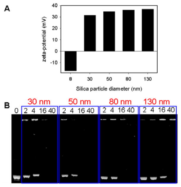
Fig. 2
(A) Zeta-potential of complexes formed by silica NPs of different sizes and DOTAP liposomes. For the 8 nm silica NPs, excess silica was used; while for the other particles, excess liposomes were used ...
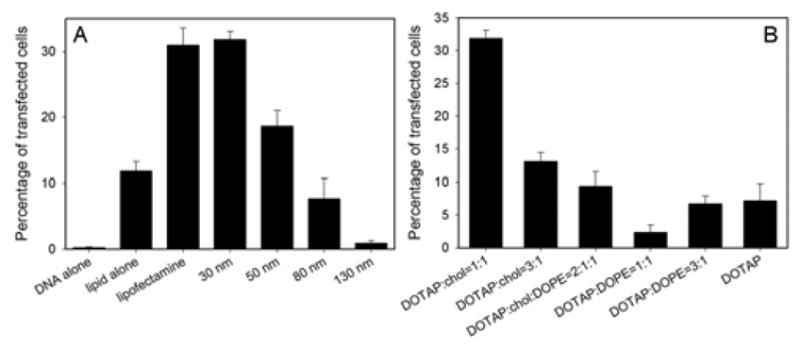
Fig. 3
Transfection efficiency as a function of silica NP core size (A) and lipid shell composition (B).
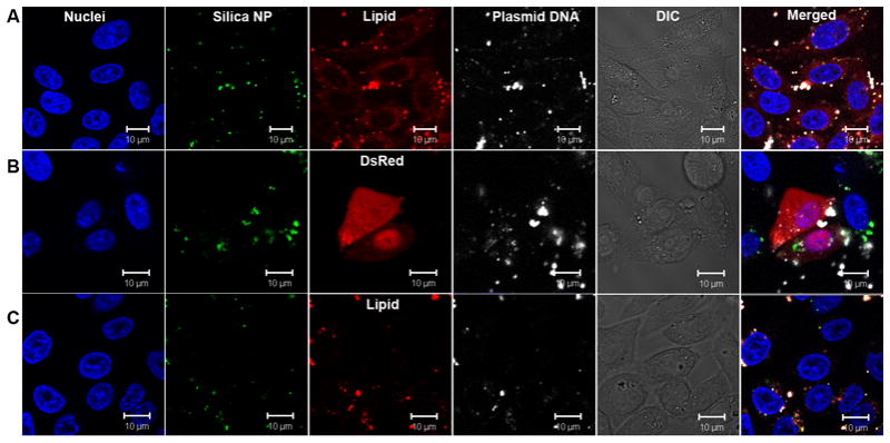
Fig. 4
DNA delivery studied by confocal fluorescence microscopy. Cell nuclei were stained with Hoechst 33342 (blue), silica NPs were labelled with a green emission fluorophore and the plasmid DNA was labelle...
Figure images are served from the NIH/NLM PubMed Central Open Access Subset or Europe PMC; copyright remains with the publishers and authors.
💬 Discussion
0 commentsNo comments yet. Be the first to start a discussion!
Leave a Comment