Abstract
Malaria remains a global health burden causing significant morbidity, yet the mechanisms underlying disease outcomes and protection are poorly understood. Herein, we analyzed the peripheral blood of a unique cohort of Malawian children with severe malaria, and performed a comprehensive overview of blood leukocytes and inflammatory mediators present in these patients. We reveal robust immune cell activation, notably of CD14+ inflammatory monocytes, NK cells and plasmacytoid dendritic cells (pDCs) that is associated with very high inflammation. Using the Plasmodium yoelii 17X YM surrogate mouse model of lethal malaria, we report a comparable pattern of immune cell activation and inflammation and found that type I IFN represents a key checkpoint for disease outcomes. Compared to wild type mice, mice lacking the type I interferon (IFN) receptor exhibited a significant decrease in immune cell activation and inflammatory response, ultimately surviving the infection. We demonstrate that pDCs were the major producers of systemic type I IFN in the bone marrow and the blood of infected mice, via TLR7/MyD88-mediated recognition of Plasmodium parasites. This robust type I IFN production required priming of pDCs by CD169+ macrophages undergoing activation upon STING-mediated sensing of parasites in the bone marrow. pDCs and macrophages displayed prolonged interactions in this compartment in infected mice as visualized by intravital microscopy. Altogether our findings describe a novel mechanism of pDC activation in vivo and precise stepwise cell/cell interactions taking place during severe malaria that contribute to immune cell activation and inflammation, and subsequent disease outcomes.
🔬 Techniques
🧬 Organisms
✨ Fluorophores
🧪 Sample Preparation
🔬 Cell Lines
🏭 Microscope Brands
🧪 Reagent Suppliers
🔴 Lasers
🎨 Filters
💻 Software Details
🏛️ Research Organizations (ROR)
Affiliated research institutions:
📋 Methods
Human studies Patient population
To characterize immune responses associated with severe human malaria we examined Malawian children with CM, enrolled in the Blantyre Malaria Research Project (BMP), an ongoing longitudinal study of CM, during the 2013 transmission season [ 78 ]. Children between the ages of 6 months and 12 years with P . falciparum infection and a Blantyre Coma Score
📊 Figures
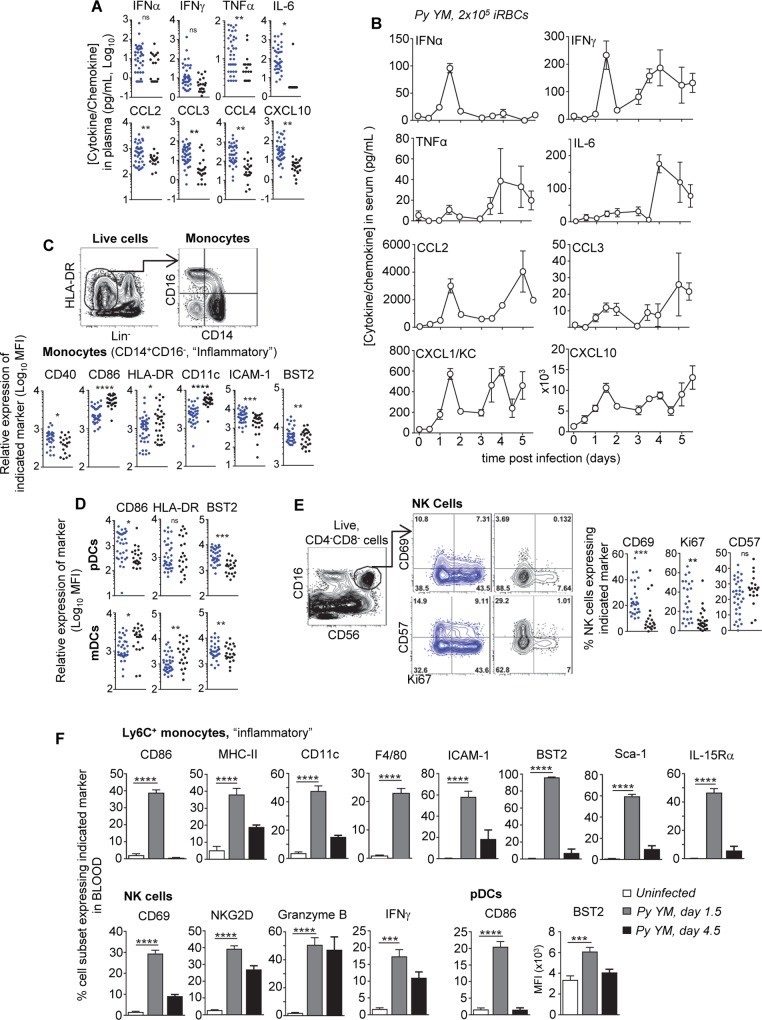
Fig 1
High inflammation and immune cell activation in the blood of severe malaria patients and in a surrogate mouse model.
( A ) Serum cytokine and chemokine levels in patients. Each point represents an individual patient, enrollment (blue), follow-up (black). ( B ) WT B6 mice were inoculated i.v. with 2x10 5 Py 17X YM iR...
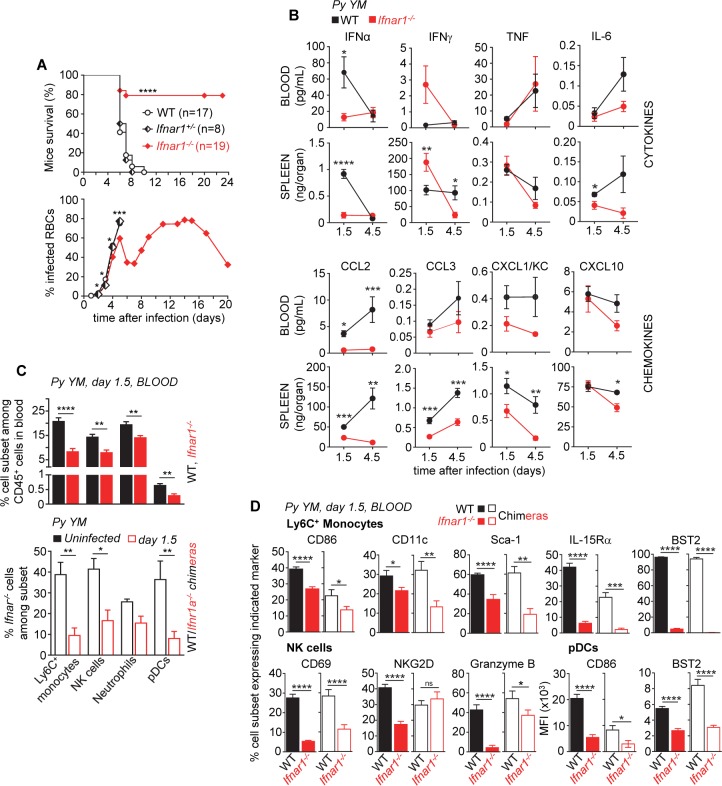
Fig 2
Type I interferon enhances immune blood leukocyte activation and lethal outcomes in severe murine malaria.
WT or Ifnar1 -/- B6 mice were inoculated i.v. with 2x10 5 Py 17X YM iRBCs. ( A ) Survival and blood parasitemia of Py -infected mice over indicated times. ( B ) Serum and spleen cytokine and chemokine...
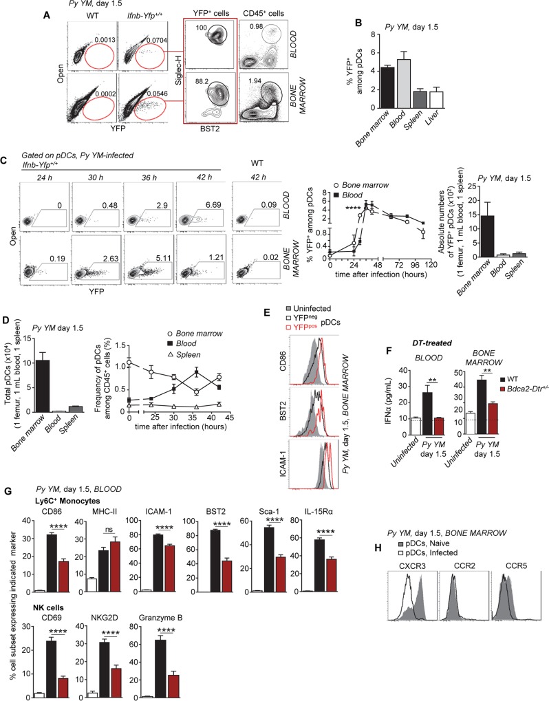
Fig 3
Plasmacytoid dendritic cells produce immune-activating type I IFN in the bone marrow and the blood of Py -infected mice.
( A ) WT Ifnb-Yfp +/+ reporter mice (n>5) were inoculated i.v. with 2x10 5 Py 17X YM iRBCs and blood and bone marrow cells were stained with the lineage markers CD11b, CD3, CD19, NK1.1, Ly6C, BST2 and...
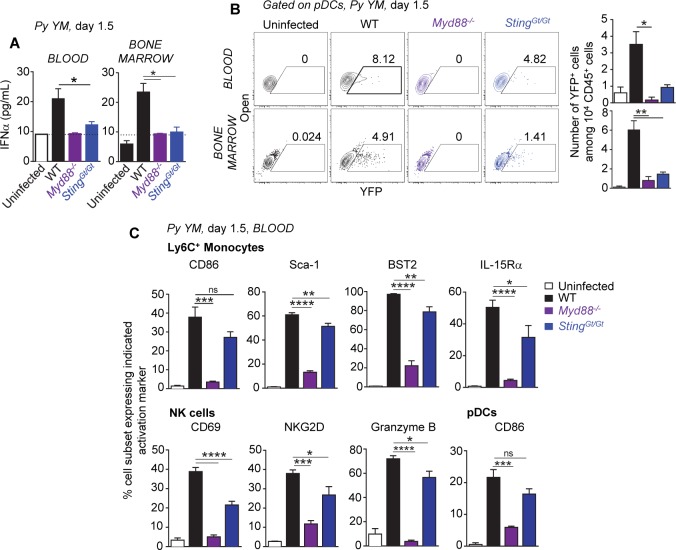
Fig 4
Production of systemic type I IFN during severe murine blood stage malaria requires both MyD88 and STING sensing pathways.
WT, Myd88 -/- or Sting Gt/Gt B6 mice crossed to Ifnb-Yfp +/+ reporter mice were inoculated i.v. with 2x10 5 Py 17X YM iRBCs. 1.5 days later, levels of IFNu03b1 ( A, n = 4u201310 mice/genotype) and fre...
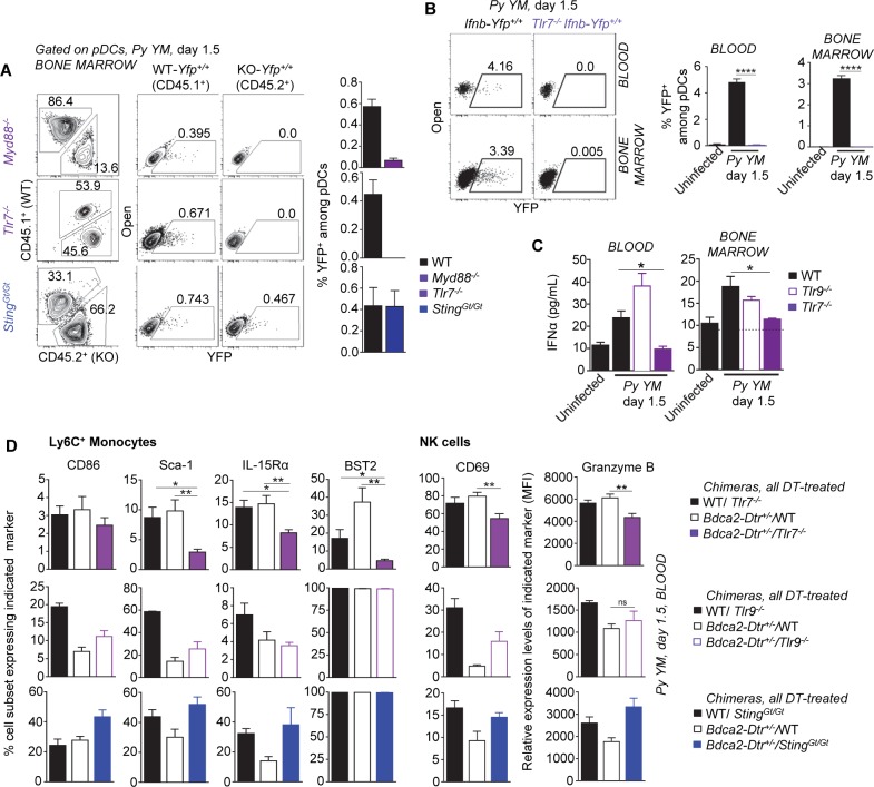
Fig 5
Plasmacytoid dendritic cells produce immune-activating type I IFN via TLR7/MyD88 but not STING.
( A ) Frequencies of WT (CD45.1 + ) or KO ( Myd88 -/- , Tlr7 -/- , Sting Gt/Gt ) YFP + pDCs in the bone marrow of Py -infected WT/KO Ifnb-Yfp +/+ mixed chimeras (n = 4u20136 mice/chimera). Bar graphs ...
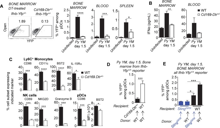
Fig 6
CD169 + macrophages control early pDC activation in the bone marrow of infected mice.
( A ) DT-treated WT and Cd169-Dtr +/- Ifnb-Yfp +/+ reporter mice (n = 7u201314 mice/genotype) were inoculated i.v. with 2x10 5 Py 17X YM iRBCs and blood and bone marrow cells were stained with the lin...
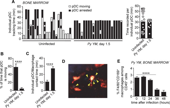
Fig 7
CD169 + macrophages form stable long-lasting interactions with pDCs in the bone marrow of P . yoelii -infected mice.
( A ) Tracking time in which individual pDCs were either moving or arrested in the bone marrow among total time monitored using intravital microscopy in Py-infected or uninfected live Ptcra-Gfp +/+ mi...
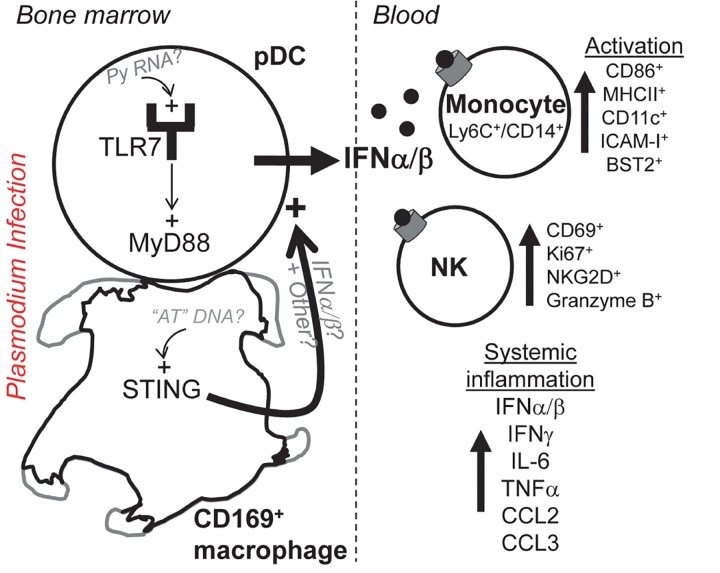
Fig 8
Mechanistic model summarizing findings.
Figure images are served from the NIH/NLM PubMed Central Open Access Subset or Europe PMC; copyright remains with the publishers and authors.
💬 Discussion
0 commentsNo comments yet. Be the first to start a discussion!
Leave a Comment