Abstract
The recently emerged severe acute respiratory syndrome coronavirus-2 (SARS-CoV-2) Beta (B.1.351) and Gamma (P.1) variants of concern (VoCs) include a key mutation (N501Y) found in the Alpha (B.1.1.7) variant that enhances affinity of the spike protein for its receptor, angiotensin-converting enzyme 2 (ACE2). Additional mutations are found in these variants at residues 417 and 484 that appear to promote antibody evasion. In contrast, the Epsilon variants (B.1.427/429) lack the N501Y mutation yet exhibit antibody evasion. We have engineered spike proteins to express these receptor binding domain (RBD) VoC mutations either in isolation or in different combinations and analyze the effects using biochemical assays and cryoelectron microscopy (cryo-EM) structural analyses. Overall, our findings suggest that the emergence of new SARS-CoV-2 variant spikes can be rationalized as the result of mutations that confer increased ACE2 affinity, increased antibody evasion, or both, providing a framework to dissect the molecular factors that drive VoC evolution.
🔬 Techniques
🔭 Microscopes
💻 Software
✨ Fluorophores
🧪 Sample Preparation
🏭 Microscope Brands
🧪 Reagent Suppliers
📷 Detectors
💻 Software Details
💻 Code & Software
💾 Data Repositories
🏷️ Research Resource Identifiers (RRIDs)
Verified research resources used in this paper:
🏛️ Research Organizations (ROR)
Affiliated research institutions:
📋 Methods
Key resources table REAGENT or RESOURCE SOURCE IDENTIFIER Antibodies
VH ab8 ( Li et al., 2020a ) N/A IgG1 ab1 ( Li et al., 2020b ) N/A IgG1 CR3022 ( Yuan et al., 2020 ) N/A Fab S309 ( Pinto et al., 2020 ) N/A Fab S2M11 ( Tortorici et al., 2020 ) N/A goat anti human IgG Jackson ImmunoReserach Cat. # 109-035-088 Chemicals, peptides, and recombinant proteins Ace2 (18-615) New England Biolabs Cat. # 73775S Linear Polyethylenimine Polysciences Cat# 23966-1 Critical commercial assays Pierce 1-Step Ultra Substrate Solution ThermoFisher Cat. # 34028 Q5 Site-Directed mutagenesis kit NEB Cat. # E0554S Experimental models: Cell lines Expi293F ThermoFisher Cat# A14527 Recombinant DNA pαH SARS-CoV-2 S HexaPro ( Hsieh et al., 2020 ) Addgene #154754 pαH HexaPro D614G This paper N/A pαH HexaPro D614G + N501Y This paper N/A pαH HexaPro D614G + K417N This paper N/A pαH HexaPro D614G + K417T This paper N/A pαH HexaPro D614G + E484K This paper N/A pαH HexaPro D614G + L452R This paper N/A pαH HexaPro D614G + N501Y + E484K This paper N/A pαH HexaPro D614G + N501Y + E484K + K417N This paper N/A pαH HexaPro D614G + N501Y + E484K + K417T This paper N/A pαH HexaPro D614G + N501Y + E484K + K417N + L452R This paper N/A pαH HexaPro D614G + N501Y + E484K + K417T + L452R This paper N/A pcDNA3.1 Fab S309 Light Chain This paper N/A pcDNA3.1 Fab S309 Heavy Chain This paper N/A pcDNA3.1 Fab S2M11 Light Chain This paper N/A pcDNA3.1 Fab S2M11 Heavy Chain This paper N/A Software and algorithms GraphPad Prism GraphPad 7.0 https://www.graphpad.com/scientific-software/prism/ EPU automated acquisition ThermoFisher Scientific https://www.thermofisher.com/us/en/home/electron-microscopy/products/software-em-3d-vis/epu-software.html UCSF Chimera ( Pettersen et al., 2004 ) https://www.cgl.ucsf.edu/chimera/ RELION 3.1 (Scheres, 2012) https://github.com/3dem/relion/releases/tag/3.1.0 crYOLO (version 1.7.4) ( Wagner et al., 2019 ) https://pypi.org/project/cryolo/ cryoSPARC live (v3.0.1) ( Punjani et al., 2017 ) https://cryosparc.com/live Deposited data S(D614G) This Paper EMDB: 25503, PDB: 7SXR S(D614G)+ACE2 This Paper global refinement: EMDB: 25509, PDB: 7SXX , focus refinement: EMDB: 25510, PDB: 7SXY S(D614G,L452R) This Paper EMDB: 25504; PDB: 7SXS S(D614G,L452R) + ACE2 This Paper global refinement: EMDB: 25511; PDB: 7SXZ , focus refinement: EMDB: 25512; PDB: 7SY0 S(D614G,N501Y) This Paper EMDB: 25505; PDB: 7SXT S(D614G,N501Y) + ACE2 This Paper global refinement: EMDB: 25513; PDB: 7SY1 , focus refinement: EMDB: 25514; PDB: 7SY2 S(D614G,N501Y,E484K) This Paper EMDB: 25506; PDB: 7SXU S(D614G,N501Y,E484K) + ACE2 This Paper global refinement: EMDB: 25515; PDB: 7SY3 , focus refinement: EMDB: 25516; PDB: 7SY4 S(D614G,N501Y,E484K,K417N) This Paper EMDB: 25507, PDB: 7SXV S(D614G,N501Y,E484K,K417N) + ACE2 This Paper global refinement: EMDB: 25517; PDB: 7SY5 , focus refinement: EMDB: 25518; PDB: 7SY6 S(D614G,N501Y,E484K,K417T) This Paper EMDB: 25508; PDB: 7SXW S(D614G,N501Y,E484K,K417T) + ACE2 This Paper global refinement: EMDB: 25519; PDB: 7SY7 , focus refinement: EMDB: 25520; PDB: 7SY8 Resource availability Lead contact Further information and requests for resources and reagents should be directed to and will be fulfilled by the Lead Contact, Sriram Subramaniam ( Sriram.Subramaniam@ubc.ca ).
Show full methods section
Key resources table REAGENT or RESOURCE SOURCE IDENTIFIER Antibodies
VH ab8 ( Li et al., 2020a ) N/A IgG1 ab1 ( Li et al., 2020b ) N/A IgG1 CR3022 ( Yuan et al., 2020 ) N/A Fab S309 ( Pinto et al., 2020 ) N/A Fab S2M11 ( Tortorici et al., 2020 ) N/A goat anti human IgG Jackson ImmunoReserach Cat. # 109-035-088 Chemicals, peptides, and recombinant proteins Ace2 (18-615) New England Biolabs Cat. # 73775S Linear Polyethylenimine Polysciences Cat# 23966-1 Critical commercial assays Pierce 1-Step Ultra Substrate Solution ThermoFisher Cat. # 34028 Q5 Site-Directed mutagenesis kit NEB Cat. # E0554S Experimental models: Cell lines Expi293F ThermoFisher Cat# A14527 Recombinant DNA pαH SARS-CoV-2 S HexaPro ( Hsieh et al., 2020 ) Addgene #154754 pαH HexaPro D614G This paper N/A pαH HexaPro D614G + N501Y This paper N/A pαH HexaPro D614G + K417N This paper N/A pαH HexaPro D614G + K417T This paper N/A pαH HexaPro D614G + E484K This paper N/A pαH HexaPro D614G + L452R This paper N/A pαH HexaPro D614G + N501Y + E484K This paper N/A pαH HexaPro D614G + N501Y + E484K + K417N This paper N/A pαH HexaPro D614G + N501Y + E484K + K417T This paper N/A pαH HexaPro D614G + N501Y + E484K + K417N + L452R This paper N/A pαH HexaPro D614G + N501Y + E484K + K417T + L452R This paper N/A pcDNA3.1 Fab S309 Light Chain This paper N/A pcDNA3.1 Fab S309 Heavy Chain This paper N/A pcDNA3.1 Fab S2M11 Light Chain This paper N/A pcDNA3.1 Fab S2M11 Heavy Chain This paper N/A Software and algorithms GraphPad Prism GraphPad 7.0 https://www.graphpad.com/scientific-software/prism/ EPU automated acquisition ThermoFisher Scientific https://www.thermofisher.com/us/en/home/electron-microscopy/products/software-em-3d-vis/epu-software.html UCSF Chimera ( Pettersen et al., 2004 ) https://www.cgl.ucsf.edu/chimera/ RELION 3.1 (Scheres, 2012) https://github.com/3dem/relion/releases/tag/3.1.0 crYOLO (version 1.7.4) ( Wagner et al., 2019 ) https://pypi.org/project/cryolo/ cryoSPARC live (v3.0.1) ( Punjani et al., 2017 ) https://cryosparc.com/live Deposited data S(D614G) This Paper EMDB: 25503, PDB: 7SXR S(D614G)+ACE2 This Paper global refinement: EMDB: 25509, PDB: 7SXX , focus refinement: EMDB: 25510, PDB: 7SXY S(D614G,L452R) This Paper EMDB: 25504; PDB: 7SXS S(D614G,L452R) + ACE2 This Paper global refinement: EMDB: 25511; PDB: 7SXZ , focus refinement: EMDB: 25512; PDB: 7SY0 S(D614G,N501Y) This Paper EMDB: 25505; PDB: 7SXT S(D614G,N501Y) + ACE2 This Paper global refinement: EMDB: 25513; PDB: 7SY1 , focus refinement: EMDB: 25514; PDB: 7SY2 S(D614G,N501Y,E484K) This Paper EMDB: 25506; PDB: 7SXU S(D614G,N501Y,E484K) + ACE2 This Paper global refinement: EMDB: 25515; PDB: 7SY3 , focus refinement: EMDB: 25516; PDB: 7SY4 S(D614G,N501Y,E484K,K417N) This Paper EMDB: 25507, PDB: 7SXV S(D614G,N501Y,E484K,K417N) + ACE2 This Paper global refinement: EMDB: 25517; PDB: 7SY5 , focus refinement: EMDB: 25518; PDB: 7SY6 S(D614G,N501Y,E484K,K417T) This Paper EMDB: 25508; PDB: 7SXW S(D614G,N501Y,E484K,K417T) + ACE2 This Paper global refinement: EMDB: 25519; PDB: 7SY7 , focus refinement: EMDB: 25520; PDB: 7SY8 Resource availability Lead contact Further information and requests for resources and reagents should be directed to and will be fulfilled by the Lead Contact, Sriram Subramaniam ( Sriram.Subramaniam@ubc.ca ).
Materials availability
All unique/stable reagents generated in this study are available from the Lead Contact with a completed Materials Transfer Agreement.
Experimental model and subject details
Expi293F cells (ThermoFisher, Cat# A14527) were grown in suspension culture using Expi293 Expression Medium (ThermoFisher, Cat# A1435102) at 37°C, 8% CO2 with agitation at 130 rpm. Method details Cloning, expression and purification of recombinant spike protein constructs The wild type SARS-CoV-2 S HexaPro expression plasmid was previously described ( Hsieh et al., 2020 ) and was a gift from Jason McLellan (Addgene plasmid #154754; http://n2t.net/addgene:154754 ; RRID:Addgene_154754). The VoC RBD mutations were introduced by site-directed mutagenesis (Q5 Site-Directed Mutagenesis Kit, New England Biolabs). Successful cloning was confirmed by Sanger sequencing (Genewiz, Inc.). Expi293 Cells were transiently transfected at a density of 3 x 10ˆ6 cells/mL using linear polyethylenimine (Polysciences Cat# 23966-1). 24-hours following transfection, media was supplemented with 2.2 mM valproic acid and expression carried out for 3-5 days at 37°C, 8% CO2. Supernatant was harvested from cells expressing spike ectodomains by centrifugation and filtered through a 0.22 μM filter prior to loading onto a 5 mL HisTrap excel column (Cytiva). The column was washed for 20 CVs with wash buffer (20 mM Tris pH 8.0, 500 mM NaCl), 5 CVs of wash buffer supplemented with 20 mM imidazole and the protein eluted with elution buffer (20 mM Tris pH 8.0, 500 mM NaCl, 500 mM imidazole). Elution fractions containing the protein were pooled and concentrated (Amicon Ultra 100 kDa cut off, Millipore Sigma) for gel filtration. Gel filtration was conducted using a Superose 6 10/300 GL column (Cytiva) pre-equilibrated with GF buffer (20 mM Tris pH 8.0, 150 mM NaCl). Fractions of the main protein peak (eluting at ∼11 mL) were pooled and concentrated to 4.5 - 5.5 mg/mL (Amicon Ultra 100 kDa cut off, Millipore Sigma). Protein samples were immediately flash-frozen in liquid nitrogen and stored at -80°C. Antibody production VH-FC ab8, IgG ab1 and IgG CR3022 were produced as previously described ( Li et al., 2020a , 2020b ).
Plasmids encoding light and heavy chains for Fab
S309 and S2M11 were synthesized (Synbio). Heavy chains were designed to incorporate a C terminal 6x histidine tag. Expi293 cells were transfected at a density of 3 x 10ˆ6 cells/mL using linear polyethylenimine (Polysciences Cat# 23966-1). 24-hours following transfection, media was supplemented with 2.2 mM valproic acid and expression carried out for 3-5 days at 37°C, 8% CO2. The supernatant was harvested by centrifugation and filtered through a 0.22 μM filter prior to loading onto a 5 mL HisTrap excel column (Cytiva). The column was washed for 20 CVs with wash buffer (20 mM Tris pH 8.0, 500 mM NaCl), 5 CVs of wash buffer supplemented with 20 mM imidazole and the protein eluted with elution buffer (20 mM Tris pH 8.0, 500 mM NaCl, 500 mM imidazole). Elution fractions containing the protein were pooled and concentrated (Amicon Ultra 10 kDa cut off, Millipore Sigma) for gel filtration. Gel filtration was conducted using a Superose 6 10/300 GL column (Cytiva) pre-equilibrated with GF buffer (20 mM Tris pH 8.0, 150 mM NaCl). Peak fractions corresponding to soluble protein were pooled and concentrated to 8 - 20 mg/mL (Amicon Ultra 10 kDa cut off, Millipore Sigma). Protein samples were stored at 4°C until use.
Electron microscopy sample preparation and data collection
S-protein samples were prepared at 2.25 mg/mL, with and without the addition of ACE2 (∼1:1.25 S-protein trimer:ACE2 molar ratio) (New England Biolabs). Vitrified samples of S-protein constructs with and without ACE2 were prepared by first glow discharging Quantifoil R1.2/1.3 300 mesh holey carbon copper grids for 1 minute using a Pelco easiGlow glow discharge unit (Ted Pella) and then applying 1.8 μL of protein suspension to the surface of the grid. Grids were blotted (12 sec, blot force -10) and plunge frozen into liquid ethane using a Vitrobot Mark IV (Thermo Fisher Scientific) at a temperature of 10°C and a humidity level of 100%. All cryo-EM samples were imaged using a 300 kV Titan Krios G4 transmission electron microscope (ThermoFisher Scientific) equipped with a Falcon4 direct electron detector in electron event registration (EER) mode. Movies were collected at 155,000x magnification (physical pixel size 0.5 Å) over a defocus range of -0.5 μm to -3 μm with a total dose of 40 e-/ Å 2 using EPU automated acquisition software.
Image processing
In general, all data processing was performed in cryoSPARC v.3.0.1 ( Punjani et al., 2017 ) unless stated otherwise. Motion correction in patch mode (EER upsampling factor 1, EER number of fractions 40), CTF estimation in patch mode, reference-free particle picking and particle extraction were performed on-the-fly in cryoSPARC. After preprocessing, particles were subjected to 2D classification and/or 3D heterogeneous classification. Final 3D refinement was done with per particle CTF estimation and aberration correction. For complex of spike protein ectodomain and human ACE2, focused refinements were performed with a soft mask covering single RBD and its bound ACE2. Global resolution and focused resolution were according to the gold-standard FSC ( Bell et al., 2016 ).
Model building and refinement
For models of spike protein ectodomain alone, SARS-CoV-2 HexaPro S trimer with N501Y mutation (PDB code 7MJG ) were docked into cryo-EM density using UCSF Chimera v.1.15 ( Pettersen et al., 2004 ). Then mutation and manual adjustment were done with COOT v.0.9.3 ( Emsley et al., 2010 ), followed by iterative rounds of refinement in COOT and Phenix v.1.19 ( Afonine et al., 2018 ). Glycans were added at N-linked glycosylation sites in COOT. For models of complex of spike protein ectodomain and human ACE2, the RBD-ACE2 subcomplex was built using coordinates of PDB code 7MJN as initial model and refined against focused refinement maps. Then it was docked into global refinement maps together with individual domains of spike protein. Model validation was performed using MolProbity ( Chen et al., 2010 ). Figures were prepared using UCSF Chimera, UCSF ChimeraX v.1.1.1 ( Goddard et al., 2018 ), and PyMOL (v.2.2 Schrodinger, LLC). Biolayer interferometry (BLI) S protein-ACE2 binding assay The binding kinetics of SARS-CoV-2 trimers and human ACE2 was analyzed with the biolayer interferometer BLItz (ForteBio, Menlo Park, CA). Protein-A biosensors (ForteBio: 18–5010) were coated with ACE2-mFc (40 μg/mL) for 2 min and incubated in DPBS (pH = 7.4) to establish baselines. Concentrations of 125, 250, 500 and 1000 nM spike trimers were used for association for 2 min followed by dissociation in DPBS for 5 min. The association (k on ) and dissociation (k off ) rates were derived from the sensorgrams fitting and used to calculate the binding equilibrium constant (K D ). Enzyme-linked immunosorbent assay (ELISA) 100 μl of wild-type or VoC RBD mutant SARS-CoV-2 S protein preparations were coated onto 96-well MaxiSorp™ plates at 2 μg/ml in PBS overnight at 4°C. All washing steps were performed 5 times with PBS + 0.05% Tween 20 (PBS-T). After washing, wells were either incubated with blocking buffer (PBS-T + 2% BSA) for 1 hr at room temperature. After washing, wells were incubated with dilutions of primary antibodies in PBS-T + 0.5% BSA buffer for 1 hr at room temperature. After washing, wells were incubated with goat anti-human IgG (Jackson ImmunoResearch) at a 1:8,000 dilution in PBS-T + 0.5% BSA buffer for 1 hr at room temperature. After washing, the substrate solution (Pierce™ 1-Step™) was used for colour development according to the manufacturer's specifications. Optical density at 450 nm was read on a Varioskan Lux plate reader (Thermo Fisher Scientific).
Analysis of convalescent patient antibody footprints
PDB entries of SARS-CoV-2 spike or RBD complexes with antibody fragments isolated from convalescent patients were selected. Antibody footprints were determined by consulting respective depositing studies along with analysis of protein-protein contacts using PDBsum ( Laskowski et al., 2018 ).
Quantification and statistical analysis The Area under the curve
(AUC) for ELISA binding data was calculated in GraphPad Prism 7. No statistical analysis was performed in this study.
Materials availability
All unique/stable reagents generated in this study are available from the Lead Contact with a completed Materials Transfer Agreement.
Experimental model and subject details
Expi293F cells (ThermoFisher, Cat# A14527) were grown in suspension culture using Expi293 Expression Medium (ThermoFisher, Cat# A1435102) at 37°C, 8% CO2 with agitation at 130 rpm.
Method details Cloning, expression and purification of recombinant spike protein constructs The wild type SARS-CoV-2 S HexaPro expression plasmid was previously described ( Hsieh et al., 2020 ) and was a gift from Jason McLellan (Addgene plasmid #154754; http://n2t.net/addgene:154754 ; RRID:Addgene_154754). The VoC RBD mutations were introduced by site-directed mutagenesis (Q5 Site-Directed Mutagenesis Kit, New England Biolabs). Successful cloning was confirmed by Sanger sequencing (Genewiz, Inc.). Expi293 Cells were transiently transfected at a density of 3 x 10ˆ6 cells/mL using linear polyethylenimine (Polysciences Cat# 23966-1). 24-hours following transfection, media was supplemented with 2.2 mM valproic acid and expression carried out for 3-5 days at 37°C, 8% CO2. Supernatant was harvested from cells expressing spike ectodomains by centrifugation and filtered through a 0.22 μM filter prior to loading onto a 5 mL HisTrap excel column (Cytiva). The column was washed for 20 CVs with wash buffer (20 mM Tris pH 8.0, 500 mM NaCl), 5 CVs of wash buffer supplemented with 20 mM imidazole and the protein eluted with elution buffer (20 mM Tris pH 8.0, 500 mM NaCl, 500 mM imidazole). Elution fractions containing the protein were pooled and concentrated (Amicon Ultra 100 kDa cut off, Millipore Sigma) for gel filtration. Gel filtration was conducted using a Superose 6 10/300 GL column (Cytiva) pre-equilibrated with GF buffer (20 mM Tris pH 8.0, 150 mM NaCl). Fractions of the main protein peak (eluting at ∼11 mL) were pooled and concentrated to 4.5 - 5.5 mg/mL (Amicon Ultra 100 kDa cut off, Millipore Sigma). Protein samples were immediately flash-frozen in liquid nitrogen and stored at -80°C. Antibody production VH-FC ab8, IgG ab1 and IgG CR3022 were produced as previously described ( Li et al., 2020a , 2020b ).
Plasmids encoding light and heavy chains for Fab
S309 and S2M11 were synthesized (Synbio). Heavy chains were designed to incorporate a C terminal 6x histidine tag. Expi293 cells were transfected at a density of 3 x 10ˆ6 cells/mL using linear polyethylenimine (Polysciences Cat# 23966-1). 24-hours following transfection, media was supplemented with 2.2 mM valproic acid and expression carried out for 3-5 days at 37°C, 8% CO2. The supernatant was harvested by centrifugation and filtered through a 0.22 μM filter prior to loading onto a 5 mL HisTrap excel column (Cytiva). The column was washed for 20 CVs with wash buffer (20 mM Tris pH 8.0, 500 mM NaCl), 5 CVs of wash buffer supplemented with 20 mM imidazole and the protein eluted with elution buffer (20 mM Tris pH 8.0, 500 mM NaCl, 500 mM imidazole). Elution fractions containing the protein were pooled and concentrated (Amicon Ultra 10 kDa cut off, Millipore Sigma) for gel filtration. Gel filtration was conducted using a Superose 6 10/300 GL column (Cytiva) pre-equilibrated with GF buffer (20 mM Tris pH 8.0, 150 mM NaCl). Peak fractions corresponding to soluble protein were pooled and concentrated to 8 - 20 mg/mL (Amicon Ultra 10 kDa cut off, Millipore Sigma). Protein samples were stored at 4°C until use.
Electron microscopy sample preparation and data collection
S-protein samples were prepared at 2.25 mg/mL, with and without the addition of ACE2 (∼1:1.25 S-protein trimer:ACE2 molar ratio) (New England Biolabs). Vitrified samples of S-protein constructs with and without ACE2 were prepared by first glow discharging Quantifoil R1.2/1.3 300 mesh holey carbon copper grids for 1 minute using a Pelco easiGlow glow discharge unit (Ted Pella) and then applying 1.8 μL of protein suspension to the surface of the grid. Grids were blotted (12 sec, blot force -10) and plunge frozen into liquid ethane using a Vitrobot Mark IV (Thermo Fisher Scientific) at a temperature of 10°C and a humidity level of 100%. All cryo-EM samples were imaged using a 300 kV Titan Krios G4 transmission electron microscope (ThermoFisher Scientific) equipped with a Falcon4 direct electron detector in electron event registration (EER) mode. Movies were collected at 155,000x magnification (physical pixel size 0.5 Å) over a defocus range of -0.5 μm to -3 μm with a total dose of 40 e-/ Å 2 using EPU automated acquisition software.
Image processing
In general, all data processing was performed in cryoSPARC v.3.0.1 ( Punjani et al., 2017 ) unless stated otherwise. Motion correction in patch mode (EER upsampling factor 1, EER number of fractions 40), CTF estimation in patch mode, reference-free particle picking and particle extraction were performed on-the-fly in cryoSPARC. After preprocessing, particles were subjected to 2D classification and/or 3D heterogeneous classification. Final 3D refinement was done with per particle CTF estimation and aberration correction. For complex of spike protein ectodomain and human ACE2, focused refinements were performed with a soft mask covering single RBD and its bound ACE2. Global resolution and focused resolution were according to the gold-standard FSC ( Bell et al., 2016 ).
Model building and refinement
For models of spike protein ectodomain alone, SARS-CoV-2 HexaPro S trimer with N501Y mutation (PDB code 7MJG ) were docked into cryo-EM density using UCSF Chimera v.1.15 ( Pettersen et al., 2004 ). Then mutation and manual adjustment were done with COOT v.0.9.3 ( Emsley et al., 2010 ), followed by iterative rounds of refinement in COOT and Phenix v.1.19 ( Afonine et al., 2018 ). Glycans were added at N-linked glycosylation sites in COOT. For models of complex of spike protein ectodomain and human ACE2, the RBD-ACE2 subcomplex was built using coordinates of PDB code 7MJN as initial model and refined against focused refinement maps. Then it was docked into global refinement maps together with individual domains of spike protein. Model validation was performed using MolProbity ( Chen et al., 2010 ). Figures were prepared using UCSF Chimera, UCSF ChimeraX v.1.1.1 ( Goddard et al., 2018 ), and PyMOL (v.2.2 Schrodinger, LLC). Biolayer interferometry (BLI) S protein-ACE2 binding assay The binding kinetics of SARS-CoV-2 trimers and human ACE2 was analyzed with the biolayer interferometer BLItz (ForteBio, Menlo Park, CA). Protein-A biosensors (ForteBio: 18–5010) were coated with ACE2-mFc (40 μg/mL) for 2 min and incubated in DPBS (pH = 7.4) to establish baselines. Concentrations of 125, 250, 500 and 1000 nM spike trimers were used for association for 2 min followed by dissociation in DPBS for 5 min. The association (k on ) and dissociation (k off ) rates were derived from the sensorgrams fitting and used to calculate the binding equilibrium constant (K D ). Enzyme-linked immunosorbent assay (ELISA) 100 μl of wild-type or VoC RBD mutant SARS-CoV-2 S protein preparations were coated onto 96-well MaxiSorp™ plates at 2 μg/ml in PBS overnight at 4°C. All washing steps were performed 5 times with PBS + 0.05% Tween 20 (PBS-T). After washing, wells were either incubated with blocking buffer (PBS-T + 2% BSA) for 1 hr at room temperature. After washing, wells were incubated with dilutions of primary antibodies in PBS-T + 0.5% BSA buffer for 1 hr at room temperature. After washing, wells were incubated with goat anti-human IgG (Jackson ImmunoResearch) at a 1:8,000 dilution in PBS-T + 0.5% BSA buffer for 1 hr at room temperature. After washing, the substrate solution (Pierce™ 1-Step™) was used for colour development according to the manufacturer's specifications. Optical density at 450 nm was read on a Varioskan Lux plate reader (Thermo Fisher Scientific).
Analysis of convalescent patient antibody footprints
PDB entries of SARS-CoV-2 spike or RBD complexes with antibody fragments isolated from convalescent patients were selected. Antibody footprints were determined by consulting respective depositing studies along with analysis of protein-protein contacts using PDBsum ( Laskowski et al., 2018 ).
Supplemental information Document S1. Figures S1–S5 and Tables S1–S3 Document S2. Article plus supplemental information
📊 Figures
Figureu00a01
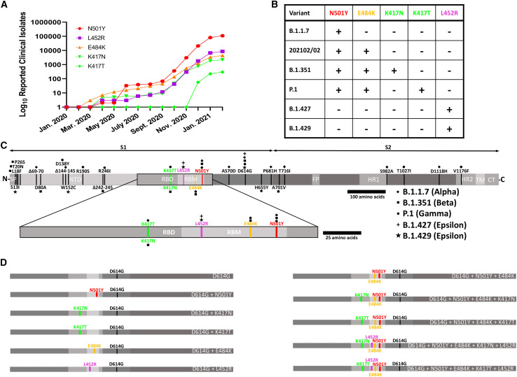
The global prevalence of SARS-CoV-2 VoC/VoI RBD mutations and their locations within the S protein (A) Global occurrences of each VoC/VoI RBD mutation over time, computed using the sum of clinical iso...
Figureu00a02
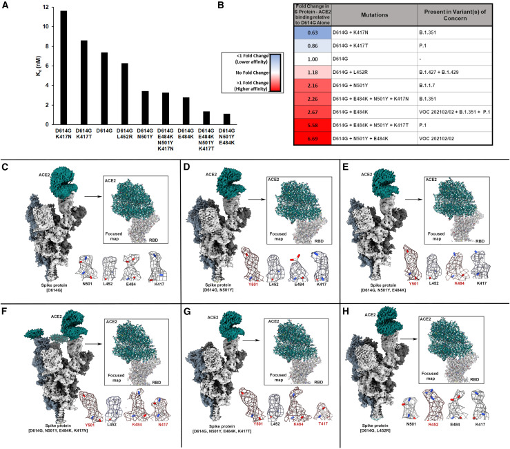
Complete sets of VoC/VoI RBD mutations increase S protein trimer-ACE2 binding affinity (A) Affinity ( K d ) measurements for VoC/VoI RBD mutant S protein-ACE2 binding as measured by biolayer interfero...
Figureu00a03
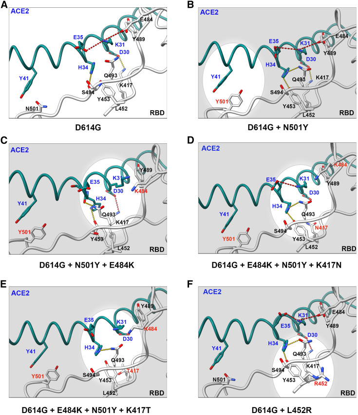
CryoEM structures of wild-type and VoC RBD-ACE2 interfaces (Au2013F) Zoomed-in views of the RBD-ACE2 binding interfaces for the six S protein-ACE2 structures. Focused refinement of the RBD-ACE2 interf...
Figureu00a04
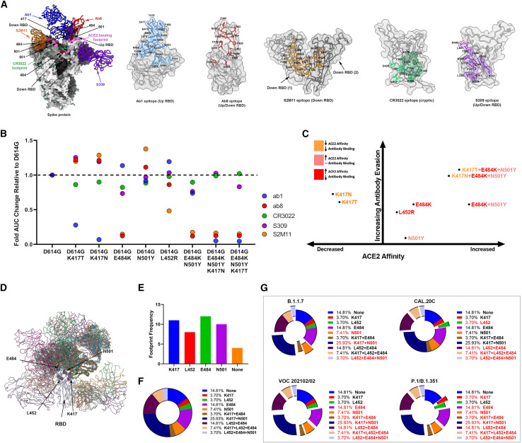
Monoclonal antibody binding against SARS-CoV-2u00a0S proteins containing VoC/VoI RBD mutations (A) Mapping of Ab1, Ab8, CR3022, S309, and S2M11 antibody footprints onto SARS-CoV-2 trimers and RBDs. Di...
Figure images are served from the NIH/NLM PubMed Central Open Access Subset or Europe PMC; copyright remains with the publishers and authors.
💬 Discussion
0 commentsNo comments yet. Be the first to start a discussion!
Leave a Comment