Abstract
Three highly pathogenic β-coronaviruses have crossed the animal-to-human species barrier in the past two decades: SARS-CoV, MERS-CoV and SARS-CoV-2. To evaluate the possibility of identifying antibodies with broad neutralizing activity, we isolated a monoclonal antibody, termed B6, that cross-reacts with eight β-coronavirus spike glycoproteins, including all five human-infecting β-coronaviruses. B6 broadly neutralizes entry of pseudotyped viruses from lineages A and C, but not from lineage B, and the latter includes SARS-CoV and SARS-CoV-2. Cryo-EM, X-ray crystallography and membrane fusion assays reveal that B6 binds to a conserved cryptic epitope located in the fusion machinery. The data indicate that antibody binding sterically interferes with the spike conformational changes leading to membrane fusion. Our data provide a structural framework explaining B6 cross-reactivity with β-coronaviruses from three lineages, along with a proof of concept for antibody-mediated broad coronavirus neutralization elicited through vaccination. This study unveils an unexpected target for next-generation structure-guided design of a pan-β-coronavirus vaccine.
🔬 Techniques
💻 Software
✨ Fluorophores
🧪 Sample Preparation
🔬 Cell Lines
🏭 Microscope Brands
🧪 Reagent Suppliers
💻 Software Details
💾 Data Repositories
🏛️ Research Organizations (ROR)
Affiliated research institutions:
📋 Methods
Workers slaughtering dromedaries in an abattoir in Kano, Nigeria, were compared with abattoir workers without direct dromedary contact, non-abattoir workers from Kano, and controls from Guangzhou, China. Exposure to dromedaries was ascertained using a questionnaire.
Serum and peripheral blood mononuclear cells
(PBMCs) were tested for MERS-CoV specific neutralising antibody and T-cell responses.
Methods Study design and participants
In this observational cohort study, workers at an abattoir in Kano, Nigeria, consenting to participate in the cohort study in March 13–26, 2018, were recruited. Non-abattoir workers were also recruited randomly from the city of Kano during the same period, and blood donors aged 18–65 years sampled in May 10–Aug 31, 2018, at Guangzhou Blood Center, Guangzhou, China, were randomly included as healthy controls from a region with no dromedary camel exposure. Convalescent blood samples collected from 14 people with symptomatic or asymptomatic virologically confirmed MERS-CoV infections detected at the King Abdulaziz Medical City, Riyadh, and King Faisal Specialist Hospital, Jeddah, Saudi Arabia, collected as part of a previously reported study 11 were included as positive controls. The clinical, serological and T-cell responses (using only interferon [IFN]-γ as a readout of positive cells) of this patient cohort have been previously reported. 11 PBMCs were collected at 6 months (patients 1–6, 8–9, 11–14 as reported in the previous publication) or 24 months (patients 18–19) after infection. Written informed consent was obtained from all study participants in Nigeria and the study was approved by the Health Research Ethics Committee of the Ministry of Health, Nigeria (MOH/Off/797/T.I/630). We obtained Institutional Review Board approval from the Health Commission of Guangdong Province to use the anonymised blood donor samples for this study. Written informed consent was obtained from all recovered patients with MERS to participate in this study and approval obtained from the Institutional Review Boards of the National Guard Hospital, Riyadh, and King Faisal Specialist Hospital, Jeddah. 12 Procedures 8 mL of blood were collected from each study participant from the abattoir and from donors from Guangzhou. PBMCs were isolated from blood using Leucosep tubes (Greiner, Kremsmünster, Austria) and Ficoll-Paque PLUS (GE Healthcare, Chicago, IL) according to the manufacturer's instructions. PBMCs were stored in liquid nitrogen and plasma at −80°C or lower before and during shipping before analysis. Plasma was heat inactivated for 30 min at 56°C before the serology testing. Anti-MERS-CoV antibody titres were determined using plaque reduction neutralisation tests. 9 , 13 A set of 20-mer peptides overlapping by ten amino acids encompassing the four MERS-CoV (HCoV-EMC/2012) structural proteins (peptides S1, S2, N, and ME encompassing the N-terminal and C-terminal portions of the spike [S] glycoprotein, the nucleocapsid [N] protein, and the transmembrane [M] and envelope [E] proteins) and five accessory proteins (ORF3, ORF4a, ORF4b, ORF5 and ORF8b) were synthesised by Sino Biological (Shanghai, China), and used for stimulation of PBMCs. T-cell responses were measured using intracellular cytokine staining assays for interferon-γ (IFN-γ) and tumour necrosis factor (TNF). Structural proteins peptide libraries of HKU1-CoV, OC43-CoV, NL63-CoV, and 229E-CoV were also synthesised by Sino Biological to detect viral-specific T-cell responses. To enhance specificity, only cells with dual expression of both IFN-γ and TNF after peptide stimulation were considered as positive. Flow cytometry was used to determine the phenotype and function of T cells. The following anti-human monoclonal antibodies were used: BV510-CD3 (HIT3a; BD, San Jose, CA), PerCP-Cy5.5-CD4 (RPA-T4; BioLegend, San Diego, CA), APC Fire750-CD8 (SK1; BioLegend), APC-IFN-γ (B27; BD), PE-TNF (MAb11; Invitrogen, Carlsbad, CA), BB515-CD45RA (HI100; BD), and PE-Cy7-CCR7 (G043H7; BioLegend). Fc receptor-blocking solution was obtained from BioLegend. For surface staining, about 1 × 10 5 cells were blocked with Fc receptor blocking solution, stained with the indicated antibodies at 4°C, and labelled with live–dead staining dye (Thermo Fisher, Waltham, MA). For in-vitro intracellular cytokine staining, 2 × 10 5 to 6 × 10 5 cells per well were cultured in 96-well round-bottom plates at 37°C for 12 h in the presence of 10 μM peptides (Sino Biological) and brefeldin A (BD Biosciences). Cells were then labelled for cell surface markers, fixed and permeabilised with Cytofix/Cytoperm solution (BD Biosciences), and stained with anti-cytokine antibodies. All flow cytometry data were acquired on a BD FACSVerse flow cytometer and analysed using FlowJo software. PBMCs were considered MERS-CoV positive if they expressed both IFN-γ and TNF in response to peptide stimulation as described previously. 14 Statistical analysis In a previous study of dromedary abattoir workers in Saudi Arabia, ten of 30 workers sampled had detectable T-cell responses to MERS-CoV. 14 On the basis of this finding, and the assumption that abattoir workers without dromedary exposure and the other control groups would have no detectable T-cell responses, eight abattoir workers would be the minimal sample size required to detect a positive result with 95% probability, where the detection probability is given by: 1 – (1 – p)n with p equivalent to 10/30 and n being the sample size. We aimed at sampling all abattoir workers who consented to participate, as long as we successfully sampled at least eight dromedary-exposed abattoir workers. Association of T-cell responses with different exposure to dromedaries was done using Fisher's exact test. In univariate analysis, we estimated the crude odds ratio (OR) for each potential epidemiological exposure factor in relation to MERS-CoV T-cell positivity using a logistic regression model. Independent risk factors for T-cell positivity were identified using multivariable logistic regression. We included a-priori variables, such as years of work in abattoir and whether other household members frequently visited camel farms, and other variables with a crude OR of more than 2 or less than 0·5 in the univariate analysis. Due to small sample size and cross-related practices of drinking camel milk and camel urine, we first fitted a logistic regression model which considered all four combinations of the two practices (eg, drinking camel milk only, drinking camel urine only, drinking both camel milk and urine or not drinking either), adjusted for potential confounding factors (Model 1). Then we further assessed the effect of drinking camel milk and camel urine separately in two models (Models 2 and 3). Missing data were handled using multiple imputation with 50 imputations by predictive mean matching using the AregImpute function in R. All statistical analyses were done using R version 3.5.1. Role of the funding source The funder of the study had no role in study design, data collection, data analysis, data interpretation, or writing of the report. The corresponding author had full access to all the data in the study and had final responsibility for the decision to submit for publication.
Show full methods section
Workers slaughtering dromedaries in an abattoir in Kano, Nigeria, were compared with abattoir workers without direct dromedary contact, non-abattoir workers from Kano, and controls from Guangzhou, China. Exposure to dromedaries was ascertained using a questionnaire.
Serum and peripheral blood mononuclear cells
(PBMCs) were tested for MERS-CoV specific neutralising antibody and T-cell responses.
Methods Study design and participants
In this observational cohort study, workers at an abattoir in Kano, Nigeria, consenting to participate in the cohort study in March 13–26, 2018, were recruited. Non-abattoir workers were also recruited randomly from the city of Kano during the same period, and blood donors aged 18–65 years sampled in May 10–Aug 31, 2018, at Guangzhou Blood Center, Guangzhou, China, were randomly included as healthy controls from a region with no dromedary camel exposure. Convalescent blood samples collected from 14 people with symptomatic or asymptomatic virologically confirmed MERS-CoV infections detected at the King Abdulaziz Medical City, Riyadh, and King Faisal Specialist Hospital, Jeddah, Saudi Arabia, collected as part of a previously reported study 11 were included as positive controls. The clinical, serological and T-cell responses (using only interferon [IFN]-γ as a readout of positive cells) of this patient cohort have been previously reported. 11 PBMCs were collected at 6 months (patients 1–6, 8–9, 11–14 as reported in the previous publication) or 24 months (patients 18–19) after infection. Written informed consent was obtained from all study participants in Nigeria and the study was approved by the Health Research Ethics Committee of the Ministry of Health, Nigeria (MOH/Off/797/T.I/630). We obtained Institutional Review Board approval from the Health Commission of Guangdong Province to use the anonymised blood donor samples for this study. Written informed consent was obtained from all recovered patients with MERS to participate in this study and approval obtained from the Institutional Review Boards of the National Guard Hospital, Riyadh, and King Faisal Specialist Hospital, Jeddah. 12 Procedures 8 mL of blood were collected from each study participant from the abattoir and from donors from Guangzhou. PBMCs were isolated from blood using Leucosep tubes (Greiner, Kremsmünster, Austria) and Ficoll-Paque PLUS (GE Healthcare, Chicago, IL) according to the manufacturer's instructions. PBMCs were stored in liquid nitrogen and plasma at −80°C or lower before and during shipping before analysis. Plasma was heat inactivated for 30 min at 56°C before the serology testing. Anti-MERS-CoV antibody titres were determined using plaque reduction neutralisation tests. 9 , 13 A set of 20-mer peptides overlapping by ten amino acids encompassing the four MERS-CoV (HCoV-EMC/2012) structural proteins (peptides S1, S2, N, and ME encompassing the N-terminal and C-terminal portions of the spike [S] glycoprotein, the nucleocapsid [N] protein, and the transmembrane [M] and envelope [E] proteins) and five accessory proteins (ORF3, ORF4a, ORF4b, ORF5 and ORF8b) were synthesised by Sino Biological (Shanghai, China), and used for stimulation of PBMCs. T-cell responses were measured using intracellular cytokine staining assays for interferon-γ (IFN-γ) and tumour necrosis factor (TNF). Structural proteins peptide libraries of HKU1-CoV, OC43-CoV, NL63-CoV, and 229E-CoV were also synthesised by Sino Biological to detect viral-specific T-cell responses. To enhance specificity, only cells with dual expression of both IFN-γ and TNF after peptide stimulation were considered as positive. Flow cytometry was used to determine the phenotype and function of T cells. The following anti-human monoclonal antibodies were used: BV510-CD3 (HIT3a; BD, San Jose, CA), PerCP-Cy5.5-CD4 (RPA-T4; BioLegend, San Diego, CA), APC Fire750-CD8 (SK1; BioLegend), APC-IFN-γ (B27; BD), PE-TNF (MAb11; Invitrogen, Carlsbad, CA), BB515-CD45RA (HI100; BD), and PE-Cy7-CCR7 (G043H7; BioLegend). Fc receptor-blocking solution was obtained from BioLegend. For surface staining, about 1 × 10 5 cells were blocked with Fc receptor blocking solution, stained with the indicated antibodies at 4°C, and labelled with live–dead staining dye (Thermo Fisher, Waltham, MA). For in-vitro intracellular cytokine staining, 2 × 10 5 to 6 × 10 5 cells per well were cultured in 96-well round-bottom plates at 37°C for 12 h in the presence of 10 μM peptides (Sino Biological) and brefeldin A (BD Biosciences). Cells were then labelled for cell surface markers, fixed and permeabilised with Cytofix/Cytoperm solution (BD Biosciences), and stained with anti-cytokine antibodies. All flow cytometry data were acquired on a BD FACSVerse flow cytometer and analysed using FlowJo software. PBMCs were considered MERS-CoV positive if they expressed both IFN-γ and TNF in response to peptide stimulation as described previously. 14 Statistical analysis In a previous study of dromedary abattoir workers in Saudi Arabia, ten of 30 workers sampled had detectable T-cell responses to MERS-CoV. 14 On the basis of this finding, and the assumption that abattoir workers without dromedary exposure and the other control groups would have no detectable T-cell responses, eight abattoir workers would be the minimal sample size required to detect a positive result with 95% probability, where the detection probability is given by: 1 – (1 – p)n with p equivalent to 10/30 and n being the sample size. We aimed at sampling all abattoir workers who consented to participate, as long as we successfully sampled at least eight dromedary-exposed abattoir workers. Association of T-cell responses with different exposure to dromedaries was done using Fisher's exact test. In univariate analysis, we estimated the crude odds ratio (OR) for each potential epidemiological exposure factor in relation to MERS-CoV T-cell positivity using a logistic regression model. Independent risk factors for T-cell positivity were identified using multivariable logistic regression. We included a-priori variables, such as years of work in abattoir and whether other household members frequently visited camel farms, and other variables with a crude OR of more than 2 or less than 0·5 in the univariate analysis. Due to small sample size and cross-related practices of drinking camel milk and camel urine, we first fitted a logistic regression model which considered all four combinations of the two practices (eg, drinking camel milk only, drinking camel urine only, drinking both camel milk and urine or not drinking either), adjusted for potential confounding factors (Model 1). Then we further assessed the effect of drinking camel milk and camel urine separately in two models (Models 2 and 3). Missing data were handled using multiple imputation with 50 imputations by predictive mean matching using the AregImpute function in R. All statistical analyses were done using R version 3.5.1. Role of the funding source The funder of the study had no role in study design, data collection, data analysis, data interpretation, or writing of the report. The corresponding author had full access to all the data in the study and had final responsibility for the decision to submit for publication.
Procedures 8 mL of blood were collected from each study participant from the abattoir and from donors from Guangzhou. PBMCs were isolated from blood using Leucosep tubes (Greiner, Kremsmünster, Austria) and Ficoll-Paque PLUS (GE Healthcare, Chicago, IL) according to the manufacturer's instructions. PBMCs were stored in liquid nitrogen and plasma at −80°C or lower before and during shipping before analysis. Plasma was heat inactivated for 30 min at 56°C before the serology testing. Anti-MERS-CoV antibody titres were determined using plaque reduction neutralisation tests. 9 , 13 A set of 20-mer peptides overlapping by ten amino acids encompassing the four MERS-CoV (HCoV-EMC/2012) structural proteins (peptides S1, S2, N, and ME encompassing the N-terminal and C-terminal portions of the spike [S] glycoprotein, the nucleocapsid [N] protein, and the transmembrane [M] and envelope [E] proteins) and five accessory proteins (ORF3, ORF4a, ORF4b, ORF5 and ORF8b) were synthesised by Sino Biological (Shanghai, China), and used for stimulation of PBMCs. T-cell responses were measured using intracellular cytokine staining assays for interferon-γ (IFN-γ) and tumour necrosis factor (TNF). Structural proteins peptide libraries of HKU1-CoV, OC43-CoV, NL63-CoV, and 229E-CoV were also synthesised by Sino Biological to detect viral-specific T-cell responses. To enhance specificity, only cells with dual expression of both IFN-γ and TNF after peptide stimulation were considered as positive. Flow cytometry was used to determine the phenotype and function of T cells. The following anti-human monoclonal antibodies were used: BV510-CD3 (HIT3a; BD, San Jose, CA), PerCP-Cy5.5-CD4 (RPA-T4; BioLegend, San Diego, CA), APC Fire750-CD8 (SK1; BioLegend), APC-IFN-γ (B27; BD), PE-TNF (MAb11; Invitrogen, Carlsbad, CA), BB515-CD45RA (HI100; BD), and PE-Cy7-CCR7 (G043H7; BioLegend). Fc receptor-blocking solution was obtained from BioLegend. For surface staining, about 1 × 10 5 cells were blocked with Fc receptor blocking solution, stained with the indicated antibodies at 4°C, and labelled with live–dead staining dye (Thermo Fisher, Waltham, MA). For in-vitro intracellular cytokine staining, 2 × 10 5 to 6 × 10 5 cells per well were cultured in 96-well round-bottom plates at 37°C for 12 h in the presence of 10 μM peptides (Sino Biological) and brefeldin A (BD Biosciences). Cells were then labelled for cell surface markers, fixed and permeabilised with Cytofix/Cytoperm solution (BD Biosciences), and stained with anti-cytokine antibodies. All flow cytometry data were acquired on a BD FACSVerse flow cytometer and analysed using FlowJo software. PBMCs were considered MERS-CoV positive if they expressed both IFN-γ and TNF in response to peptide stimulation as described previously. 14
Supplementary Material Supplementary appendix
📊 Figures
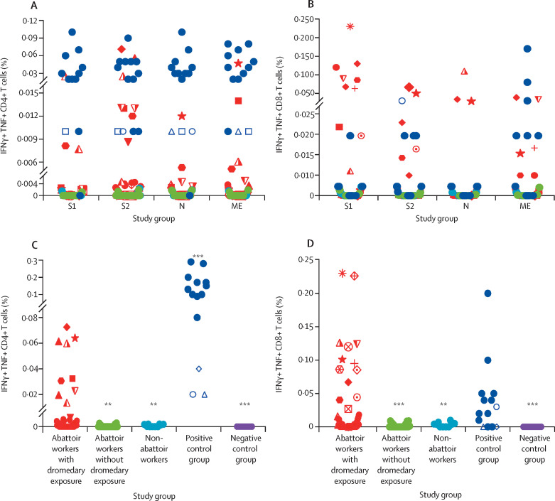
Figure 1
MERS-CoV-specific CD4 + and CD8 + T-cell responses in camel workers and controls (A) Frequencies of MERS-CoV-specific CD4+ T cells. (B) Frequencies of MERS-CoV-specific CD8+ T cells. (C) Summary of ag...
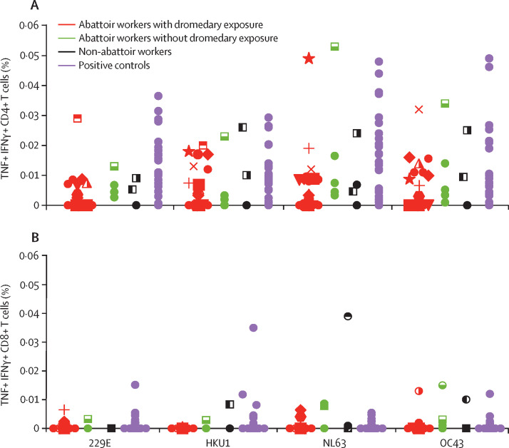
Figure 2
T-cell responses against endemic human coronaviruses in the study cohort Aggregate CD4+ (A) and CD8+ (B) T-cell responses to the structural peptide pools from human coronaviruses (229E, HKU1, NL63, an...
Figure images are served from the NIH/NLM PubMed Central Open Access Subset or Europe PMC; copyright remains with the publishers and authors.
💬 Discussion
0 commentsNo comments yet. Be the first to start a discussion!
Leave a Comment