Abstract
Tetraspanin uroplakins (UPs) Ia and Ib, together with their single-spanning transmembrane protein partners UP II and IIIa, form a unique crystalline 2D array of 16-nm particles covering almost the entire urothelial surface. A 6 Å–resolution cryo-EM structure of the UP particle revealed that the UP tetraspanins have a rod-shaped structure consisting of four closely packed transmembrane helices that extend into the extracellular loops, capped by a disulfide-stabilized head domain. The UP tetraspanins form the primary complexes with their partners through tight interactions of the transmembrane domains as well as the extracellular domains, so that the head domains of their tall partners can bridge each other at the top of the heterotetramer. The secondary interactions between the primary complexes and the tertiary interaction between the 16-nm particles contribute to the formation of the UP tetraspanin network. The rod-shaped tetraspanin structure allows it to serve as stable pilings in the lipid sea, ideal for docking partner proteins to form structural/signaling networks.
🔬 Techniques
🔭 Microscopes
🧬 Organisms
💻 Software
🧪 Sample Preparation
🏭 Microscope Brands
💻 Software Details
💾 Data Repositories
🏛️ Research Organizations (ROR)
Affiliated research institutions:
📋 Methods
Preparation and EM examination of mouse urothelial plaques
Mouse urothelial plaques were isolated by sucrose density gradient and differential detergent wash as described previously ( Wu et al., 1990 ; Liang et al., 1999 ); the quality of the purified urothelial plaques was assessed by negative staining and EM. For cryo-EM, 5 μl of the purified plaques, adjusted to ∼0.1 mg/ml, was applied to a molybdenum grid (300 meshes) with a layer of newly prepared carbon film and transferred to a tannic acid (0.75%) solution. After the excess liquid was blotted with filter paper, the sample was quickly immersed into liquid nitrogen. The frozen sample was loaded onto a Gatan cryo-holder and transferred to an electron microscope (CM200 FEG; Philips) operated at a voltage of 200 kV.
Image recording and processing
Electron micrographs were taken at a magnification of 50,000 in low-dose mode and with 0.5–1.7 μm defocus, at up to 50° tilt angles. The micrographs were screened using an optical diffractometer to select the regions with the best diffraction spots. Because of the heterogeneity of the sample, 1,000 micrographs, and the unbending of the images was performed using MRC software suites ( Crowther et al., 1996 ). The 3D density map was visualized using O ( Jones et al., 1991 ) or AMIRA (Mercury Computer System, Inc.) software packages. Density segmentation and model building Segmentation of the density map of the 16-nm particle was performed using AMIRA. For model building of the UP tetraspanins, transmembrane poly-alanine helices were first built into the electron density using O and then the poly-alanine model of EC1 (based on the homology region of casein kinase-1; Protein Data Bank accession no. 2CSN) and EC2 (based on the structure of CD81 large extracellular loop; Protein Data Bank accession no. 1G8Q) were manually docked into the electron density map. The modeled extracellular loops were connected to the TMs following the density map and were locally adjusted using O. Online supplemental material Fig. S1 shows calculated diffractions and several lattice lines of 2D crystals of mouse UPs. Fig. S2 is a hypothetical model of the FimH–uroplakin interaction, illustrating that FimH has to reach into the crevice formed by two neighboring joints. Online supplemental material is available at http://www.jcb.org/cgi/content/full/jcb.200602086/DC1 .
Show full methods section
Preparation and EM examination of mouse urothelial plaques
Mouse urothelial plaques were isolated by sucrose density gradient and differential detergent wash as described previously ( Wu et al., 1990 ; Liang et al., 1999 ); the quality of the purified urothelial plaques was assessed by negative staining and EM. For cryo-EM, 5 μl of the purified plaques, adjusted to ∼0.1 mg/ml, was applied to a molybdenum grid (300 meshes) with a layer of newly prepared carbon film and transferred to a tannic acid (0.75%) solution. After the excess liquid was blotted with filter paper, the sample was quickly immersed into liquid nitrogen. The frozen sample was loaded onto a Gatan cryo-holder and transferred to an electron microscope (CM200 FEG; Philips) operated at a voltage of 200 kV.
Image recording and processing
Electron micrographs were taken at a magnification of 50,000 in low-dose mode and with 0.5–1.7 μm defocus, at up to 50° tilt angles. The micrographs were screened using an optical diffractometer to select the regions with the best diffraction spots. Because of the heterogeneity of the sample, 1,000 micrographs, and the unbending of the images was performed using MRC software suites ( Crowther et al., 1996 ). The 3D density map was visualized using O ( Jones et al., 1991 ) or AMIRA (Mercury Computer System, Inc.) software packages. Density segmentation and model building Segmentation of the density map of the 16-nm particle was performed using AMIRA. For model building of the UP tetraspanins, transmembrane poly-alanine helices were first built into the electron density using O and then the poly-alanine model of EC1 (based on the homology region of casein kinase-1; Protein Data Bank accession no. 2CSN) and EC2 (based on the structure of CD81 large extracellular loop; Protein Data Bank accession no. 1G8Q) were manually docked into the electron density map. The modeled extracellular loops were connected to the TMs following the density map and were locally adjusted using O. Online supplemental material Fig. S1 shows calculated diffractions and several lattice lines of 2D crystals of mouse UPs. Fig. S2 is a hypothetical model of the FimH–uroplakin interaction, illustrating that FimH has to reach into the crevice formed by two neighboring joints. Online supplemental material is available at http://www.jcb.org/cgi/content/full/jcb.200602086/DC1 .
Online supplemental material Fig. S1 shows calculated diffractions and several lattice lines of 2D crystals of mouse UPs. Fig. S2 is a hypothetical model of the FimH–uroplakin interaction, illustrating that FimH has to reach into the crevice formed by two neighboring joints. Online supplemental material is available at http://www.jcb.org/cgi/content/full/jcb.200602086/DC1 .
Supplementary Material [Supplemental Material Index]
📊 Figures
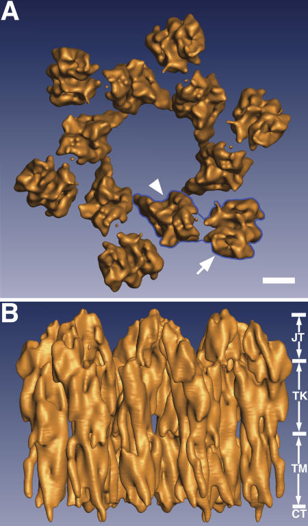
Figure 1.
The 3D structure of the mouse UP tetraspanin complexes at 6-u00c5 resolution. (A) The top view of the hexagonal 16-nm particle shows that it consists of six subunits (one of them is outlined in blue) ...
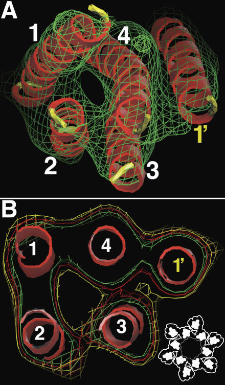
Figure 2.
The TM of the primary UP tetraspanin pair as a five-TM helix bundle. (A) The transmembrane helices of the UP Ia/II tetraspanin pair viewed from the cytoplasmic side. The electron density is represente...
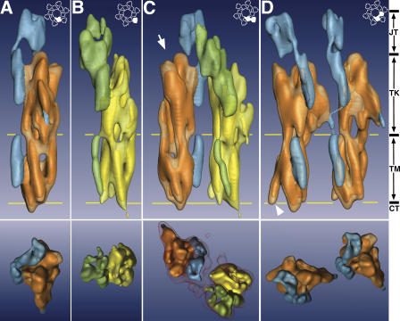
Figure 3.
The subdomains of the 16-nm particle and the relation between the UP tetraspanins and their partners. The top and bottom panels represent the side and top views, respectively, of the electron densitie...
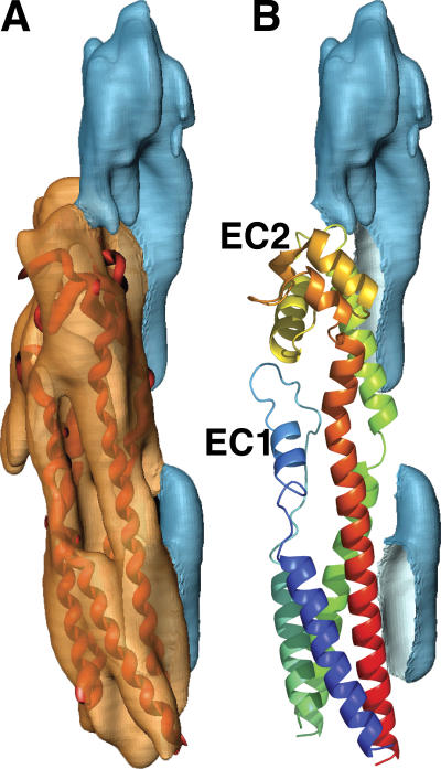
Figure 4.
Molecular model of the UP tetraspanins. (A) A molecular model of UP Ia tetraspanin is fitted into the electron density map. (B) The density of the UP Ia tetraspanin is removed to show the model's rela...
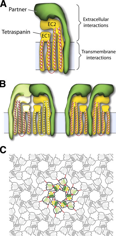
Figure 5.
Interactions in the UP complexes and the formation of the tetraspanin networks. (A) Primary interaction. A UP tetraspanin (UP Ia or Ib; yellow) interacts with its partner (UP II or IIIa; green) via bo...
Figure images are served from the NIH/NLM PubMed Central Open Access Subset or Europe PMC; copyright remains with the publishers and authors.
💬 Discussion
0 commentsNo comments yet. Be the first to start a discussion!
Leave a Comment