Abstract
The Hepatitis B Virus (HBV) double-stranded DNA genome is reverse transcribed from its RNA pregenome (pgRNA) within the virus core (or capsid). Phosphorylation of the arginine-rich carboxy-terminal domain (CTD) of the HBV capsid protein (Cp183) is essential for pgRNA encapsidation and reverse transcription. However, the structure of the CTD remains poorly defined. Here we report sub-nanometer resolution cryo-EM structures of in vitro assembled empty and pgRNA-filled Cp183 capsids in unphosphorylated and phosphorylation-mimic states. In empty capsids, we found unexpected evidence of surface accessible CTD density partially occluding pores in the capsid surface. We also observed that CTD organization changed substantively as a function of phosphorylation. In RNA-filled capsids, unphosphorylated CTDs favored thick ropes of RNA, while the phosphorylation-mimic favored a mesh of thin, high-density strands suggestive of single stranded RNA. These results demonstrate that the CTD can regulate nucleic acid structure, supporting the hypothesis that the HBV capsid has a functional role as a nucleic acid chaperone.
🔬 Techniques
🧬 Organisms
✨ Fluorophores
🏭 Microscope Brands
🧪 Reagent Suppliers
📷 Detectors
💻 Software Details
💾 Data Repositories
🏛️ Research Organizations (ROR)
Affiliated research institutions:
📋 Methods
Purification of HBV capsids
The pgRNA production, the plasmids coded for HBV Cp183-SSS and Cp183-EEE, and the capsid purification were described previously [15] . Capsids stored at −80°C were disassembled by dialysis at 4°C in the disassembly buffers (1.5 M guanidine HCl, 0.5 M LiCl, 50 mM HEPES at pH 7.5, 10 mM DTT for Cp183-SSS, and 1.5 M guanidine HCl, 1.5 M LiCl, 50 mM Tris at pH 9.5, 10 mM DTT for Cp183-EEE). The encapsidated heterogeneous RNA packaged from E. Coli cells was precipitated by a spin of 20,000× g for 15 min at 4°C. Protein dimers were recovered from the supernatant and purified by size exclusion chromatography using an analytical grade Superose 6 column (GE Lifesciences) equilibrated in disassembly buffer. Fractions containing core protein were identified by SDS-PAGE. Cp183-SSS and Cp183-EEE dimers were either used for the reassembly experiments immediately or stored at 4°C for a short period. Formation of the empty capsids (Cp183 e -SSS and Cp183 e -EEE) was approached by dialyzing the purified dimers in the reassembly buffer (250 mM NaCl, 50 mM HEPES at pH 7.5, 2 mM DTT for Cp183-SSS and 250 mM NaCl, 50 mM Tris pH 7.4, 2 mM DTT for Cp183-EEE). The pgRNA-filled capsids (Cp183 RNA -SSS and Cp183 RNA -EEE) were prepared by reassembling the purified dimers with in vitro transcribed HBV pgRNA at a molar ratio of protein dimer to RNA polymer = 120∶1 in the reassembly buffer (150 mM NaCl, 50 mM HEPES at pH 7.5, 2 mM DTT for Cp183-SSS and 150 mM NaCl, 50 mM Tris at pH 7.4, 2 mM DTT for Cp183-EEE) overnight. Samples for cryo-EM were further concentrated by Amicon Ultra centrifugal filter units (Millipore, MA). The quality and the concentration of the sample were routinely checked by negative stained EM using 2% uranyl acetate.
Show full methods section
Purification of HBV capsids
The pgRNA production, the plasmids coded for HBV Cp183-SSS and Cp183-EEE, and the capsid purification were described previously [15] . Capsids stored at −80°C were disassembled by dialysis at 4°C in the disassembly buffers (1.5 M guanidine HCl, 0.5 M LiCl, 50 mM HEPES at pH 7.5, 10 mM DTT for Cp183-SSS, and 1.5 M guanidine HCl, 1.5 M LiCl, 50 mM Tris at pH 9.5, 10 mM DTT for Cp183-EEE). The encapsidated heterogeneous RNA packaged from E. Coli cells was precipitated by a spin of 20,000× g for 15 min at 4°C. Protein dimers were recovered from the supernatant and purified by size exclusion chromatography using an analytical grade Superose 6 column (GE Lifesciences) equilibrated in disassembly buffer. Fractions containing core protein were identified by SDS-PAGE. Cp183-SSS and Cp183-EEE dimers were either used for the reassembly experiments immediately or stored at 4°C for a short period. Formation of the empty capsids (Cp183 e -SSS and Cp183 e -EEE) was approached by dialyzing the purified dimers in the reassembly buffer (250 mM NaCl, 50 mM HEPES at pH 7.5, 2 mM DTT for Cp183-SSS and 250 mM NaCl, 50 mM Tris pH 7.4, 2 mM DTT for Cp183-EEE). The pgRNA-filled capsids (Cp183 RNA -SSS and Cp183 RNA -EEE) were prepared by reassembling the purified dimers with in vitro transcribed HBV pgRNA at a molar ratio of protein dimer to RNA polymer = 120∶1 in the reassembly buffer (150 mM NaCl, 50 mM HEPES at pH 7.5, 2 mM DTT for Cp183-SSS and 150 mM NaCl, 50 mM Tris at pH 7.4, 2 mM DTT for Cp183-EEE) overnight. Samples for cryo-EM were further concentrated by Amicon Ultra centrifugal filter units (Millipore, MA). The quality and the concentration of the sample were routinely checked by negative stained EM using 2% uranyl acetate.
Cryo-electron microscopy
The sample preparation and cryo-EM operation were followed well established procedures described previously [28] . Briefly, a drop of 3.5 µl sample solution was applied on a glow-discharged Quantifoil holey-carbon grid (R2/2), blotted with filter paper from both sides for 4 s to produce a thin layer of specimen solution across the holes. The grids were quickly plunged into liquid ethane bath cooled by liquid nitrogen in a cryo-container. All processes described above were performed by a FEI Vitrobot. The vitrified specimen on the grid was then transferred to a Gatan 626DH cryo-holder and kept at the low temperature environment (
📊 Figures
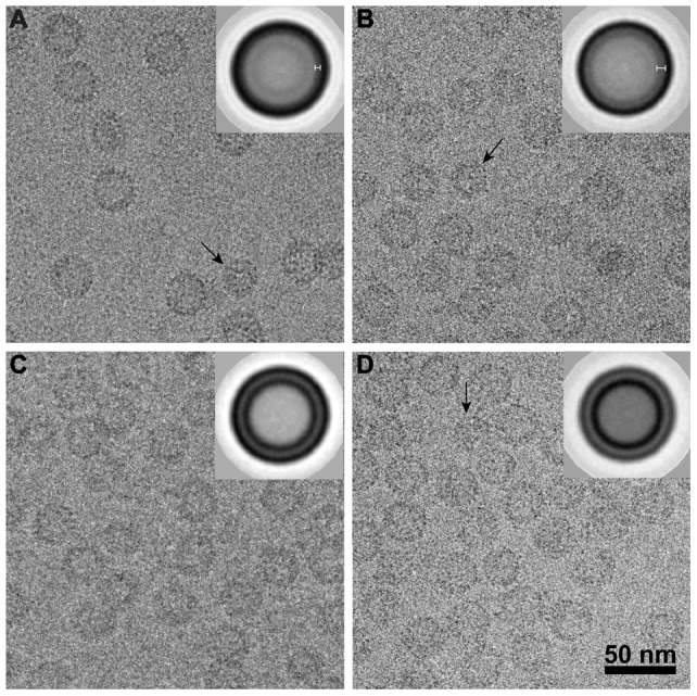
Figure 1
Cryo-micrographs of frozen-hydrated HBV capsids.
(A) Cp183 e -SSS (e for empty), (B) Cp183 e -EEE, (C) Cp183 RNA -SSS, and (D) Cp183 RNA -EEE particles are shown, frozen hydrated in vitreous ice. These particles show the typical morphology of HBV ca...
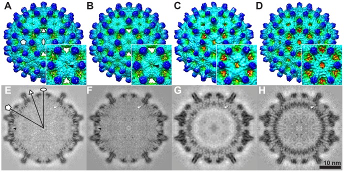
Figure 2
Cryo-EM 3D reconstructions of empty and pgRNA-filled Cp183 capsids.
Surface shaded exterior maps of Tu200a=u200a4 (A) Cp183 e -SSS, (B) Cp183 e -EEE, (C) Cp183 RNA -SSS, (D) Cp183 RNA -EEE and their related central sections (Eu2013H). Insets show enlarged views of the...
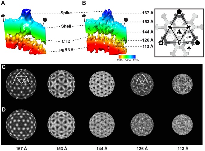
Figure 3
Interaction between the HBV capsid and pgRNA.
A radially color-coded isosurface rendering of AB dimers and related pgRNA of (A) Cp183 RNA -SSS and (B) Cp183 RNA -EEE viewed from a 90u00b0 rotation of the region identified by an arrow in the right...
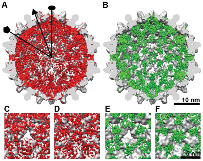
Figure 4
Spatial organization of the CTDs.
Difference maps of CTD density were calculated by subtracting the low-pass filtered atomic model of Cp149 from the (A) Cp183 e -SSS and (B) Cp183 e -EEE. The resulting CTD density (red and green, resp...
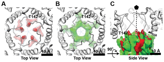
Figure 5
The phosphorylation-mimic EEE mutation alters CTD structure.
Viewed from the capsid exterior, along a fivefold axis, the Cp149 atomic model (PDB entry 1QGT, gray) fits into cryo-EM density of (A, red) Cp183 e -SSS and (B, green) Cp183 e -EEE. The last visible r...
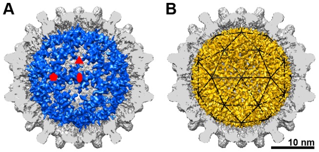
Figure 6
Structural organization of pgRNA.
The difference maps of the pgRNA from (A) Cp183 RNA -SSS (blue) and (B) Cp183 RNA -EEE (gold) superimposed on cutaway of their respective empty capsids. By subtracting the empty Cp183 capsids from the...
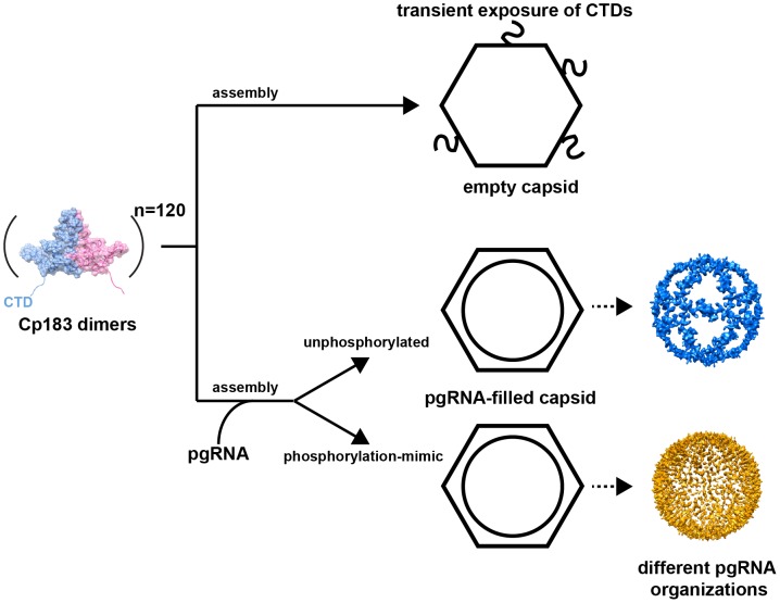
Figure 7
An assembly schema for HBV Cp183 capsids.
Assembly of Cp183 dimers in the absence of RNA results in empty capsids with CTDs transiently exposed through the pores on the icosahedral twofold axes. Co-assembly of Cp183 dimers with pgRNA results ...
Figure images are served from the NIH/NLM PubMed Central Open Access Subset or Europe PMC; copyright remains with the publishers and authors.
💬 Discussion
0 commentsNo comments yet. Be the first to start a discussion!
Leave a Comment