Abstract
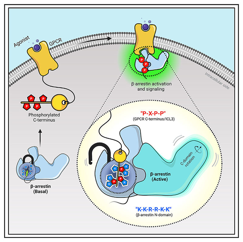
Agonist-induced GPCR phosphorylation is a key determinant for the binding and activation of β-arrestins (βarrs). However, it is not entirely clear how different GPCRs harboring divergent phosphorylation patterns impart converging active conformation on βarrs leading to broadly conserved functional responses such as desensitization, endocytosis, and signaling. Here, we present multiple cryo-EM structures of activated βarrs in complex with distinct phosphorylation patterns derived from the carboxyl terminus of different GPCRs. These structures help identify a P-X-P-P type phosphorylation motif in GPCRs that interacts with a spatially organized K-K-R-R-K-K sequence in the N-domain of βarrs. Sequence analysis of the human GPCRome reveals the presence of this phosphorylation pattern in a large number of receptors, and its contribution in βarr activation is demonstrated by targeted mutagenesis experiments combined with an intrabody-based conformational sensor. Taken together, our findings provide important structural insights into the ability of distinct GPCRs to activate βarrs through a significantly conserved mechanism.
🔬 Techniques
🔭 Microscopes
🧬 Organisms
✨ Fluorophores
🧪 Sample Preparation
🔬 Cell Lines
🏭 Microscope Brands
🧪 Reagent Suppliers
📷 Detectors
💻 Software Details
💾 Data Repositories
🏷️ Research Resource Identifiers (RRIDs)
Verified research resources used in this paper:
🏛️ Research Organizations (ROR)
Affiliated research institutions:
📋 Methods
Star+Methods Detailed methods are provided in the online version of this paper and include the following: KEY RESOURCES TABLE RESOURCE AVAILABILITY ○ Lead contact ○ Materials availability ○ Data and code availability EXPERIMENTAL MODEL AND SUBJECT DETAILS ○ Human cell lines ○ Insect cells METHOD DETAILS ○ General reagents, plasmids, and cell culture ○ Expression and purification of βarrs ○ Expression and purification of Fabs ○ Co-immunoprecipitation assay ○ Reconstitution of phosphopeptide-βarr-Fab complexes ○ Negative-staining EM ○ Cryo-EM sample preparation and data acquisition ○ Cryo-EM data processing and model building ○ Model building and refinement ○ NanoBiT assay for βarr2 WT and βarr2 DM recruitment ○ NanoBiT assay for βarr trafficking ○ NanoBiT assay for Ib30 reactivity B Receptor surface expression QUANTIFICATION AND STATISTICAL ANALYSIS
Star+Methods Key Resources Table
REAGENT or RESOURCE SOURCE IDENTIFIER Antibodies Monoclonal ANTI-FLAG
M2-HRP antibody Sigma-Aldrich Cat# A8592; RRID: AB_439702 Chemicals, peptides, and recombinant proteins TRIS SRL Cat# 71033 HEPES SRL Cat# 63732 NaCl SRL Cat# 41721 EDTA SRL Cat# 12070 Phenylmethanesulfonyl Fluoride (PMSF) SRL Cat# 84375 (84375) Benzamidine Hydrochloride SRL Cat# 93014 (0248255) Lysozyme SRL Cat# 45822 Glycerol SRL Cat# 77453 Dithiothreitol HiMedia Cat# MB070 Lauryl Maltose Neopentyl Glycol (MNG) Anatrace Cat# NG310, CAS no.1257852-96-2 Paraformaldehyde (PFA) Sigma Aldrich Cat# P6148, CAS no. 30525-89-4 Poly-D-lysine Sigma Aldrich Cat# P0899 TMB (Tetramethylbenzidine) Thermo Fisher Scientific Cat# 34028 Janus Green B Sigma Aldrich Cat# 201677 PEI (Polyethylenimine) Polysciences Cat# 23966 Bovine Serum Albumin, BSA SRL Cat# 83803 (0140105) HBSS - Hank’s Balanced Salt Solution Thermo Fisher Scientific Cat# 14065 GIBCO Fetal Bovine Serum Thermo Fisher Scientific Cat# 10270-106 DMEM Cellclone Cat# CC3004 Phosphate-buffered saline (PBS) Sigma Aldrich Cat# D1283 GIBCO Penicillin-Streptomycin Thermo Fisher Scientific Cat# 15140122 Coelenterazine Goldbio Cat# CZ05 Glyco-diosgenin (GDN) Anatrace GDN101 Cholesteryl Hemisuccinate Sigma C6512 Coomassie briliiant Blue SRL Cat# 64222 Uranyl formate Polysciences Cat# 24762-1 Recombinant rat β-arrestin1 Purified N/A Recombinant bovine β-arrestin2 Purified N/A C5aR1pp1 Chemically synthesized N/A C5aR1pp2 Chemically synthesized N/A C5aR1pp3 Chemically synthesized N/A V2Rpp Chemically synthesized N/A CXCR4pp1 Chemically synthesized N/A CXCR4pp2 Chemically synthesized N/A CXCR4pp3 Chemically synthesized N/A CXCR4pp4 Chemically synthesized N/A Recombinant human C5a Purified N/A Bradykinin Genscript N/A Arginine Vasopressin Peptide (AVP) Genscript N/A Uranyl formate Polysciences Cat# 24762-1 Formvar/carbon coated 300 mesh copper grids PELCO (Ted Pella) Cat# 01753-F Critical commercial assays Site directed mutagenesis kit NEB Cat# E0554 NanoBiT assay Promega N/A Deposited data C5aR1pp-βarr1-Fab30 This study PDB: 8GO8, EMD-34173 V2Rpp-βarr2-Fab30 This study PDB: 8GOC, EMD-34175 C5aR1pp-βarr2-Fab30 This study PDB: 8GOO, EMD-34178 CXCR4pp-βarr1-Fab30 This study PDB: 8GP3, EMD-34188 C5aR1pp-βarr1-Fab30-Local-refine This study PDB: 8I0N, EMD-35104 V2Rpp-βarr2-Fab30-Local-refine This study PDB: 8I10, EMD-35115 C5aR1pp-βarr2-Fab30-Local-refine This study PDB: 8I0Z, EMD-35114 CXCR4pp-βarr1-Fab30-Local-refine This study PDB: 8I0Q, EMD-35106 Experimental models: Cell lines Human: HEK293 ATCC Cat# CRL-3216 Oligonucleotides C5aR1_R-insertion SDM primer Forward: CGCTCCACAGTGGACACTATGG This study N/A C5aR1_R-insertion SDM primer Reverse: GCGCGTGAATGACTTGCT This study N/A B2R mut Forward: AACACGGACCTCCATCTCCGTG This study N/A B2R mut Reverse GTCATGGAGTTCTCCATCTGAATGGG This study N/A M2R mut _Forward: AAGTCTACTTCACTGGGCCAC This study N/A M2R mut _Reverse: GACGGTGTTTTCGTCCTG This study N/A β-arrestin2 Double mutant (βarr2DM) Fw: ggcgAACAACCGTGAAAAACGTG This study N/A β-arrestin2 Double mutant (βarr2DM) Rv: aggaaCGGGGTGATGGTGTAAAC This study N/A Recombinant DNA PcDNA_3.1 (empty vector) Dr. Arun K Shukla N/A pcDNA3.1_V2R-WT Dr. Arun K Shukla N/A pcDNA3.1_C5aR1-WT Dr. Arun K Shukla N/A pcDNA3.1_CXCR3-WT Dr. Arun K Shukla N/A pcDNA3.1_CXCR7-WT Dr. Arun K Shukla N/A pcDNA3.1_B2R-WT Dr. Arun K Shukla N/A pcDNA3.1_M2R-WT Dr. Arun K Shukla N/A pcDNA3.1_C5aR1-mut TRRST This study N/A pcDNA3.1_B2R-mut Δ G368/L370T Dr. Arun K Shukla N/A pcDNA3.1_M2R-mut TVKST This study N/A pCAGGS_V2R-WT-SmBiT Dr. Arun K Shukla N/A pCAGGS _C5aR1-SmBiT Dr. Arun K Shukla N/A pCAGGS_SmBiT-βarr2 WT Dr. Asuka Inoue N/A pCAGGS_SmBiT-βarr2 DM This study N/A pCAGGS_LgBiT-βarr2 WT Dr. Asuka Inoue N/A pCAGGS_LgBiT-βarr2 DM This study N/A pCAGGS_LgBiT-FYVE Dr. Asuka Inoue N/A pCAGGS_LgBiT-Ib30 Dr. Asuka Inoue N/A Software and algorithms UCSF Chimera Pettersen et al. 64 https://www.cgl.ucsf.edu/chimera/ UCSF Chimera X Pettersen et al. 65 https://www.rbvi.ucsf.edu/chimerax/ COOT Emsley et al. 66 https://www2.mrc-lmb.cam.ac.uk/personal/pemsley/coot/ cryoSPARC Punjani et al. 67 https://cryosparc.com/ PDBsum Laskowski et al. 68 http://www.ebi.ac.uk/thornton-srv/databases/pdbsum/ Phenix Liebschner et al. 69 https://www.phenix-online.org/ PyMol Schrodinger https://pymol.org/2/ Prism 8 GraphPad Software https://www.graphpad.com/scientific-software/prism/ Relion3.1.2 Zivanov et al. 70 https://www3.mrc-lmb.cam.ac.uk/relion/index.php?title=Main_Page SerialEM Mastronarde 71 https://bio3d.colorado.edu/SerialEM/ Graphpad Prism 9 GraphPad Software, San Diego, California USA https://www.graphpad.com/scientific-software/prism/ ImageJ Schneider et al. 72 https://imagej.nih.gov/ij/download.html Resource Availability Lead contact Further information and requests for reagents should be addressed to the lead contact, Dr. Arun K. Shukla ( arshukla@iitk.ac.in ).
Show full methods section
Star+Methods Detailed methods are provided in the online version of this paper and include the following: KEY RESOURCES TABLE RESOURCE AVAILABILITY ○ Lead contact ○ Materials availability ○ Data and code availability EXPERIMENTAL MODEL AND SUBJECT DETAILS ○ Human cell lines ○ Insect cells METHOD DETAILS ○ General reagents, plasmids, and cell culture ○ Expression and purification of βarrs ○ Expression and purification of Fabs ○ Co-immunoprecipitation assay ○ Reconstitution of phosphopeptide-βarr-Fab complexes ○ Negative-staining EM ○ Cryo-EM sample preparation and data acquisition ○ Cryo-EM data processing and model building ○ Model building and refinement ○ NanoBiT assay for βarr2 WT and βarr2 DM recruitment ○ NanoBiT assay for βarr trafficking ○ NanoBiT assay for Ib30 reactivity B Receptor surface expression QUANTIFICATION AND STATISTICAL ANALYSIS
Star+Methods Key Resources Table
REAGENT or RESOURCE SOURCE IDENTIFIER Antibodies Monoclonal ANTI-FLAG
M2-HRP antibody Sigma-Aldrich Cat# A8592; RRID: AB_439702 Chemicals, peptides, and recombinant proteins TRIS SRL Cat# 71033 HEPES SRL Cat# 63732 NaCl SRL Cat# 41721 EDTA SRL Cat# 12070 Phenylmethanesulfonyl Fluoride (PMSF) SRL Cat# 84375 (84375) Benzamidine Hydrochloride SRL Cat# 93014 (0248255) Lysozyme SRL Cat# 45822 Glycerol SRL Cat# 77453 Dithiothreitol HiMedia Cat# MB070 Lauryl Maltose Neopentyl Glycol (MNG) Anatrace Cat# NG310, CAS no.1257852-96-2 Paraformaldehyde (PFA) Sigma Aldrich Cat# P6148, CAS no. 30525-89-4 Poly-D-lysine Sigma Aldrich Cat# P0899 TMB (Tetramethylbenzidine) Thermo Fisher Scientific Cat# 34028 Janus Green B Sigma Aldrich Cat# 201677 PEI (Polyethylenimine) Polysciences Cat# 23966 Bovine Serum Albumin, BSA SRL Cat# 83803 (0140105) HBSS - Hank’s Balanced Salt Solution Thermo Fisher Scientific Cat# 14065 GIBCO Fetal Bovine Serum Thermo Fisher Scientific Cat# 10270-106 DMEM Cellclone Cat# CC3004 Phosphate-buffered saline (PBS) Sigma Aldrich Cat# D1283 GIBCO Penicillin-Streptomycin Thermo Fisher Scientific Cat# 15140122 Coelenterazine Goldbio Cat# CZ05 Glyco-diosgenin (GDN) Anatrace GDN101 Cholesteryl Hemisuccinate Sigma C6512 Coomassie briliiant Blue SRL Cat# 64222 Uranyl formate Polysciences Cat# 24762-1 Recombinant rat β-arrestin1 Purified N/A Recombinant bovine β-arrestin2 Purified N/A C5aR1pp1 Chemically synthesized N/A C5aR1pp2 Chemically synthesized N/A C5aR1pp3 Chemically synthesized N/A V2Rpp Chemically synthesized N/A CXCR4pp1 Chemically synthesized N/A CXCR4pp2 Chemically synthesized N/A CXCR4pp3 Chemically synthesized N/A CXCR4pp4 Chemically synthesized N/A Recombinant human C5a Purified N/A Bradykinin Genscript N/A Arginine Vasopressin Peptide (AVP) Genscript N/A Uranyl formate Polysciences Cat# 24762-1 Formvar/carbon coated 300 mesh copper grids PELCO (Ted Pella) Cat# 01753-F Critical commercial assays Site directed mutagenesis kit NEB Cat# E0554 NanoBiT assay Promega N/A Deposited data C5aR1pp-βarr1-Fab30 This study PDB: 8GO8, EMD-34173 V2Rpp-βarr2-Fab30 This study PDB: 8GOC, EMD-34175 C5aR1pp-βarr2-Fab30 This study PDB: 8GOO, EMD-34178 CXCR4pp-βarr1-Fab30 This study PDB: 8GP3, EMD-34188 C5aR1pp-βarr1-Fab30-Local-refine This study PDB: 8I0N, EMD-35104 V2Rpp-βarr2-Fab30-Local-refine This study PDB: 8I10, EMD-35115 C5aR1pp-βarr2-Fab30-Local-refine This study PDB: 8I0Z, EMD-35114 CXCR4pp-βarr1-Fab30-Local-refine This study PDB: 8I0Q, EMD-35106 Experimental models: Cell lines Human: HEK293 ATCC Cat# CRL-3216 Oligonucleotides C5aR1_R-insertion SDM primer Forward: CGCTCCACAGTGGACACTATGG This study N/A C5aR1_R-insertion SDM primer Reverse: GCGCGTGAATGACTTGCT This study N/A B2R mut Forward: AACACGGACCTCCATCTCCGTG This study N/A B2R mut Reverse GTCATGGAGTTCTCCATCTGAATGGG This study N/A M2R mut _Forward: AAGTCTACTTCACTGGGCCAC This study N/A M2R mut _Reverse: GACGGTGTTTTCGTCCTG This study N/A β-arrestin2 Double mutant (βarr2DM) Fw: ggcgAACAACCGTGAAAAACGTG This study N/A β-arrestin2 Double mutant (βarr2DM) Rv: aggaaCGGGGTGATGGTGTAAAC This study N/A Recombinant DNA PcDNA_3.1 (empty vector) Dr. Arun K Shukla N/A pcDNA3.1_V2R-WT Dr. Arun K Shukla N/A pcDNA3.1_C5aR1-WT Dr. Arun K Shukla N/A pcDNA3.1_CXCR3-WT Dr. Arun K Shukla N/A pcDNA3.1_CXCR7-WT Dr. Arun K Shukla N/A pcDNA3.1_B2R-WT Dr. Arun K Shukla N/A pcDNA3.1_M2R-WT Dr. Arun K Shukla N/A pcDNA3.1_C5aR1-mut TRRST This study N/A pcDNA3.1_B2R-mut Δ G368/L370T Dr. Arun K Shukla N/A pcDNA3.1_M2R-mut TVKST This study N/A pCAGGS_V2R-WT-SmBiT Dr. Arun K Shukla N/A pCAGGS _C5aR1-SmBiT Dr. Arun K Shukla N/A pCAGGS_SmBiT-βarr2 WT Dr. Asuka Inoue N/A pCAGGS_SmBiT-βarr2 DM This study N/A pCAGGS_LgBiT-βarr2 WT Dr. Asuka Inoue N/A pCAGGS_LgBiT-βarr2 DM This study N/A pCAGGS_LgBiT-FYVE Dr. Asuka Inoue N/A pCAGGS_LgBiT-Ib30 Dr. Asuka Inoue N/A Software and algorithms UCSF Chimera Pettersen et al. 64 https://www.cgl.ucsf.edu/chimera/ UCSF Chimera X Pettersen et al. 65 https://www.rbvi.ucsf.edu/chimerax/ COOT Emsley et al. 66 https://www2.mrc-lmb.cam.ac.uk/personal/pemsley/coot/ cryoSPARC Punjani et al. 67 https://cryosparc.com/ PDBsum Laskowski et al. 68 http://www.ebi.ac.uk/thornton-srv/databases/pdbsum/ Phenix Liebschner et al. 69 https://www.phenix-online.org/ PyMol Schrodinger https://pymol.org/2/ Prism 8 GraphPad Software https://www.graphpad.com/scientific-software/prism/ Relion3.1.2 Zivanov et al. 70 https://www3.mrc-lmb.cam.ac.uk/relion/index.php?title=Main_Page SerialEM Mastronarde 71 https://bio3d.colorado.edu/SerialEM/ Graphpad Prism 9 GraphPad Software, San Diego, California USA https://www.graphpad.com/scientific-software/prism/ ImageJ Schneider et al. 72 https://imagej.nih.gov/ij/download.html Resource Availability Lead contact Further information and requests for reagents should be addressed to the lead contact, Dr. Arun K. Shukla ( arshukla@iitk.ac.in ).
Materials availability
Reagents described in this manuscript are available upon reasonable request from the lead contact .
Experimental Model and Subject Details Human cell lines
HEK-293 cells were purchased from ATCC for all the cellular experiments performed in the study. The cell line was examined frequently under the microscope for proper morphology, but they were not authenticated. They were cultured in DMEM with fetal bovine serum (FBS) at 37°C in 5% CO 2 . In this study, any stable, knockout, or knockdown cell lines were not generated, and the details of previously generated cell lines are referenced in the manuscript.
Insect cells
Sf9 cells were obtained from Expression systems, and they were routinely monitored under the microscope for proper morphology. These cells were maintained in a shaker incubator at 27°C with 135rpm shaking, and sub-cultured in protein-free insect cell medium purchased from Expression Systems. Method Details General reagents, plasmids, and cell culture Most of the general reagents were purchased from Sigma Aldrich unless mentioned otherwise. Dulbecco’s Modified Eagle’s Medium (DMEM), Dulbecco’s Phosphate buffer saline (PBS), Fetal-Bovine Serum (FBS), Trypsin-EDTA, Hank’s balanced salt solution (HBSS), and penicillin-streptomycin solution were purchased from Thermo Fisher Scientific. HEK-293 cells were obtained from ATCC and maintained in DMEM (Gibco, Cat no. 12800-017) supplemented with 10% FBS (Gibco, Cat no. 10270-106) and 100U ml -1 penicillin (Gibco, Cat no. 15140122) and 100μg ml -1 streptomycin (Gibco, Cat no. 15140-122) at 37°C under 5% CO 2 . The cDNA coding region for the mentioned receptors namely, V2R, C5aR1, B2R, M2R, CXCR3, and CXCR7 were cloned in pcDNA3.1 consist of HA signal sequence followed by FLAG tag at the N-terminus of the receptor. The mutants generated for the study are as follows: deletion of G368 and a substitution L370T in B2R; insertion of an Arg residue between R337 and S338 in C5aR1; insertion of a Lys residue between V308 and S309 in M2R; using Q5 site-directed mutagenesis kit (NEB, Cat. no. E0554S). For the NanoBiT assay, receptors harboring SmBiT at the C-terminus were generated by subcloning in the lab, and other constructs have been described previously. 41 , 43 , 73 All the constructs were verified by DNA sequencing (Macrogen).
Expression and purification of βarrs
Full length rat βarr1, βarr2 WT and bovine βarr2 DM were cloned into pGEX-4T3 vector with thrombin cleavage site between GST tag and βarr. Similar protocol was followed for purifying all three forms of βarr. βarrs were expressed in E. coli BL21 cells and grown in Terrific broth media supplemented with 100μg ml -1 ampicillin. A primary culture of 50ml volume was inoculated with an isolated colony from freshly transformed LB-amp plate. Primary culture was grown till a cell optical density at 600nm (OD 600 ) of 0.8-1 and further inoculated into a secondary culture of TB-Amp of 1.5L volume till OD 600 0.8-1. The expression of βarrs was then induced with 25μM IPTG concentration and cells were allowed to grow till 16h at 18°C. Cultures were harvested and stored at -80°C until further use. Harvested pellets were of 12-15g in mass. For purification, cells were lysed by sonication in lysis buffer; 25mM Tris, pH 8.5, 150mM NaCl, 1mM PMSF (phenylmethylsulfonyl fluoride), 2mM Benzamidine, 1mM EDTA (Ethylenediaminetetraacetic acid), 5% Glycerol, 2mM Dithiothreitol (DTT) and 1mg ml -1 Lysozyme. The lysate was centrifuged at 18,000-20,000rpm at 4°C and supernatant was allowed to bind to Glutathione resin (GS resin) (Glutathione Sepharose™ 4 Fast Flow, GE Healthcare Cat. no. 17-5132-02) in a batch binding mode for overnight at 4°C. GS-resin bound GST-βarr was transferred into Econo columns (Biorad, Cat. no. 7372512) and washed rigorously with wash buffer (25mM Tris, pH 8.5, 150mM NaCl, 2mM DTT and 0.02% n-dodecyl-β-D-maltopyranoside (DDM). Afterward, on-column cleavage was set up by adding thrombin to 1:1 resin:buffer slurry at room temperature for 2h. βarrs were then eluted with gravity flow and further with buffer 25mM Tris, pH 8.5, 350mM NaCl and 0.02% DDM and 2mM DTT. Eluted proteins were concentrated and further purified on a HiLoad 16/600 Superdex column in buffer 25mM Tris, pH 8.5, 350mM NaCl, 2mM DTT and 0.02% DDM. Fractions corresponding to pure βarr were flash frozen with 10% glycerol and stored at -80°C until further use.
Expression and purification of Fabs
A similar protocol for expression and purification was followed for all the Fabs and they were purified as previously mentioned. 39 Briefly, Fabs were expressed in the periplasmic fraction of E. coli M55244 cells (ATCC) and purified using Protein L resin (GE Healthcare Cat. no. 17547802) with gravity flow affinity chromatography. Cells transformed with Fab plasmid were grown in 50ml 2xYT media and allowed to grow overnight at 30°C. 1L 2xYT media was inoculated with 5% volume of initial inoculum and grown for an additional 8h at 30°C. Cells were collected and resuspended in an equal volume of CRAP medium supplemented with 100μg ml -1 ampicillin, and grown further for 16h at 30°C. For purification, cells were lysed in lysis buffer (50mM HEPES-Na + , pH 8.0, 0.5M NaCl, 0.5% (v/v) Triton X-100, 0.5mM MgCl 2 ) by sonication. Cell lysate was heated in a 65°C water bath for 30min and cooled immediately on ice for 5min. Lysate was centrifuged at 20,000rpm and passed through pre-equilibrated Protein L resin packed gravity flow affinity columns. Binding was performed at room temperature and beads were washed extensively with wash buffer (50mM HEPES-Na + , pH 8.0, 0.5M NaCl). Fabs were eluted with 100mM acetic acid into tubes containing 10% volume of 1M HEPES, pH 8.0 for neutralization. Eluted samples were desalted into buffer (20mM HEPES-Na + , pH 8.0, 0.1M NaCl) using a pre-packed PD-10 column (GE Healthcare Cat. no. 17085101). Purified Fabs were flash-frozen and stored at -80°C supplemented with 10% (v/v) glycerol until further use.
Co-immunoprecipitation assay
For co-immunoprecipitation assay, 2.5μg of β-arrestins were incubated with different phosphopeptides at 10-fold molar excess in binding buffers (20mM HEPES, pH 7.4, 150mM NaCl) for 1h at room temperature for activation. Post peptide-induced activation, 5μg Fab30 was added, and reaction was incubated for an additional 1h at room temperature. After 1h, 25μl of pre-equilibrated protein L beads (Capto™ L resin, GE Healthcare Cat. no. 17547802) was added and reaction was incubated for 90min at room temperature, followed by five washes with binding buffer containing 0.01% LMNG. Bound protein was eluted with 2X SDS loading buffer and 15μl sample was analyzed on 12% SDS-PAGE. For statistical analyses, protein bands were quantified using ImageJ software suite 72 and the values were plotted using GraphPad Prism software v 9.5. The data were normalized with respect to their respective experimental control and appropriate statistical analyses were performed as indicated in the corresponding figure legend. Reconstitution of phosphopeptide-βarr-Fab complexes A previously published protocol was followed for complex purification with minor modifications. 31 Briefly, βarrs were activated with corresponding phosphopeptides at a 1:3 molar ratios of βarr:phosphopeptide for 30-40min at room temperature. Respective Fabs were added to the phosphopeptide-βarr mixture at 1:1.5 molar ratio of βarr:Fab and incubated for 1h at room temperature. To remove excess Fabs, the phosphopeptide-βarr-Fab complexes were concentrated with 30,000 MWCO concentrators (Vivaspin, Cytiva Cat. no. 28932361) and injected into Superose 6 Increase 10/300 GL (Cytiva Cat. no. 29091596) gel-filtration column. Fractions were further analyzed on SDS-PAGE and selected fractions were pooled and concentrated for structural studies. Negative-staining EM Complex formation, homogeneity, and particle quality of the samples were judged through negative staining of the samples prior to data collection under cryogenic conditions for high resolution reconstructions. Negative staining of the samples was performed with uranyl formate in accordance with the previously published protocols. 74 For imaging, 3.5μl of the samples were dispensed on glow discharged carbon/formvar coated 300 mesh Cu grids (PELCO, Ted Pella) and allowed to adsorb for 1min, followed by blotting off the sample using a filter paper. The grid was then touched on a first drop of freshly prepared 0.75% (w/v) uranyl formate stain and immediately blotted off, followed by staining for 30sec on a second drop of stain. Imaging of the negatively stained samples were performed on a FEI Tecnai G2 12 Twin TEM (LaB6) operating at 120kV and equipped with a Gatan CCD camera (4k x 4k) at 30,000x magnification. Data processing of the collected micrographs for the individual samples were performed with Relion 3.1.2. 70 Approximately 10,000 particles were autopicked using the gaussian blob picker within Relion and the extracted particles were subjected to reference free 2D classification.
Cryo-EM sample preparation and data acquisition Quantifoil holey carbon grids
(Cu or Au, R2/1 or R2/2) were glow discharged for 45sec with a Glocube glow discharge system (Quorum technologies Ltd, UK). 3μl of the complex was dispensed on the glow discharged grid, blotted for 3sec with a Whatman paper filter no. 1 at 10°C and maintained at 90% humidity and then plunge frozen into liquid ethane (−180 °C) using a Leica GP plunger (Leica Microsystems, Austria). For C5aR1pp-βarr1-Fab30 complex, cryo-EM data collection was performed on R2/2 Cu 300 mesh grid using a Titan Krios electron microscope (Thermofisher Scientific, USA) operating at 300kV equipped with the Gatan Energy Filter. Movies were recorded in counting mode with a Gatan K2 Summit DED (Gatan, USA) using the automated SerialEM software 71 at a nominal magnification of 165,000x and a pixel size of 0.82Å at sample level. 6,212 movie stacks consisting of 40 frames were recorded over a defocus range of 0.5 to 2.5μm with a total dose of 49e - /A 2 and total exposure time of 5sec. For CXCR4pp-βarr1-Fab30 complex, cryo-EM data collection was performed on R2/2 Cu 300 mesh grid using a Glacios electron microscope (Thermofisher Scientific, USA) operating at 200kV. Movies were recorded in counting mode with a Gatan K3 DED (Gatan, USA) using the automated SerialEM software at a nominal magnification of 46,000x and a pixel size of 0.878Å at sample level. 5,637 movie stacks consisting of 40 frames were recorded over a defocus range of 0.5 to 2.5μm with a total dose of 49.3e - /A 2 and total exposure time of 2.9sec. For V2Rpp-βarr2-Fab30 complex, cryo-EM data collection was performed on R2/2 Au 200 mesh grid using a Titan Krios electron microscope (Thermofisher Scientific, USA) operating at 300kV equipped with the Gatan Energy Filter. Movies were recorded in counting mode with a Gatan K2 Summit DED (Gatan, USA) using the automated SerialEM software at a nominal magnification of 165,000x and a pixel size of 0.82Å at sample level. 9,720 movie stacks consisting of 40 frames were recorded over a defocus range of 0.5 to 2.5μm with a total dose of 48.7e - /A 2 and total exposure time of 4sec. For C5aR1pp-βarr2-Fab30 complex, cryo-EM data collection was performed on R2/2 Cu 300 mesh grid using a Glacios electron microscope (Thermofisher Scientific, USA) operating at 200kV. Movies were recorded in counting mode with a Gatan K3 DED (Gatan, USA) using the automated SerialEM software at a nominal magnification of 46,000x and a pixel size of 0.878Å at sample level. 8,614 movie stacks consisting of 40 frames were recorded over a defocus range of 0.5 to 2.5μm with a total dose of 51e - /A 2 and total exposure time of 3sec.
Cryo-EM data processing and model building
All image processing steps were performed in cryoSPARC version 3.3.2 67 unless otherwise stated. In brief, for the C5aR1pp-βarr1-Fab30 complex, 6,212 movie stacks were subjected to patch motion correction (multi), followed by CTF refinement with patch CTF multi. 5,790 motion corrected micrographs with CTF fit resolution better than 4.5Å were selected for further processing. 4,304,237 particle projections were automatically picked with blob picker, extracted with a box size of 480pixels and fourier cropped to 64pixels. The particle stack so obtained was subjected to multiple rounds of 2D classification. The class averages with clear secondary structural features were selected and re-extracted with a box size of 480pixels and fourier cropped to 256pixels resulting in a pixel size of 1.5375Å. 295,922 re-extracted particles were then subjected to Ab-initio reconstruction and 3D classification/Heterogeneous refinement with C1 symmetry yielding 4 models. 80,437 particles corresponding to a dimer and containing 47.9% of the total particles were subjected to non-uniform refinement with C2 symmetry to yield a map with an estimated resolution of 3.41Å (voxel size of 1.5375Å). Further, local refinement was performed by masking out the variable domains of Fab30 resulting into an estimated resolution of 3.26Å. Local resolution of all reconstructions was estimated using the Blocres within cryoSPARC version 3.3.2. For the CXCR4pp-βarr1-Fab30 complex data set, 5,637 movies were motion corrected using a patch of 5x5 patch within patch motion correction (multi). Following CTF estimation, 5,236 motion corrected micrographs with CTF fit resolution better than 6Å were curated for further processing. 3,236,193 particles were automatically picked using the blob-picker sub-program and subsequently extracted with a box size of 480pixels and fourier cropped to 64pixels. The extracted particles were subjected to several rounds of 2D classification to remove junk particles. 104,707 particles corresponding to the clean class averages were selected, re-extracted with a box size of 480pixels and fourier cropped to 256pixels (pixel size of 1.65) and used to produce two ab-initio models. The particles corresponding to the two ab-initio models were subjected to heterogenous refinement/3D classification which produced a 3D class with clear dimeric conformation and a particle count of 86,525. This particle set was re-extracted with full box size of 480pixels (pixel size of 0.878) and subjected to non-uniform refinement with C2 symmetry which converge to a map with 4.81Å resolution as estimated using the gold standard Fourier Shell Correlation (GFSC) using the 0.143 criterion. Local refinement was performed by masking out the variable domains of Fab30 which resulted into an estimated resolution of 4.45Å. For the V2Rpp-βarr2-Fab30 complex, 9,720 movies were motion corrected with 5x5 patches followed by CTF estimation with patch CTF (multi). Following CTF refinement, 8,295 movies with CTF fit resolution better than 4.5Å were used for further processing. Particle picking from the curated micrographs was performed automatically with the blob picker sub-program to obtain an initial stack of 2,444,407 particles. The particles were then extracted with a box size of 512pixels and fourier cropped to a box size of 64pixels. The extracted particles were subjected to several rounds of reference free 2D classification. 2D class averages with evident secondary features containing 161,436 particles were extracted with a box size of 512pixels and fourier cropped to a box size of 256 (pixel size of 1.64). This sub-set of particles was used for ab-initio reconstruction and subsequent rounds of 3D/Heterogeneous classification with C1 symmetry to obtain 2 models. 92,018 particles corresponding to a trimer were re-extracted with full box size of 512pixels which refined to an overall resolution of 4.18Å (voxel size of 0.82Å) with NU refinement (C3 symmetry) according to the gold standard Fourier shell correlation (FSC) criterion of 0.143. Subsequently, local refinement was performed on βarr and the variable domains of Fab30 and resulted into an estimated resolution of 3.96Å. For the C5aR1pp-βarr2-Fab30 complex data set, 8,614 movies were motion corrected using patch motion correction (multi) and subsequent CTF estimation was performed through patch CTF (multi). 8,157 micrographs with CTF fit resolution better than 6Å were curated for particle picking using the blob picker sub-program. 4,012,616 particles were automatically picked and extracted with a box size of 512pixels and fourier cropped to 64pixels. Reasonable class averages after several rounds of reference free 2D classification yielded a particle set containing 54,193 particle projections, which was re-extracted with a box size of 512pixels and fourier cropped to 360pixels (pixel size of 1.2487Å) for subsequent used for ab-initio reconstruction generating two ab-initio models. Following heterogenous refinement/3D classification, the 3D class with evident features of a trimer and containing 38,206 particles was subjected to non-uniform refinement with C3 symmetry to yield a reconstruction at 4.41Å (final voxel size of 1.2487Å) as determined by gold standard Fourier Shell Correlation (FSC) using the 0.143 criterion. Local refinement was performed masking out the variable domains of Fab30 which improved the resolution to 4.33Å.
Model building and refinement
Coordinates from a previously solved V2Rpp bound βarr1 structure (PDB 4JQI) was used to dock the model into the EM density map of C5aR1pp-βarr1-Fab30 using Chimera. 64 The EM map was then used for manual rebuilding of the βarr1 residues and placing the phosphopeptide in COOT. 66 The rebuilt model was subjected to real space refinement in Phenix 69 to obtain a model with 97.23% of the residues in most favored region and 2.77% in the allowed region of the Ramachandran plot. The protomeric structure from the IP6-βarr2 (PDB 5TV1) complex solved in a previous study was used as an initial model to dock into the density map of V2Rpp-βarr2-Fab30 complex and regenerate the trimeric complex with C3 symmetry. The rigid body fitted trimeric model and the phosphopeptides were then rebuilt manually into the EM density map. The rebuilt trimeric coordinates with the phosphopeptides were subsequently subjected to real space refinement in Phenix to reach a final model with 95.05% in the favored region and 4.76% in the allowed region of the Ramachandran plot. For model building into the 4.33Å C5aR1pp-βarr2-Fab30 coulombic map, the co-ordinates corresponding to V2Rpp peptide were deleted from the trimeric co-ordinates of V2Rpp-βarr2-Fab30 complex (PDB 8GOC), and the resulting model was docked into the EM map in Chimera. The “all atom refine” sub-module within the “refine” module in COOT was used for initial fitting of the model into the EM map, followed by manual rebuilding of the phosphopeptides. Multiple rounds of Phenix real space refinement combined with iterative model building yielded a model with 94.9% of the residues residing in the most favored region of the Ramachandran plot. The dimeric co-ordinates from the cryo-EM structure of C5aR1pp-βarr1-Fab30 (PDB 8GO8) without the phosphopeptide was used as an initial model to dock into the CXCR4pp-βarr1-Fab30 EM map using Chimera. The docked model along with the coulombic map were imported into COOT and the model was subjected to “all atom refine” for fitting the atoms into the density. The phosphopeptide was manually built into the density to yield a complete model, which was subsequently used to refine the model against the EM map with Phenix real space refinement. The final refined model had 96.62% residues in the most favored regions and 3.38% in the allowed regions of the Ramachandran plot. For model building into the locally refined maps, coordinates of the individual full-length structures were used to dock into the corresponding maps followed by iterative rounds of manual adjustments in COOT and real space refinement in Phenix. All the refined models were validated using “Comprehensive Validation (cryo-EM)” sub-module in Phenix. 3D reconstruction and model refinement statistics for both full and local-refined structures are provided as Table 1 . Figures in the manuscript have been prepared with Chimera 64 and ChimeraX 65 software. Domain rotation analysis was performed with PyMOL. 75 The interaction interface of βarr oligomers in the cryo-EM structures were identified using PDBSum. 68 NanoBiT assay for βarr2 WT and βarr2 DM recruitment βarr2 WT and βarr2 DM recruitment downstream of V2R and C5aR1 in response to AVP and C5a, respectively, was measured using NanoBiT (Enzyme linked complementation-based assay) assay following the protocol described earlier. 76 Receptor constructs were tagged with SmBiT at the carboxyl-terminus, and βarr2 constructs were N-terminally tagged with LgBiT. Briefly, HEK-293 cells were transfected with indicated receptor constructs (3.57#x03BC;g) and βarr2 (βarr2 WT/DM ) constructs (3.5μg) using polyethylenimine (PEI) linear (Polysciences, Cat. no. 19850) at a ratio of 1:3 (DNA:PEI linear). After 16-18h of transfection, cells were trypsinized, harvested, and resuspended in assay buffer (1XHBSS, 5mM HEPES, pH 7.4, 0.01% BSA) containing 10μM coelenterazine (GoldBio, Cat. no. CZ05). Resuspended cells were seeded in a white flat bottom 96-well plate (100μl well -1 ). After 2h of incubation (90min at 37°C and 30min at room temperature), basal luminescence was recorded using a multimode plate reader (FLUOstar Omega, BMG Lab-tech). Later, cells were stimulated with varying doses of indicated ligands followed by measurement of luminescence signal for 20 cycles. For data analysis, ligand induced change in signals were taken and normalized with the lowest ligand dose luminescence value, and fold normalized data was plotted using nonlinear regression three-parameter sigmoidal concentration-response curve in GraphPad Prism v 9.5 software. NanoBiT assay for βarr trafficking Agonist-induced βarr2 WT and βarr2 DM endosomal trafficking downstream of the receptors mentioned above was studied using NanoBiT assay as described in the recruitment experiment. The only exception from the recruitment assay was that the receptor constructs were not tagged with SmBiT, but rather βarr2 (βarr2 WT/DM ) and FYVE constructs N-terminally fused with SmBiT and LgBiT respectively were used for enzyme complementation. For each experiment, 3μg of indicated receptors, 2μg of SmBiT-βarr2 WT/DM , and 5μg of LgBiT-FYVE were used. Fold normalized change in signals were plotted using nonlinear regression three-parameter sigmoidal concentration-response curve in GraphPad Prism v 9.5 software. NanoBiT assay for Ib30 reactivity To assess Ib30 reactivity in response to an agonist for the mentioned receptors, NanoBiT assay was used following the same protocol as discussed in the βarr2 WT and βarr2 DM recruitment assay. 77 For enzyme complementation, N-terminally SmBiT fused βarr1 and N-terminally LgBiT fused Ib30 were used. For transfection, 3μg receptor except for M2R, CXCR7 (5μg), and CXCR3 (7μg), 2μg SmBiT-βarr1, and 5μg LgBiT-Ib30 were used. Transfected cells were stimulated with varying doses of respective ligands (mentioned in corresponding figures). Fold normalized change in luminescence were plotted using nonlinear regression three/four-parameter sigmoidal concentration-response curve in GraphPad Prism v 9.5 software.
Receptor surface expression
Receptor surface expression in various assays was measured using a previously described whole cell-based surface ELISA assay. 78 To study the surface expression of the receptor, cells transfected with a particular receptor were seeded into a 0.01% poly-D-Lysine pre-coated 24-well plate at a density of 2x10 5 cells well -1 . Post 24h of seeding cells were washed once with ice-cold 1XTBS, fixed with 4% PFA (w/v in 1XTBS) on ice for 20min, washed again three times with 1XTBS, and blocked with 1% BSA (prepared in 1XTBS) at room temperature for 1.5h. Afterward, cells were incubated with anti-FLAG M2-HRP antibody at 1:5000 dilution (Sigma, Cat. no. A8592) for 1.5h, which was followed by three washes in 1% BSA. Subsequently, incubated with TMB-ELISA substrate (Thermo Fisher Scientific, Cat. no. 34028) until a light blue color appeared. To quench the reaction, 100μl of the colored solution was transferred to another 96-well plate containing 100μl of 1M H 2 SO 4 , and the absorbance was measured at 450nm. Afterward, the TMB substrate was removed, washed twice with 1XTBS, and incubated with 0.2% (w/v) Janus Green (Sigma; Cat. no. 201677) for 15min at room temperature. Later, cells were washed with water to remove the excess stain, followed by the addition of 800μl of 0.5N HCl in each well. Thereupon, the colored solution was transferred to a 96-well plate for measuring the absorbance at 595nm. The signal intensity was normalized by calculating the ratio of A450/A595 values followed by quantifying fold increase with respect to the A450/A595nm value of negative control (mock transfection) and plotted using the GraphPad Prism v 9.5.
Quantification And Statistical Analysis GraphPad
Prism v9.5 was used to plot and analyze all the functional data presented in this manuscript, and all the relevant details such as number of replicates, data normalization, mean±sem, and statistical analyses are mentioned in the corresponding figure legends.
Materials availability
Reagents described in this manuscript are available upon reasonable request from the lead contact .
Experimental Model and Subject Details Human cell lines
HEK-293 cells were purchased from ATCC for all the cellular experiments performed in the study. The cell line was examined frequently under the microscope for proper morphology, but they were not authenticated. They were cultured in DMEM with fetal bovine serum (FBS) at 37°C in 5% CO 2 . In this study, any stable, knockout, or knockdown cell lines were not generated, and the details of previously generated cell lines are referenced in the manuscript.
Insect cells
Sf9 cells were obtained from Expression systems, and they were routinely monitored under the microscope for proper morphology. These cells were maintained in a shaker incubator at 27°C with 135rpm shaking, and sub-cultured in protein-free insect cell medium purchased from Expression Systems.
Method Details General reagents, plasmids, and cell culture Most of the general reagents were purchased from Sigma Aldrich unless mentioned otherwise. Dulbecco’s Modified Eagle’s Medium (DMEM), Dulbecco’s Phosphate buffer saline (PBS), Fetal-Bovine Serum (FBS), Trypsin-EDTA, Hank’s balanced salt solution (HBSS), and penicillin-streptomycin solution were purchased from Thermo Fisher Scientific. HEK-293 cells were obtained from ATCC and maintained in DMEM (Gibco, Cat no. 12800-017) supplemented with 10% FBS (Gibco, Cat no. 10270-106) and 100U ml -1 penicillin (Gibco, Cat no. 15140122) and 100μg ml -1 streptomycin (Gibco, Cat no. 15140-122) at 37°C under 5% CO 2 . The cDNA coding region for the mentioned receptors namely, V2R, C5aR1, B2R, M2R, CXCR3, and CXCR7 were cloned in pcDNA3.1 consist of HA signal sequence followed by FLAG tag at the N-terminus of the receptor. The mutants generated for the study are as follows: deletion of G368 and a substitution L370T in B2R; insertion of an Arg residue between R337 and S338 in C5aR1; insertion of a Lys residue between V308 and S309 in M2R; using Q5 site-directed mutagenesis kit (NEB, Cat. no. E0554S). For the NanoBiT assay, receptors harboring SmBiT at the C-terminus were generated by subcloning in the lab, and other constructs have been described previously. 41 , 43 , 73 All the constructs were verified by DNA sequencing (Macrogen).
Expression and purification of βarrs
Full length rat βarr1, βarr2 WT and bovine βarr2 DM were cloned into pGEX-4T3 vector with thrombin cleavage site between GST tag and βarr. Similar protocol was followed for purifying all three forms of βarr. βarrs were expressed in E. coli BL21 cells and grown in Terrific broth media supplemented with 100μg ml -1 ampicillin. A primary culture of 50ml volume was inoculated with an isolated colony from freshly transformed LB-amp plate. Primary culture was grown till a cell optical density at 600nm (OD 600 ) of 0.8-1 and further inoculated into a secondary culture of TB-Amp of 1.5L volume till OD 600 0.8-1. The expression of βarrs was then induced with 25μM IPTG concentration and cells were allowed to grow till 16h at 18°C. Cultures were harvested and stored at -80°C until further use. Harvested pellets were of 12-15g in mass. For purification, cells were lysed by sonication in lysis buffer; 25mM Tris, pH 8.5, 150mM NaCl, 1mM PMSF (phenylmethylsulfonyl fluoride), 2mM Benzamidine, 1mM EDTA (Ethylenediaminetetraacetic acid), 5% Glycerol, 2mM Dithiothreitol (DTT) and 1mg ml -1 Lysozyme. The lysate was centrifuged at 18,000-20,000rpm at 4°C and supernatant was allowed to bind to Glutathione resin (GS resin) (Glutathione Sepharose™ 4 Fast Flow, GE Healthcare Cat. no. 17-5132-02) in a batch binding mode for overnight at 4°C. GS-resin bound GST-βarr was transferred into Econo columns (Biorad, Cat. no. 7372512) and washed rigorously with wash buffer (25mM Tris, pH 8.5, 150mM NaCl, 2mM DTT and 0.02% n-dodecyl-β-D-maltopyranoside (DDM). Afterward, on-column cleavage was set up by adding thrombin to 1:1 resin:buffer slurry at room temperature for 2h. βarrs were then eluted with gravity flow and further with buffer 25mM Tris, pH 8.5, 350mM NaCl and 0.02% DDM and 2mM DTT. Eluted proteins were concentrated and further purified on a HiLoad 16/600 Superdex column in buffer 25mM Tris, pH 8.5, 350mM NaCl, 2mM DTT and 0.02% DDM. Fractions corresponding to pure βarr were flash frozen with 10% glycerol and stored at -80°C until further use.
Expression and purification of Fabs
A similar protocol for expression and purification was followed for all the Fabs and they were purified as previously mentioned. 39 Briefly, Fabs were expressed in the periplasmic fraction of E. coli M55244 cells (ATCC) and purified using Protein L resin (GE Healthcare Cat. no. 17547802) with gravity flow affinity chromatography. Cells transformed with Fab plasmid were grown in 50ml 2xYT media and allowed to grow overnight at 30°C. 1L 2xYT media was inoculated with 5% volume of initial inoculum and grown for an additional 8h at 30°C. Cells were collected and resuspended in an equal volume of CRAP medium supplemented with 100μg ml -1 ampicillin, and grown further for 16h at 30°C. For purification, cells were lysed in lysis buffer (50mM HEPES-Na + , pH 8.0, 0.5M NaCl, 0.5% (v/v) Triton X-100, 0.5mM MgCl 2 ) by sonication. Cell lysate was heated in a 65°C water bath for 30min and cooled immediately on ice for 5min. Lysate was centrifuged at 20,000rpm and passed through pre-equilibrated Protein L resin packed gravity flow affinity columns. Binding was performed at room temperature and beads were washed extensively with wash buffer (50mM HEPES-Na + , pH 8.0, 0.5M NaCl). Fabs were eluted with 100mM acetic acid into tubes containing 10% volume of 1M HEPES, pH 8.0 for neutralization. Eluted samples were desalted into buffer (20mM HEPES-Na + , pH 8.0, 0.1M NaCl) using a pre-packed PD-10 column (GE Healthcare Cat. no. 17085101). Purified Fabs were flash-frozen and stored at -80°C supplemented with 10% (v/v) glycerol until further use.
Co-immunoprecipitation assay
For co-immunoprecipitation assay, 2.5μg of β-arrestins were incubated with different phosphopeptides at 10-fold molar excess in binding buffers (20mM HEPES, pH 7.4, 150mM NaCl) for 1h at room temperature for activation. Post peptide-induced activation, 5μg Fab30 was added, and reaction was incubated for an additional 1h at room temperature. After 1h, 25μl of pre-equilibrated protein L beads (Capto™ L resin, GE Healthcare Cat. no. 17547802) was added and reaction was incubated for 90min at room temperature, followed by five washes with binding buffer containing 0.01% LMNG. Bound protein was eluted with 2X SDS loading buffer and 15μl sample was analyzed on 12% SDS-PAGE. For statistical analyses, protein bands were quantified using ImageJ software suite 72 and the values were plotted using GraphPad Prism software v 9.5. The data were normalized with respect to their respective experimental control and appropriate statistical analyses were performed as indicated in the corresponding figure legend. Reconstitution of phosphopeptide-βarr-Fab complexes A previously published protocol was followed for complex purification with minor modifications. 31 Briefly, βarrs were activated with corresponding phosphopeptides at a 1:3 molar ratios of βarr:phosphopeptide for 30-40min at room temperature. Respective Fabs were added to the phosphopeptide-βarr mixture at 1:1.5 molar ratio of βarr:Fab and incubated for 1h at room temperature. To remove excess Fabs, the phosphopeptide-βarr-Fab complexes were concentrated with 30,000 MWCO concentrators (Vivaspin, Cytiva Cat. no. 28932361) and injected into Superose 6 Increase 10/300 GL (Cytiva Cat. no. 29091596) gel-filtration column. Fractions were further analyzed on SDS-PAGE and selected fractions were pooled and concentrated for structural studies. Negative-staining EM Complex formation, homogeneity, and particle quality of the samples were judged through negative staining of the samples prior to data collection under cryogenic conditions for high resolution reconstructions. Negative staining of the samples was performed with uranyl formate in accordance with the previously published protocols. 74 For imaging, 3.5μl of the samples were dispensed on glow discharged carbon/formvar coated 300 mesh Cu grids (PELCO, Ted Pella) and allowed to adsorb for 1min, followed by blotting off the sample using a filter paper. The grid was then touched on a first drop of freshly prepared 0.75% (w/v) uranyl formate stain and immediately blotted off, followed by staining for 30sec on a second drop of stain. Imaging of the negatively stained samples were performed on a FEI Tecnai G2 12 Twin TEM (LaB6) operating at 120kV and equipped with a Gatan CCD camera (4k x 4k) at 30,000x magnification. Data processing of the collected micrographs for the individual samples were performed with Relion 3.1.2. 70 Approximately 10,000 particles were autopicked using the gaussian blob picker within Relion and the extracted particles were subjected to reference free 2D classification.
Cryo-EM sample preparation and data acquisition Quantifoil holey carbon grids
(Cu or Au, R2/1 or R2/2) were glow discharged for 45sec with a Glocube glow discharge system (Quorum technologies Ltd, UK). 3μl of the complex was dispensed on the glow discharged grid, blotted for 3sec with a Whatman paper filter no. 1 at 10°C and maintained at 90% humidity and then plunge frozen into liquid ethane (−180 °C) using a Leica GP plunger (Leica Microsystems, Austria). For C5aR1pp-βarr1-Fab30 complex, cryo-EM data collection was performed on R2/2 Cu 300 mesh grid using a Titan Krios electron microscope (Thermofisher Scientific, USA) operating at 300kV equipped with the Gatan Energy Filter. Movies were recorded in counting mode with a Gatan K2 Summit DED (Gatan, USA) using the automated SerialEM software 71 at a nominal magnification of 165,000x and a pixel size of 0.82Å at sample level. 6,212 movie stacks consisting of 40 frames were recorded over a defocus range of 0.5 to 2.5μm with a total dose of 49e - /A 2 and total exposure time of 5sec. For CXCR4pp-βarr1-Fab30 complex, cryo-EM data collection was performed on R2/2 Cu 300 mesh grid using a Glacios electron microscope (Thermofisher Scientific, USA) operating at 200kV. Movies were recorded in counting mode with a Gatan K3 DED (Gatan, USA) using the automated SerialEM software at a nominal magnification of 46,000x and a pixel size of 0.878Å at sample level. 5,637 movie stacks consisting of 40 frames were recorded over a defocus range of 0.5 to 2.5μm with a total dose of 49.3e - /A 2 and total exposure time of 2.9sec. For V2Rpp-βarr2-Fab30 complex, cryo-EM data collection was performed on R2/2 Au 200 mesh grid using a Titan Krios electron microscope (Thermofisher Scientific, USA) operating at 300kV equipped with the Gatan Energy Filter. Movies were recorded in counting mode with a Gatan K2 Summit DED (Gatan, USA) using the automated SerialEM software at a nominal magnification of 165,000x and a pixel size of 0.82Å at sample level. 9,720 movie stacks consisting of 40 frames were recorded over a defocus range of 0.5 to 2.5μm with a total dose of 48.7e - /A 2 and total exposure time of 4sec. For C5aR1pp-βarr2-Fab30 complex, cryo-EM data collection was performed on R2/2 Cu 300 mesh grid using a Glacios electron microscope (Thermofisher Scientific, USA) operating at 200kV. Movies were recorded in counting mode with a Gatan K3 DED (Gatan, USA) using the automated SerialEM software at a nominal magnification of 46,000x and a pixel size of 0.878Å at sample level. 8,614 movie stacks consisting of 40 frames were recorded over a defocus range of 0.5 to 2.5μm with a total dose of 51e - /A 2 and total exposure time of 3sec.
Cryo-EM data processing and model building
All image processing steps were performed in cryoSPARC version 3.3.2 67 unless otherwise stated. In brief, for the C5aR1pp-βarr1-Fab30 complex, 6,212 movie stacks were subjected to patch motion correction (multi), followed by CTF refinement with patch CTF multi. 5,790 motion corrected micrographs with CTF fit resolution better than 4.5Å were selected for further processing. 4,304,237 particle projections were automatically picked with blob picker, extracted with a box size of 480pixels and fourier cropped to 64pixels. The particle stack so obtained was subjected to multiple rounds of 2D classification. The class averages with clear secondary structural features were selected and re-extracted with a box size of 480pixels and fourier cropped to 256pixels resulting in a pixel size of 1.5375Å. 295,922 re-extracted particles were then subjected to Ab-initio reconstruction and 3D classification/Heterogeneous refinement with C1 symmetry yielding 4 models. 80,437 particles corresponding to a dimer and containing 47.9% of the total particles were subjected to non-uniform refinement with C2 symmetry to yield a map with an estimated resolution of 3.41Å (voxel size of 1.5375Å). Further, local refinement was performed by masking out the variable domains of Fab30 resulting into an estimated resolution of 3.26Å. Local resolution of all reconstructions was estimated using the Blocres within cryoSPARC version 3.3.2. For the CXCR4pp-βarr1-Fab30 complex data set, 5,637 movies were motion corrected using a patch of 5x5 patch within patch motion correction (multi). Following CTF estimation, 5,236 motion corrected micrographs with CTF fit resolution better than 6Å were curated for further processing. 3,236,193 particles were automatically picked using the blob-picker sub-program and subsequently extracted with a box size of 480pixels and fourier cropped to 64pixels. The extracted particles were subjected to several rounds of 2D classification to remove junk particles. 104,707 particles corresponding to the clean class averages were selected, re-extracted with a box size of 480pixels and fourier cropped to 256pixels (pixel size of 1.65) and used to produce two ab-initio models. The particles corresponding to the two ab-initio models were subjected to heterogenous refinement/3D classification which produced a 3D class with clear dimeric conformation and a particle count of 86,525. This particle set was re-extracted with full box size of 480pixels (pixel size of 0.878) and subjected to non-uniform refinement with C2 symmetry which converge to a map with 4.81Å resolution as estimated using the gold standard Fourier Shell Correlation (GFSC) using the 0.143 criterion. Local refinement was performed by masking out the variable domains of Fab30 which resulted into an estimated resolution of 4.45Å. For the V2Rpp-βarr2-Fab30 complex, 9,720 movies were motion corrected with 5x5 patches followed by CTF estimation with patch CTF (multi). Following CTF refinement, 8,295 movies with CTF fit resolution better than 4.5Å were used for further processing. Particle picking from the curated micrographs was performed automatically with the blob picker sub-program to obtain an initial stack of 2,444,407 particles. The particles were then extracted with a box size of 512pixels and fourier cropped to a box size of 64pixels. The extracted particles were subjected to several rounds of reference free 2D classification. 2D class averages with evident secondary features containing 161,436 particles were extracted with a box size of 512pixels and fourier cropped to a box size of 256 (pixel size of 1.64). This sub-set of particles was used for ab-initio reconstruction and subsequent rounds of 3D/Heterogeneous classification with C1 symmetry to obtain 2 models. 92,018 particles corresponding to a trimer were re-extracted with full box size of 512pixels which refined to an overall resolution of 4.18Å (voxel size of 0.82Å) with NU refinement (C3 symmetry) according to the gold standard Fourier shell correlation (FSC) criterion of 0.143. Subsequently, local refinement was performed on βarr and the variable domains of Fab30 and resulted into an estimated resolution of 3.96Å. For the C5aR1pp-βarr2-Fab30 complex data set, 8,614 movies were motion corrected using patch motion correction (multi) and subsequent CTF estimation was performed through patch CTF (multi). 8,157 micrographs with CTF fit resolution better than 6Å were curated for particle picking using the blob picker sub-program. 4,012,616 particles were automatically picked and extracted with a box size of 512pixels and fourier cropped to 64pixels. Reasonable class averages after several rounds of reference free 2D classification yielded a particle set containing 54,193 particle projections, which was re-extracted with a box size of 512pixels and fourier cropped to 360pixels (pixel size of 1.2487Å) for subsequent used for ab-initio reconstruction generating two ab-initio models. Following heterogenous refinement/3D classification, the 3D class with evident features of a trimer and containing 38,206 particles was subjected to non-uniform refinement with C3 symmetry to yield a reconstruction at 4.41Å (final voxel size of 1.2487Å) as determined by gold standard Fourier Shell Correlation (FSC) using the 0.143 criterion. Local refinement was performed masking out the variable domains of Fab30 which improved the resolution to 4.33Å.
Model building and refinement
Coordinates from a previously solved V2Rpp bound βarr1 structure (PDB 4JQI) was used to dock the model into the EM density map of C5aR1pp-βarr1-Fab30 using Chimera. 64 The EM map was then used for manual rebuilding of the βarr1 residues and placing the phosphopeptide in COOT. 66 The rebuilt model was subjected to real space refinement in Phenix 69 to obtain a model with 97.23% of the residues in most favored region and 2.77% in the allowed region of the Ramachandran plot. The protomeric structure from the IP6-βarr2 (PDB 5TV1) complex solved in a previous study was used as an initial model to dock into the density map of V2Rpp-βarr2-Fab30 complex and regenerate the trimeric complex with C3 symmetry. The rigid body fitted trimeric model and the phosphopeptides were then rebuilt manually into the EM density map. The rebuilt trimeric coordinates with the phosphopeptides were subsequently subjected to real space refinement in Phenix to reach a final model with 95.05% in the favored region and 4.76% in the allowed region of the Ramachandran plot. For model building into the 4.33Å C5aR1pp-βarr2-Fab30 coulombic map, the co-ordinates corresponding to V2Rpp peptide were deleted from the trimeric co-ordinates of V2Rpp-βarr2-Fab30 complex (PDB 8GOC), and the resulting model was docked into the EM map in Chimera. The “all atom refine” sub-module within the “refine” module in COOT was used for initial fitting of the model into the EM map, followed by manual rebuilding of the phosphopeptides. Multiple rounds of Phenix real space refinement combined with iterative model building yielded a model with 94.9% of the residues residing in the most favored region of the Ramachandran plot. The dimeric co-ordinates from the cryo-EM structure of C5aR1pp-βarr1-Fab30 (PDB 8GO8) without the phosphopeptide was used as an initial model to dock into the CXCR4pp-βarr1-Fab30 EM map using Chimera. The docked model along with the coulombic map were imported into COOT and the model was subjected to “all atom refine” for fitting the atoms into the density. The phosphopeptide was manually built into the density to yield a complete model, which was subsequently used to refine the model against the EM map with Phenix real space refinement. The final refined model had 96.62% residues in the most favored regions and 3.38% in the allowed regions of the Ramachandran plot. For model building into the locally refined maps, coordinates of the individual full-length structures were used to dock into the corresponding maps followed by iterative rounds of manual adjustments in COOT and real space refinement in Phenix. All the refined models were validated using “Comprehensive Validation (cryo-EM)” sub-module in Phenix. 3D reconstruction and model refinement statistics for both full and local-refined structures are provided as Table 1 . Figures in the manuscript have been prepared with Chimera 64 and ChimeraX 65 software. Domain rotation analysis was performed with PyMOL. 75 The interaction interface of βarr oligomers in the cryo-EM structures were identified using PDBSum. 68 NanoBiT assay for βarr2 WT and βarr2 DM recruitment βarr2 WT and βarr2 DM recruitment downstream of V2R and C5aR1 in response to AVP and C5a, respectively, was measured using NanoBiT (Enzyme linked complementation-based assay) assay following the protocol described earlier. 76 Receptor constructs were tagged with SmBiT at the carboxyl-terminus, and βarr2 constructs were N-terminally tagged with LgBiT. Briefly, HEK-293 cells were transfected with indicated receptor constructs (3.57#x03BC;g) and βarr2 (βarr2 WT/DM ) constructs (3.5μg) using polyethylenimine (PEI) linear (Polysciences, Cat. no. 19850) at a ratio of 1:3 (DNA:PEI linear). After 16-18h of transfection, cells were trypsinized, harvested, and resuspended in assay buffer (1XHBSS, 5mM HEPES, pH 7.4, 0.01% BSA) containing 10μM coelenterazine (GoldBio, Cat. no. CZ05). Resuspended cells were seeded in a white flat bottom 96-well plate (100μl well -1 ). After 2h of incubation (90min at 37°C and 30min at room temperature), basal luminescence was recorded using a multimode plate reader (FLUOstar Omega, BMG Lab-tech). Later, cells were stimulated with varying doses of indicated ligands followed by measurement of luminescence signal for 20 cycles. For data analysis, ligand induced change in signals were taken and normalized with the lowest ligand dose luminescence value, and fold normalized data was plotted using nonlinear regression three-parameter sigmoidal concentration-response curve in GraphPad Prism v 9.5 software. NanoBiT assay for βarr trafficking Agonist-induced βarr2 WT and βarr2 DM endosomal trafficking downstream of the receptors mentioned above was studied using NanoBiT assay as described in the recruitment experiment. The only exception from the recruitment assay was that the receptor constructs were not tagged with SmBiT, but rather βarr2 (βarr2 WT/DM ) and FYVE constructs N-terminally fused with SmBiT and LgBiT respectively were used for enzyme complementation. For each experiment, 3μg of indicated receptors, 2μg of SmBiT-βarr2 WT/DM , and 5μg of LgBiT-FYVE were used. Fold normalized change in signals were plotted using nonlinear regression three-parameter sigmoidal concentration-response curve in GraphPad Prism v 9.5 software. NanoBiT assay for Ib30 reactivity To assess Ib30 reactivity in response to an agonist for the mentioned receptors, NanoBiT assay was used following the same protocol as discussed in the βarr2 WT and βarr2 DM recruitment assay. 77 For enzyme complementation, N-terminally SmBiT fused βarr1 and N-terminally LgBiT fused Ib30 were used. For transfection, 3μg receptor except for M2R, CXCR7 (5μg), and CXCR3 (7μg), 2μg SmBiT-βarr1, and 5μg LgBiT-Ib30 were used. Transfected cells were stimulated with varying doses of respective ligands (mentioned in corresponding figures). Fold normalized change in luminescence were plotted using nonlinear regression three/four-parameter sigmoidal concentration-response curve in GraphPad Prism v 9.5 software.
Receptor surface expression
Receptor surface expression in various assays was measured using a previously described whole cell-based surface ELISA assay. 78 To study the surface expression of the receptor, cells transfected with a particular receptor were seeded into a 0.01% poly-D-Lysine pre-coated 24-well plate at a density of 2x10 5 cells well -1 . Post 24h of seeding cells were washed once with ice-cold 1XTBS, fixed with 4% PFA (w/v in 1XTBS) on ice for 20min, washed again three times with 1XTBS, and blocked with 1% BSA (prepared in 1XTBS) at room temperature for 1.5h. Afterward, cells were incubated with anti-FLAG M2-HRP antibody at 1:5000 dilution (Sigma, Cat. no. A8592) for 1.5h, which was followed by three washes in 1% BSA. Subsequently, incubated with TMB-ELISA substrate (Thermo Fisher Scientific, Cat. no. 34028) until a light blue color appeared. To quench the reaction, 100μl of the colored solution was transferred to another 96-well plate containing 100μl of 1M H 2 SO 4 , and the absorbance was measured at 450nm. Afterward, the TMB substrate was removed, washed twice with 1XTBS, and incubated with 0.2% (w/v) Janus Green (Sigma; Cat. no. 201677) for 15min at room temperature. Later, cells were washed with water to remove the excess stain, followed by the addition of 800μl of 0.5N HCl in each well. Thereupon, the colored solution was transferred to a 96-well plate for measuring the absorbance at 595nm. The signal intensity was normalized by calculating the ratio of A450/A595 values followed by quantifying fold increase with respect to the A450/A595nm value of negative control (mock transfection) and plotted using the GraphPad Prism v 9.5.
📊 Figures
Graphical abstract
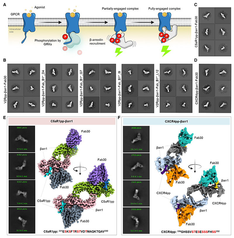
Figure 1
Reconstitution and structure determination of C5aR1pp/CXCR4pp-u03b2arr1 complexes
(A) Agonist stimulation of GPCRs leads to receptor phosphorylation by GPCR kinases (GRKs) followed by the recruitment and activation of u03b2arrs governed through the phosphorylated residues and activ...
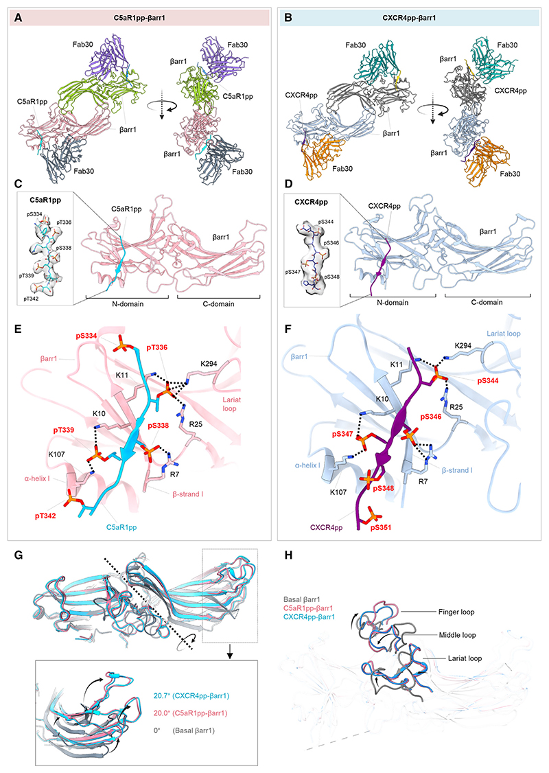
Figure 2
Overall structures and key structural features of C5aR1pp/CXCR4pp-u03b2arr1 complexes
(A and B) Overall structures of C5aR1pp-u03b2arr1-Fab30 and CXCR4pp-u03b2arr1-Fab30 complexes shown with ribbon representation. The constant domains of Fab30 were masked out during refinement. (C and ...
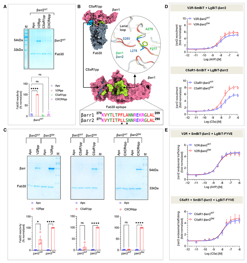
Figure 3
Generation and characterization of u03b2arr2 DM for structure determination
(A) Fab30 reactivity to C5aR1pp and CXCR4pp activated u03b2arr2 WT was measured by co-immunoprecipitation (coIP) assay. C5aR1pp and CXCR4pp activated u03b2arr2 WT were not recognized by Fab30 (top). D...
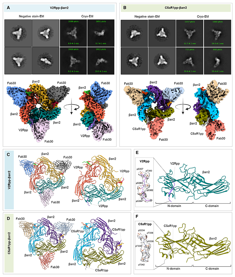
Figure 4
Structures of V2Rpp/C5aR1pp-u03b2arr2 complexes
(A and B) Overall cryo-EM structures of V2Rpp-u03b2arr2-Fab30 (left) and C5aR1pp-u03b2arr2-Fab30 complexes (right), respectively, in a trimeric assembly with u03b2arr2 and Fab30 molecules colored as i...
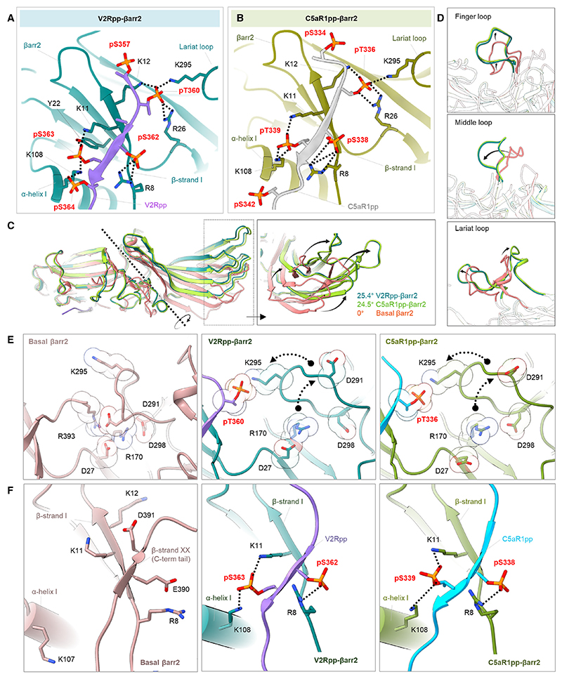
Figure 5
Active conformations of phosphopeptide-bound u03b2arr2
(A and B) Extensive charge-charge interactions between the phosphate residues in V2Rpp/C5aR1pp with Lys/Arg in the N-domain (represented as black dotted lines) stabilize the V2Rpp and C5aR1pp into the...
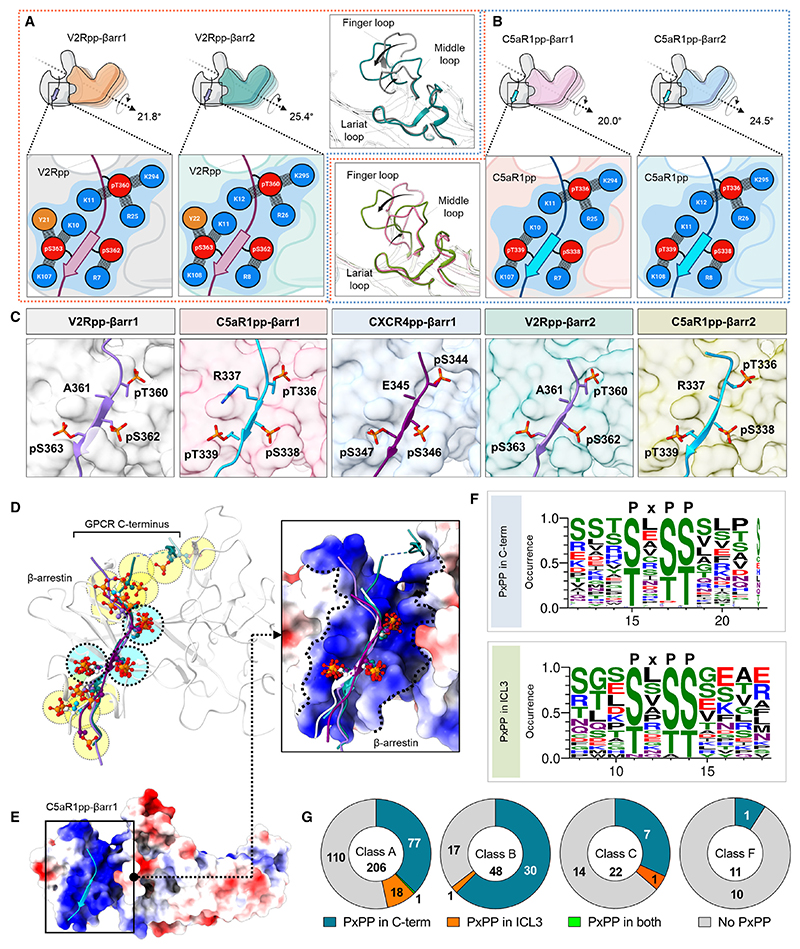
Figure 6
Identification of a key phosphorylation motif in GPCRs driving u03b2arr activation
(A) Comparison of the V2Rpp-bound u03b2arr1 and u03b2arr2 structures reveals similar interactions of V2Rpp with both isoforms of u03b2arrs although a slightly higher inter-domain rotation is observed ...
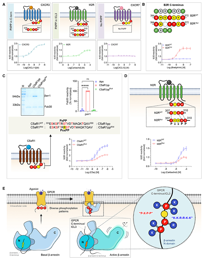
Figure 7
A key phosphorylation motif for u03b2arr activation
(A) NanoBiT-based assay for assessing Ib30 reactivity to CXCR3 (left), M2R (middle), and CXCR7 (right) activated u03b2arr1 (Receptor+SmBiT-u03b2arr1+LgBiT-Ib30) (mean u00b1 SEM; n = 3; normalized as f...
Figure images are served from the NIH/NLM PubMed Central Open Access Subset or Europe PMC; copyright remains with the publishers and authors.
💬 Discussion
0 commentsNo comments yet. Be the first to start a discussion!
Leave a Comment