Abstract
Eukaryotic translation initiation is tightly regulated, requiring a set of conserved initiation factors (eIFs). Translation of a capped mRNA depends on the trimeric eIF4F complex and eIF4B to load the mRNA onto the 43S pre-initiation complex comprising 40S and initiation factors 1, 1A, 2, 3 and 5 as well as initiator-tRNA. Binding of the mRNA is followed by mRNA scanning in the 48S pre-initiation complex, until a start codon is recognised. Here, we use a reconstituted system to prepare human 48S complexes assembled on capped mRNA in the presence of eIF4B and eIF4F. The highly purified h-48S complexes are used for cross-linking/mass spectrometry, revealing the protein interaction network in this complex. We report the electron cryo-microscopy structure of the h-48S complex at 6.3 Å resolution. While the majority of eIF4B and eIF4F appear to be flexible with respect to the ribosome, additional density is detected at the entrance of the 40S mRNA channel which we attribute to the RNA-recognition motif of eIF4B. The eight core subunits of eIF3 are bound at the 40S solvent-exposed side, as well as the subunits eIF3d, eIF3b and eIF3i. elF2 and initiator-tRNA bound to the start codon are present at the 40S intersubunit side. This cryo-EM structure represents a molecular snap-shot revealing the h-48S complex following start codon recognition.
🔬 Techniques
🔭 Microscopes
🧬 Organisms
💻 Software
🧪 Sample Preparation
🔬 Cell Lines
🏭 Microscope Brands
🧪 Reagent Suppliers
💻 Software Details
💾 Data Repositories
🏛️ Research Organizations (ROR)
Affiliated research institutions:
📋 Methods
Plasmids
The plasmid pET28a-MVHL-STOP2 is derived from pET28a-MVHL-STOP ( 23 ). It contains four CAA repeats followed by 52 nucleotides β-globin 5′-UTR, the coding region for Met, Val, His, Leu (MVHL), the stop codon UAA, the rest of the β-globin ORF followed by downstream sequences complementary to a DNA oligonucleotide for RNaseH cleavage and a biotinylated oligonucleotide for affinity purification ( Supplementary Figure S1A ). Plasmids for expression of eIF1, eIF1A, eIF4A, and eIF5 are described in ( 13 , 24 ). The gene encoding human eIF4B was sub-cloned from the plasmid pET(His6-eIF4B) ( 24 ) into the pACEBac1 vector via Rsr II and Sal I restriction sites for insect cell expression. In vitro transcription and mRNA capping The plasmid pET28a-MVHL-STOP2 was amplified by PCR using specific oligonucleotides (forward primer 5′-TCCGGCGTAGAGGATCGAGATC-3′, reverse primer 5′-GACTCGAGCAGATCTATTAAGAGCGGTCGGTAAAACTTCGGCCAGTGAATTTCAGTGGTATTTGTG-3′). The PCR product was transcribed in vitro using T7 RNA polymerase and purified by LiCl/EtOH precipitations. The purified mRNA was capped using Vaccinia Virus Capping Enzyme and the ScriptCap m7G Capping System (CellScript). Human initiation factors, 40S ribosome purification and tRNA aminoacylation Native human factors eIF2, eIF3, eIF4F and human 40S ribosomal subunits were purified from HeLa cytoplasmic lysate (Cilbiotech SA, Belgium) as described in ( 23 ). Human eIF1, eIF1A, eIF4A, and eIF5 were expressed as recombinant proteins in Escherichia coli and purified as described ( 23 , 25 ). His-tagged human eIF4B was expressed using the Multibac/insect cell expression system ( 26 ). For eIF4B purification, insect cell pellets from ∼1 l of culture were dissolved in the protein buffer L (20 mM Tris–HCl, pH 7.5, 100 mM KOAc, 5% glycerol, 2 mM DTT). The cells were lysed by four freeze-thaw cycles. Subsequently, the cleared cell lysate was applied onto a HisTrap column (GE Healthcare) equilibrated with buffer L. After washing with buffer L supplemented with 10 mM imidazole, eIF4B was eluted by an imidazole gradient from 10 mm to 200 mM in buffer L. The fractions containing eIF4B were applied onto a MonoQ column (GE Healthcare) equilibrated with buffer L. eIF4B was eluted by a KCl gradient from 100 to 500 mM in buffer L. Met-tRNAi was amino-acylated using recombinant E. coli methionyl-tRNA synthetase ( 23 ). 48S complex preparation Human 48S complexes were assembled as described in Ref. ( 23 ) with the following modifications: The 48S assembly was performed in reaction buffer A (20 mM Tris–HCl, pH 7.5, 50 mM KOAc, 2.5 mM MgCl 2 , 2 mM DTT, 0.25 mM spermidine) supplemented with 200 U RiboLock RNase inhibitor (Thermo), 1 mM ATP, 0.2 mM GMPPNP, 35 pmol of capped MVHL-stop mRNA, 35 pmol Met-tRNAi, 50 pmol purified human small ribosomal subunits (h-40S), 100 pmol eIF2, 50 pmol eIF3, 80 pmol eIF4F, eIF4A, eIF4B, eIF1, eIF1A, eIF5 each, in a volume of 500 μl. The reaction mix was incubated for 30 min at 37°C. Subsequently, a biotinylated 2′- O -methyl-RNA oligonucleotide (IBA GmbH) with the sequence 5′-dT*dT*dT*CAGAUCUAUUAA GAGCGGUCGGdT*dT*dT*-3′ and Streptavidin High-Capacity Agarose (Thermo) were used for affinity purification of the h-48S complexes. The streptavidin beads were washed in buffer B (20 mM Tris–HCl, pH 7.5, 40 mM KOAc, 7.5 mM MgCl 2 , 2 mM DTT, 0.25 mM spermidine) until the wash fractions were protein-free (based on OD 280 nm measurements). Subsequently, the oligonucleotide 5′-TTCGGCCAGTGAATTTC-3′ was allowed to anneal to the mRNA. The mRNA-bound complexes were eluted from the streptavidin beads by addition of 140U RNaseH (New England Biolabs) in reaction buffer A supplemented with 0.25 mM GMPPNP. The ribosomal complexes were analysed by toe-print assays (primer extension inhibition) using AMV reverse transcriptase (Promega) and a 6-carboxyfluorescein (FAM)-labeled oligonucleotide (5′-GCAATGAAAATAAATTTCC-3′) complementary to the 3′-UTR region of the MVHL-stop mRNA. The resulting fragments were analyzed by commercial sequencing services (capillary sequencing, FASTERIS SA, Switzerland). Based on the intensity of toe-print signals, around one third of the h-48S complexes were retained after elution from the beads. Electron cryo-microscopy Data were collected on a FEI Titan Krios microscope (MPI Göttingen) operated at 300 kV under low-dose conditions (30 ± 5 e − /Å 2 ) using a defocus range of 1.5–4 μm. Single-frame images were recorded on a Falcon II detector at a calibrated magnification of 112,000 (yielding a pixel size of ∼1.25 Å). We discarded micrographs that showed noticeable signs of astigmatism or drift. 2,380 micrographs were used for image processing. Image processing 192,543 particles were picked semi-automatically using EMAN2 ( 27 ). The parameters for the contrast transfer function were estimated for the micrographs using CTFFIND4 ( 28 ). 2D class averaging, 3D classifications, and refinements were performed with RELION ( 29 ). After 2D class averaging, classes with aberrant particles were discarded. 165,372 particles belonging to the best 2D classes were selected for 3D reconstruction. To obtain an initial reconstruction, the 40S + HCV IRES cryo-EM reconstruction (EMD-3019) ( 30 ) was low-pass filtered to 60 Å and used as an input model. Subsequently, all selected particles were classified into four classes by 3D classification. Two of the four classes could be refined to high resolution: Class 1 (30.6% particles; 6.3 Å; h-48S complex) and Class 3 (42.5% particles; 6.6 Å; 40S with TC). Class 2 (5.5% particles) contained dimers of 40S + eIF3. Class 4 (21.4% particles) was similar to Class 1. The resolutions reported are after gold-standard refinement and using the FSC = 0.143 criterion ( 31 ). The local resolution was estimated using RESMAP ( 32 ). All maps were further processed for the modulation transfer function of the detector and sharpened by applying a negative B factors (–91 Å 2 for the h-48S map and –165 Å 2 for the 40S-TC map; estimated as in ( 33 )). Fitting of atomic structures into the cryo-EM maps Volumes obtained by RELION were segmented in Chimera using the SEGGER tool ( 34 , 35 ). The atomic models of 40S, tRNAi and eIF2 subunits from the mammalian 48S model ( 20 ), atomic models of different eIF3 subunits ( 15 ) as well as the crystal structure of Nasonia vitripennis eIF3d ( 22 ) and the NMR structure of the eIF4B RRM ( 36 ) were fitted into the h-48S map using Chimera. The figures were generated by PyMOL (The PyMOL Molecular Graphics System, Version 1.8 Schrödinger, LLC) and by Chimera ( 34 ). Cross-linking/mass spectrometry analysis Affinity purified h-48S complexes were cross-linked with 1 mM DSS-d0/d12 (Creative Molecules) in buffer C (20 mM HEPES–KOH pH 8, 100 mM KCl, 5 mM MgCl 2 , 0.25 mM GMPPNP), dialyzed against water and lyophilized. Processing of the cross-linked complexes, LC–MS analysis and data processing with xQuest was essentially performed as described in ( 37 ), with the exception that all MS data was acquired on a Thermo Orbitrap Fusion Lumos mass spectrometer connected to a Thermo Easy-nLC 1200 HPLC system. The Lumos instrument was operated in data-dependent top speed acquisition mode with a cycle time of 3 s and using collision-induced dissociation in the linear ion trap for fragmentation. One data set was acquired in high/low resolution mode with precursor ion detection in the Orbitrap analyser (resolution = 120 000) and fragment ion detection in the linear ion trap (rapid scan mode setting). A second data set was acquired in high/high resolution mode with both precursor and fragment ion detection in the Orbitrap (at 60 000 and 30 000 resolution, respectively). MS/MS spectra were searched against a database containing all 40S ribosomal subunits, initiation factors and nine contaminant proteins (including keratins). xProphet ( 38 ) was used to adjust the false discovery rates (FDR) to
Show full methods section
Plasmids
The plasmid pET28a-MVHL-STOP2 is derived from pET28a-MVHL-STOP ( 23 ). It contains four CAA repeats followed by 52 nucleotides β-globin 5′-UTR, the coding region for Met, Val, His, Leu (MVHL), the stop codon UAA, the rest of the β-globin ORF followed by downstream sequences complementary to a DNA oligonucleotide for RNaseH cleavage and a biotinylated oligonucleotide for affinity purification ( Supplementary Figure S1A ). Plasmids for expression of eIF1, eIF1A, eIF4A, and eIF5 are described in ( 13 , 24 ). The gene encoding human eIF4B was sub-cloned from the plasmid pET(His6-eIF4B) ( 24 ) into the pACEBac1 vector via Rsr II and Sal I restriction sites for insect cell expression. In vitro transcription and mRNA capping The plasmid pET28a-MVHL-STOP2 was amplified by PCR using specific oligonucleotides (forward primer 5′-TCCGGCGTAGAGGATCGAGATC-3′, reverse primer 5′-GACTCGAGCAGATCTATTAAGAGCGGTCGGTAAAACTTCGGCCAGTGAATTTCAGTGGTATTTGTG-3′). The PCR product was transcribed in vitro using T7 RNA polymerase and purified by LiCl/EtOH precipitations. The purified mRNA was capped using Vaccinia Virus Capping Enzyme and the ScriptCap m7G Capping System (CellScript). Human initiation factors, 40S ribosome purification and tRNA aminoacylation Native human factors eIF2, eIF3, eIF4F and human 40S ribosomal subunits were purified from HeLa cytoplasmic lysate (Cilbiotech SA, Belgium) as described in ( 23 ). Human eIF1, eIF1A, eIF4A, and eIF5 were expressed as recombinant proteins in Escherichia coli and purified as described ( 23 , 25 ). His-tagged human eIF4B was expressed using the Multibac/insect cell expression system ( 26 ). For eIF4B purification, insect cell pellets from ∼1 l of culture were dissolved in the protein buffer L (20 mM Tris–HCl, pH 7.5, 100 mM KOAc, 5% glycerol, 2 mM DTT). The cells were lysed by four freeze-thaw cycles. Subsequently, the cleared cell lysate was applied onto a HisTrap column (GE Healthcare) equilibrated with buffer L. After washing with buffer L supplemented with 10 mM imidazole, eIF4B was eluted by an imidazole gradient from 10 mm to 200 mM in buffer L. The fractions containing eIF4B were applied onto a MonoQ column (GE Healthcare) equilibrated with buffer L. eIF4B was eluted by a KCl gradient from 100 to 500 mM in buffer L. Met-tRNAi was amino-acylated using recombinant E. coli methionyl-tRNA synthetase ( 23 ). 48S complex preparation Human 48S complexes were assembled as described in Ref. ( 23 ) with the following modifications: The 48S assembly was performed in reaction buffer A (20 mM Tris–HCl, pH 7.5, 50 mM KOAc, 2.5 mM MgCl 2 , 2 mM DTT, 0.25 mM spermidine) supplemented with 200 U RiboLock RNase inhibitor (Thermo), 1 mM ATP, 0.2 mM GMPPNP, 35 pmol of capped MVHL-stop mRNA, 35 pmol Met-tRNAi, 50 pmol purified human small ribosomal subunits (h-40S), 100 pmol eIF2, 50 pmol eIF3, 80 pmol eIF4F, eIF4A, eIF4B, eIF1, eIF1A, eIF5 each, in a volume of 500 μl. The reaction mix was incubated for 30 min at 37°C. Subsequently, a biotinylated 2′- O -methyl-RNA oligonucleotide (IBA GmbH) with the sequence 5′-dT*dT*dT*CAGAUCUAUUAA GAGCGGUCGGdT*dT*dT*-3′ and Streptavidin High-Capacity Agarose (Thermo) were used for affinity purification of the h-48S complexes. The streptavidin beads were washed in buffer B (20 mM Tris–HCl, pH 7.5, 40 mM KOAc, 7.5 mM MgCl 2 , 2 mM DTT, 0.25 mM spermidine) until the wash fractions were protein-free (based on OD 280 nm measurements). Subsequently, the oligonucleotide 5′-TTCGGCCAGTGAATTTC-3′ was allowed to anneal to the mRNA. The mRNA-bound complexes were eluted from the streptavidin beads by addition of 140U RNaseH (New England Biolabs) in reaction buffer A supplemented with 0.25 mM GMPPNP. The ribosomal complexes were analysed by toe-print assays (primer extension inhibition) using AMV reverse transcriptase (Promega) and a 6-carboxyfluorescein (FAM)-labeled oligonucleotide (5′-GCAATGAAAATAAATTTCC-3′) complementary to the 3′-UTR region of the MVHL-stop mRNA. The resulting fragments were analyzed by commercial sequencing services (capillary sequencing, FASTERIS SA, Switzerland). Based on the intensity of toe-print signals, around one third of the h-48S complexes were retained after elution from the beads. Electron cryo-microscopy Data were collected on a FEI Titan Krios microscope (MPI Göttingen) operated at 300 kV under low-dose conditions (30 ± 5 e − /Å 2 ) using a defocus range of 1.5–4 μm. Single-frame images were recorded on a Falcon II detector at a calibrated magnification of 112,000 (yielding a pixel size of ∼1.25 Å). We discarded micrographs that showed noticeable signs of astigmatism or drift. 2,380 micrographs were used for image processing. Image processing 192,543 particles were picked semi-automatically using EMAN2 ( 27 ). The parameters for the contrast transfer function were estimated for the micrographs using CTFFIND4 ( 28 ). 2D class averaging, 3D classifications, and refinements were performed with RELION ( 29 ). After 2D class averaging, classes with aberrant particles were discarded. 165,372 particles belonging to the best 2D classes were selected for 3D reconstruction. To obtain an initial reconstruction, the 40S + HCV IRES cryo-EM reconstruction (EMD-3019) ( 30 ) was low-pass filtered to 60 Å and used as an input model. Subsequently, all selected particles were classified into four classes by 3D classification. Two of the four classes could be refined to high resolution: Class 1 (30.6% particles; 6.3 Å; h-48S complex) and Class 3 (42.5% particles; 6.6 Å; 40S with TC). Class 2 (5.5% particles) contained dimers of 40S + eIF3. Class 4 (21.4% particles) was similar to Class 1. The resolutions reported are after gold-standard refinement and using the FSC = 0.143 criterion ( 31 ). The local resolution was estimated using RESMAP ( 32 ). All maps were further processed for the modulation transfer function of the detector and sharpened by applying a negative B factors (–91 Å 2 for the h-48S map and –165 Å 2 for the 40S-TC map; estimated as in ( 33 )). Fitting of atomic structures into the cryo-EM maps Volumes obtained by RELION were segmented in Chimera using the SEGGER tool ( 34 , 35 ). The atomic models of 40S, tRNAi and eIF2 subunits from the mammalian 48S model ( 20 ), atomic models of different eIF3 subunits ( 15 ) as well as the crystal structure of Nasonia vitripennis eIF3d ( 22 ) and the NMR structure of the eIF4B RRM ( 36 ) were fitted into the h-48S map using Chimera. The figures were generated by PyMOL (The PyMOL Molecular Graphics System, Version 1.8 Schrödinger, LLC) and by Chimera ( 34 ). Cross-linking/mass spectrometry analysis Affinity purified h-48S complexes were cross-linked with 1 mM DSS-d0/d12 (Creative Molecules) in buffer C (20 mM HEPES–KOH pH 8, 100 mM KCl, 5 mM MgCl 2 , 0.25 mM GMPPNP), dialyzed against water and lyophilized. Processing of the cross-linked complexes, LC–MS analysis and data processing with xQuest was essentially performed as described in ( 37 ), with the exception that all MS data was acquired on a Thermo Orbitrap Fusion Lumos mass spectrometer connected to a Thermo Easy-nLC 1200 HPLC system. The Lumos instrument was operated in data-dependent top speed acquisition mode with a cycle time of 3 s and using collision-induced dissociation in the linear ion trap for fragmentation. One data set was acquired in high/low resolution mode with precursor ion detection in the Orbitrap analyser (resolution = 120 000) and fragment ion detection in the linear ion trap (rapid scan mode setting). A second data set was acquired in high/high resolution mode with both precursor and fragment ion detection in the Orbitrap (at 60 000 and 30 000 resolution, respectively). MS/MS spectra were searched against a database containing all 40S ribosomal subunits, initiation factors and nine contaminant proteins (including keratins). xProphet ( 38 ) was used to adjust the false discovery rates (FDR) to
📊 Figures
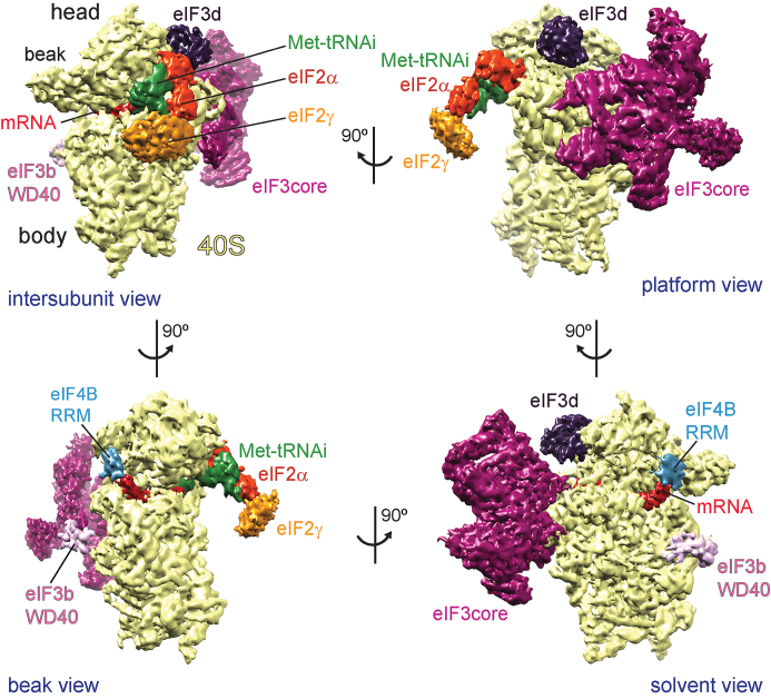
Figure 1.
Segmented cryo-EM structure of the human 48S complex shown in four different views. The h-48S cryo-EM map was segmented for the 40S ribosomal subunit (yellow), the different initiation factors, initia...
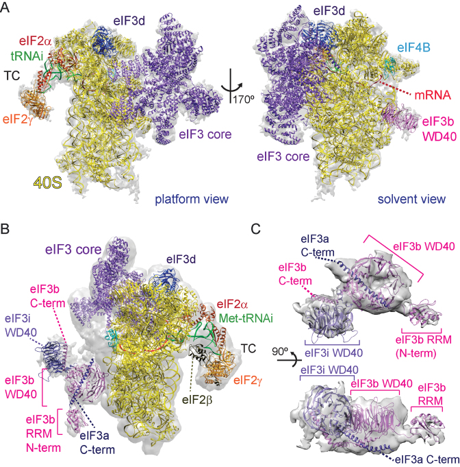
Figure 2.
Initiation factor 3 core and YLC subunits are bound to the solvent-exposed side of 40S in the human 48S complex. ( A ) Fitting of the atomic structures of the eIF3 PCI/MPN core (purple), eIF3d (dark p...
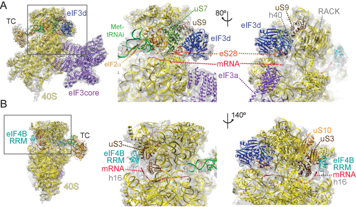
Figure 3.
Factors eIF3d and eIF4B are bound at the mRNA exit and mRNA entry channel of 40S in the h-48S complex. ( A) The subunit eIF3d is bound to the 40S head at the mRNA exit channel. The crystal structure o...
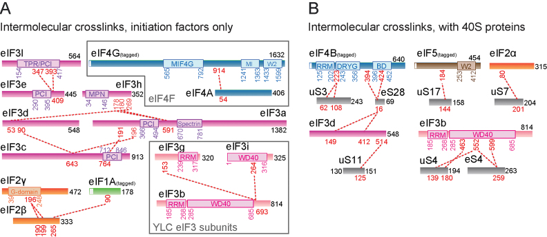
Figure 4.
Cross-linking/mass spectrometry analysis of human 48S complex. ( A ) Cross-links identified between different eukaryotic initiation factors and their subunits are shown as red dashed lines. Cross-link...
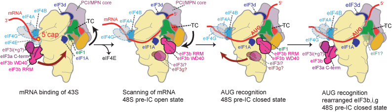
Figure 5.
Model for h-48S conformational changes during 5u2032cap-dependent translation initiation. Left: 43S (comprising 40S-eIF3-eIF1-eIF1A, TC and eIF5) attachment to capped mRNA is supported by eIF4F (trans...
Figure images are served from the NIH/NLM PubMed Central Open Access Subset or Europe PMC; copyright remains with the publishers and authors.
💬 Discussion
0 commentsNo comments yet. Be the first to start a discussion!
Leave a Comment