Abstract
Dynein-2 assembles with polymeric intraflagellar transport (IFT) trains to form a transport machinery that is crucial for cilia biogenesis and signaling. Here we recombinantly expressed the ~1.4-MDa human dynein-2 complex and solved its cryo-EM structure to near-atomic resolution. The two identical copies of the dynein-2 heavy chain are contorted into different conformations by a WDR60-WDR34 heterodimer and a block of two RB and six LC8 light chains. One heavy chain is steered into a zig-zag conformation, which matches the periodicity of the anterograde IFT-B train. Contacts between adjacent dyneins along the train indicate a cooperative mode of assembly. Removal of the WDR60-WDR34-light chain subcomplex renders dynein-2 monomeric and relieves autoinhibition of its motility. Our results converge on a model in which an unusual stoichiometry of non-motor subunits controls dynein-2 assembly, asymmetry, and activity, giving mechanistic insight into the interaction of dynein-2 with IFT trains and the origin of diverse functions in the dynein family.
🔬 Techniques
🧬 Organisms
💻 Software
✨ Fluorophores
🧪 Sample Preparation
🏭 Microscope Brands
🧪 Reagent Suppliers
📷 Detectors
💻 Software Details
💾 Data Repositories
🏛️ Research Organizations (ROR)
Affiliated research institutions:
📋 Methods
Expression of the dynein-2 complex
To generate a plasmid for expression of the human dynein-2 complex, the following genes were synthesised with codon-optimisation for insect cells (Epoch): the dynein-2 heavy chain ( DYNC2H1 ), two intermediate chains ( WDR60 and WDR34 ), light-intermediate chain ( DYNC2LI1 ), light chains ( DYNLRB1/2 , DYNLL1/2 , DYNLT1/3 , and TCTEX1D2 ), and one component (LC8-like; LOC392067 ) that proved not to be part of dynein-2 and thus served as a control for the specificity of the complex. See Supplementary Table 1 for alternate subunit nomenclature. Using Gibson assembly, each subunit was inserted into one of a family of plasmids derived from pACEBac1 (Geneva Biotech). Each plasmid had an expression cassette containing a polH promoter and a SV40 terminator, followed by a SwaI site. Using flanking PmeI sites, expression cassettes were excised from the parent plasmid and inserted into the SwaI site of another plasmid in the family using Gibson assembly. Using this strategy in a pyramid fashion ( Supplementary Fig. 1a ), a plasmid containing all subunits was assembled. DHC2 has an N-terminal His tag, ZZ tag, TEV cleavage sites, and SNAP f tag and WDR34 has a C-terminal Strep tag. A construct lacking the intermediate and light chains (ΔIC-LC) was made by excising their expression cassettes using restriction enzymes. DNA sequences were verified by complete plasmid next-generation sequencing (Massachusetts General Hospital Center for Computational & Integrative Biology). For baculovirus-based expression in insect cells, constructs were inserted into the Tn7 site of the EMBacY bacmid 58 . DH10EMBacY E. coli (Geneva Biotech) were transformed with plasmids via electroporation, followed by selection on antibiotic plates and blue/white screening. Successful transposants (white colonies) were used to inoculate 3 ml cultures for overnight growth, followed by purification of bacmid DNA using alkaline lysis and isopropanol precipitation as described 59 . For baculovirus production and protein expression, Sf9 insect cells were grown in Insect-XPRESS Medium + L-glutamine (Lonza) at 27 °C. Non-adherent cultures were maintained at 1 – 2 x 10 6 cells/ml in flasks with shaking at 100 rpm. Baculoviruses were prepared as described 25 . Medium (100 μl), bacmid DNA (2 μg), and FuGene HD transfection reagent (3 μl) were mixed and incubated for 15 min, then added drop-wise to 1 x 10 6 Sf9 cells growing adherently in a 6-well plate with 2 ml of medium. After 3 days, the efficiency of transfection was assessed by monitoring YFP expression from EMBacY using a Countess II FL cell counter with an EVOS light cube (Thermo Fisher Scientific). The supernatant (V 0 virus) was added to a 50 ml Sf9 culture and incubated for 3 days. The resulting supernatant (V 1 virus) was stored at 4 °C, and used to infect 0.25 – 4 L cultures at a ratio of 1% (v/v) for protein production. Three-days after infection, cell pellets were harvested by centrifugation, washed in 1x PBS, flash frozen in liquid nitrogen, and stored at -80 °C.
Show full methods section
Expression of the dynein-2 complex
To generate a plasmid for expression of the human dynein-2 complex, the following genes were synthesised with codon-optimisation for insect cells (Epoch): the dynein-2 heavy chain ( DYNC2H1 ), two intermediate chains ( WDR60 and WDR34 ), light-intermediate chain ( DYNC2LI1 ), light chains ( DYNLRB1/2 , DYNLL1/2 , DYNLT1/3 , and TCTEX1D2 ), and one component (LC8-like; LOC392067 ) that proved not to be part of dynein-2 and thus served as a control for the specificity of the complex. See Supplementary Table 1 for alternate subunit nomenclature. Using Gibson assembly, each subunit was inserted into one of a family of plasmids derived from pACEBac1 (Geneva Biotech). Each plasmid had an expression cassette containing a polH promoter and a SV40 terminator, followed by a SwaI site. Using flanking PmeI sites, expression cassettes were excised from the parent plasmid and inserted into the SwaI site of another plasmid in the family using Gibson assembly. Using this strategy in a pyramid fashion ( Supplementary Fig. 1a ), a plasmid containing all subunits was assembled. DHC2 has an N-terminal His tag, ZZ tag, TEV cleavage sites, and SNAP f tag and WDR34 has a C-terminal Strep tag. A construct lacking the intermediate and light chains (ΔIC-LC) was made by excising their expression cassettes using restriction enzymes. DNA sequences were verified by complete plasmid next-generation sequencing (Massachusetts General Hospital Center for Computational & Integrative Biology). For baculovirus-based expression in insect cells, constructs were inserted into the Tn7 site of the EMBacY bacmid 58 . DH10EMBacY E. coli (Geneva Biotech) were transformed with plasmids via electroporation, followed by selection on antibiotic plates and blue/white screening. Successful transposants (white colonies) were used to inoculate 3 ml cultures for overnight growth, followed by purification of bacmid DNA using alkaline lysis and isopropanol precipitation as described 59 . For baculovirus production and protein expression, Sf9 insect cells were grown in Insect-XPRESS Medium + L-glutamine (Lonza) at 27 °C. Non-adherent cultures were maintained at 1 – 2 x 10 6 cells/ml in flasks with shaking at 100 rpm. Baculoviruses were prepared as described 25 . Medium (100 μl), bacmid DNA (2 μg), and FuGene HD transfection reagent (3 μl) were mixed and incubated for 15 min, then added drop-wise to 1 x 10 6 Sf9 cells growing adherently in a 6-well plate with 2 ml of medium. After 3 days, the efficiency of transfection was assessed by monitoring YFP expression from EMBacY using a Countess II FL cell counter with an EVOS light cube (Thermo Fisher Scientific). The supernatant (V 0 virus) was added to a 50 ml Sf9 culture and incubated for 3 days. The resulting supernatant (V 1 virus) was stored at 4 °C, and used to infect 0.25 – 4 L cultures at a ratio of 1% (v/v) for protein production. Three-days after infection, cell pellets were harvested by centrifugation, washed in 1x PBS, flash frozen in liquid nitrogen, and stored at -80 °C.
Purification of the dynein-2 complex
All steps were performed at 4 °C. In a typical preparation, frozen cell pellets from 4 L of Sf9 culture were resuspended in purification buffer (50 mM HEPES [pH 7.5], 100 mM KCl, 150 mM K-acetate, 2 mM Mg-acetate, 1 mM EGTA, 10% [v/v] glycerol, 1 mM DTT, 0.2 mM Mg-ATP, 1 mM PMSF, Roche cOmplete™ EDTA-free Protease Inhibitor Cocktail) to a total volume of 200 ml. Cells were lysed in batches using a Dounce homogenizer with 10 – 20 strokes with a tight clearance pestle. Lysates were clarified by ultracentrifugation in a Type 70 Ti rotor at 360,562 g for 45 min. The supernatant was incubated for 1 h on a roller with 10 ml IgG Sepharose 6 resin (GE Healthcare) that had been pre-washed in purification buffer. The resin was then transferred to a glass column, washed with 2 x 100 ml volumes of purification buffer and 1x 25 ml volume of TEV buffer (as purification buffer, lacking KCl), and transferred to two 15 ml plastic conical tubes. Each tube was supplemented with 400 μg TEV protease, filled with TEV buffer, capped and and incubated overnight on a roller. TEV-cleaved proteins were separated from the resin using an empty column, concentrated to ~2 mg/ml using Amicon Ultra centrifugal filter devices (100 kDa cutoff). Aliquots of 50 – 100 μl were flash frozen in liquid nitrogen and stored at -80 °C. Finally, before use, aliquots were thawed and loaded onto a Superose 6 Increase 3.2/300 column using an ÄKTAmicro system (GE Healthcare) pre-equilibrated with gel filtration buffer (50 mM Tris [pH 7.5], 150 mM K-acetate, 2 mM Mg-acetate, 1 mM EGTA, 1 mM DTT, 0.1 mM MgATP). Fractions (50 μl) were collected and analyzed by SDS-PAGE (4 – 12% Tris-Bis gels with Sypro Red staining (Thermo Frisher Scientific)). Subunits (except LC8-like) were detected in the peak fraction by mass spectrometry (BSRC Mass Spectrometry and Proteomics Facility, University of St Andrews).
Negative stain electron microscopy
Immediately following gel filtration, samples were diluted to 5 – 130 nM and applied to EM grids using the rapid staining method. By pipetting ~4 μl of 2% uranyl acetate, a small air gap and then ~4 μl of sample into a single tip, the contents were rapidly deposited onto a glow-discharged continuous carbon grid (Electron Microscopy Sciences). Grids were then stained in three sequential drops of 75 μl 2% uranyl acetate, blotted and air dried. Micrographs were collected using a Tecnai T12 microscope (Thermo Fisher Scientific) operating at 120 keV with a tungsten electron source and a 4k x 4k CCD camera (US4000, Thermo Fisher Scientific). Nominal magnification was 67,000X, giving a 1.67 Å/pixel sampling at the object level. Images were collected with a dose of 40 − 50 e - /Å 2 and a nominal defocus of -1 μm. In total 458 and 415 micrographs were collected for the dynein-2 holoenzyme and ΔIC-LC complexes respectively. Dynein-2 holoenzyme particles were manually picked and subjected to initial 2D classification using Relion v2.1 60 . Flexibility between the tail and motor domains was further analysed using multivariate statistical analysis in Imagic 61 . A total of 4,465 particles were aligned and classified with a mask encompassing the entire molecule. By inspection of the 2D class averages, a subset of particles corresponding to the major tail view was selected and re-aligned based on the motor domain region. The aligned particles were then subjected to further classification using a mask that encompassed all observed tail positions but excluded the motor domains. To quantify the tail positions in the resulting 27 class averages (1,261 particles), the angle between the C2 symmetry axis in the motor domains and the dimerization domain in the tail was measured using ImageJ. A polar plot histogram of tail angles was generated using MatLab R2017b. Particles of the ΔIC-LC sample were picked manually and subjected to 2D classification using Relion. To analyze flexibility in the tail, Imagic was used to align and classify 2,827 ΔIC-LC particles based on the tail region. By inspection of the 2D class averages, a subset of particles corresponding to the major tail view was selected and subjected to further classification to analyse tail flexibility (25 class averages, 1,116 particles).
Cryo-electron microscopy
Immediately following gel filtration, the dynein-2 complex was diluted to 60 – 70 nM and vitrified using a Vitrobot Mark IV system (Thermo Fisher Scientific) set to 4 °C and 95% humidity. Sample (3 μl) was applied to glow-discharged EM grids containing a lacey carbon support film covered with a 3 nm layer of continuous carbon (Agar Scientific). Following 10 s incubation, the grid was manually blotted within the Vitrobot chamber, and a second 3 μl aliquot of sample was then added and incubated for 20 s. The grid was blotted for 5 – 6 s using a force setting of 0 before vitrification in liquid ethane and storage under liquid nitrogen.
Data collection
An initial cryo-EM dataset of dynein-2 holoenzyme was collected on a Tecnai G2 Polara instrument (Thermo Fisher Scientific) operating at 300 keV, equipped with a K2 Summit direct electron detector and GIF Quantum energy filter (slit width 20 eV). Using SerialEM software, micrographs were collected in counting mode with a nominal magnification of 125,000 X (1.39 Å/pixel sampling), 18 s exposures, and 60 frame movies. The total dose per movie was 47.5 e - /Å 2 . Three further datasets were collected on a Titan Krios instrument equipped with a K2 Summit direct electron detector and GIF Quantum energy filter (slit width 20 eV) at the eBIC imaging facility (Diamond Light Source, Didcot). Data collection parameters are shown in Table 1 . Image pre-processing Movies were aligned, dose-weighted and summed using MotionCor2 62 . Micrographs were then visually screened and mis-targeted images containing thick carbon support in more than ~50% of the field of view and empty areas were discarded. Gctf 63 was used to determine CTF parameters.
Tail reconstruction
Negative stain classification of the dynein-2 holoenzyme complex revealed extreme flexibility between the tail and motor domains ( Supplementary Video 1 ). These two portions of the molecule were processed separately. An initial reconstruction of the tail was generated from the Polara dataset. A total of 3,236 particles were picked manually using Boxer 64 and subjected to 2D classification using Relion (v2.0 or v2.1, used for all subsequent image processing steps unless stated). Five of the resulting 2D averages were used as templates for automated particle picking using Gautomatch ( http://www.mrc-lmb.cam.ac.uk/kzhang/ ). The resulting 21,191 picks were subjected to 2D classification and 11,095 particles from well-defined averages were subjected to 3D classification, using a 60 Å low-pass-filtered dynein-1 tail map 53 (EMDB-3707) as a starting model, and refinement to a 10 Å resolution initial map. Projections of this 10 Å tail map were generated using SPIDER 65 and eight views were used as templates for automated particle picking of Krios Dataset 1 using Gautomatch. Class averages from Dataset 1 were used as templates for automated particle picking of Datasets 2 and 3. Permissive parameters were used to ‘over pick’ the micrographs, yielding 757,402 initial picks, from which 461,684 putative particles were isolated. Following extensive rounds of 2D and 3D classification, 68,623 particles were refined to yield a 4.5 Å tail reconstruction, which was sharpened using a B-factor of -125 Å. Further focused refinements were used to determine maps of three tail sub-regions with improved local density ( Supplementary Fig. 2h–j ). The first, encompassing DHC2 TAIL -A, LIC3-A, WDR60, WDR34, RB and LC8, refined to 4.4 Å. The second, encompassing DHC2 N-terminal domain (ND), DHC2 TAIL -A (bundles 1–5), DHC2 TAIL -B (bundles 1–3), WDR60, WDR34 and RB subunits, also refined to 4.4 Å. The third, encompassing DHC2 TAIL -A (bundles 5–8), DHC2 TAIL -B (bundles 3–8), LIC3-A and –B, and LC8 subunits, was subjected to 3D classification, revealing flexibility in the position of TCTEX/TCTEX1D2 density ( Supplementary Fig. 4c ). Particles from class 1 containing the best defined TCTEX/TCTEX1D2 and DHC2 TAIL -B densities were refined to 7.5 Å. Half maps, refined maps, sharpened and masked maps, and mask used have been deposited to the EMDB.
Motor domain reconstruction
A model of the autoinhibited state of dynein-2 motor domains 25 , derived from PDB 4RH7 45 , was low-pass filtered and projected in SPIDER. Eight distinct views were used as templates for automated particle picking of the initial Polara dataset. The resulting picks were subjected to 2D classification using Relion and 8,313 particles from well-defined averages were selected for 3D classification. Particles from the best-defined 3D class were refined to give an 11 Å initial map of the motor domains from 5,133 particles. Six 2D averages from the Polara dataset were used as templates for automated particle picking of Krios Datasets 1 – 3 ( Table 1 ), from which 229,969 putative particles were isolated. Following extensive rounds of 2D and 3D classification, 57,265 particles were refined with C2 symmetry to yield a 3.9 Å motor domain reconstruction, which was sharpened using a B-factor of -100 Å. Half maps, refined map, sharpened and masked map, and mask used have been deposited to the EMDB. Local resolutions in the tail and motor domain maps were calculated using Relion. Map visualization was carried out in UCSF Chimera 66 and UCSF Chimera X 67 . Model building Tail domain RosettaCM 44 , COOT 68 , iMOD fit 69 , SWISS-MODEL 70 , RaptorX Contact 71 , and PSI-PRED 72 software were used to build an atomic model of the tail as detailed in Supplementary Table 2 . RosettaCM was used for density-guided rebuilding, completion, and refinement of homology models. Template-target alignments were generated using HHpred 73 and templates were aligned to the cryo-EM density using Chimera’s ‘Fit in Map’ command. A total of 100-300 Rosetta models were generated and the conformation with the lowest energy (including fit-to-density energy) was used. Coordinates for all subunits were combined in the final tail model and subjected to rounds of relaxation into the density using Rosetta, real-space refinement using Phenix (phenix.real_space_refine, default parameters) 74 , and manual adjustments. For deposition to wwPDB, the tail model was truncated to poly-alanine (phenix.pdbtools) to denote that the majority of side chains positions are not experimentally determined. Register shifts cannot be excluded in the peripheral lower-resolution regions of the cryo-EM map ( Supplementary Fig. 2f ) and none of our conclusions rest on the sequence register in these regions. Motor domains The crystal structure of the dynein-2 motor domain as a monomer (PDB 4RH7) 45 was separated into rigid bodies and docked into the motor domain EM density using UCSF Chimera’s ‘Fit in Map’ command, followed by manual adjustment using COOT. A second copy of the motor domain was then generated and fit into the second motor domain EM density using ‘Fit in Map’. Real-space refinement was performed using Phenix (phenix.real_space_refine, default parameters) 74 . Holoenzyme The dynein-2 tail and motor domain structures were fitted into the sub-tomogram average of anterograde IFT-B trains from C. reinhardtii cilia 29 using Chimera’s ‘Fit in Map’ command. The two DHC2 bundles connecting the tail and motor domain were modelled using SWISS-MODEL [template PDB 3VKG, Chain A 75 ] and RaptorX Contact (deposition as UNK).
Microtubule gliding assays
Dynein-2 holoenzyme and ΔIC-LC samples were biotinylated for microtubule gliding assays via their N-terminal SNAP f tag as described 25 . Flow chambers were made between glass slides, biotin-PEG coverslips, and double-sided tape. Fluorescently-labelled microtubules were visualized on an Eclipse Ti-E inverted microscope with a CFI Apo TIRF 1.49 N.A. oil objective, Perfect Focus System, H-TIRF module, LU-N4 laser unit (Nikon) and a quad band filter set (Chroma). Images were recorded with 100 ms exposures on an iXon DU888 Ultra EMCCD camera (Andor), controlled with NIS-Elements AR Software (Nikon). Temperature of the flow chamber was maintained at 25ºC by an objective heater (Okolab). Chambers were sequentially incubated with 1) blocking solution (0.75% Pluronic F-127, 5 mg/ml casein) for 5 min, followed by two washes with gel filtration buffer containing taxol, SEC-T (50 mM Tris [pH 7.5], 150 mM K-acetate, 2 mM Mg-acetate, 1 mM EGTA, 1 mM DTT, 20 μM taxol); 2) 0.5 mg/ml neutravidin for 2 min, followed by two washes with SEC-T supplemented with 1 mg/ml casein; 3) biotinylated motor protein (~40 μg/ml) for 2 min, followed by two washes with SEC-T supplemented with 1 mg/ml casein; 4) 0.1 μM Alexa-488 microtubules in assay solution (SEC-T supplemented with 1 mg/ml casein, 1 mM Mg-ATP, 71 mM β-mercaptoethanol, 20 mM glucose, 300 μg/ml glucose oxidase, 60 μg/ml catalase). Microtubule lengths and gliding velocities were calculated from kymographs generated in FIJI 76 , 77 . Graphing was performed in Prism5 (GraphPad). Reporting Summary Further information on experimental design is available in the Nature Research Reporting Summary linked to this article.
Data availability
Cryo-EM maps are available from the EMDB under accession codes EMD-4918 (dynein-2 tail domain) and EMD-4917 (dynein-2 motor domains). Coordinates are available from the RCSB Protein Data Bank under accession codes 6RLB (dynein-2 tail domain), 6RLA (dynein-2 motor domains), and 6SC2 (dynein-2, docked into sub-tomogram average of the anterograde IFT-B train 29 [EMDB-4303]). All other data supporting the conclusions of this manuscript are available from the corresponding author upon reasonable request.
📊 Figures
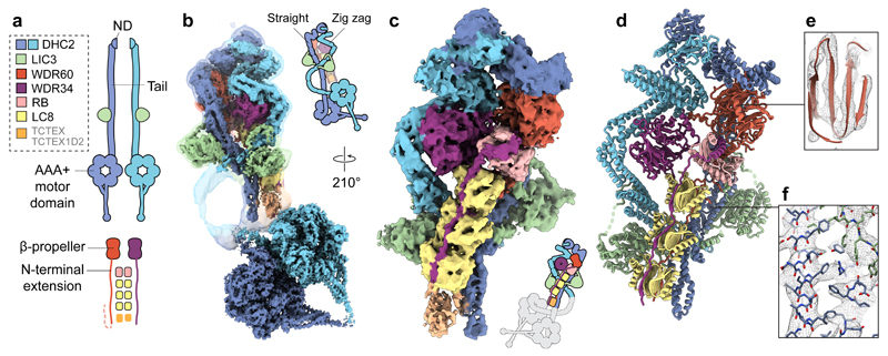
Figure 1
Cryo-EM structure of the dynein-2 complex.
( a ) Overview of the human dynein-2 subunits and their stoichiometry in the structure, coloured according to the code in the upper left. The two copies of DHC2 are coloured in different shades of blu...
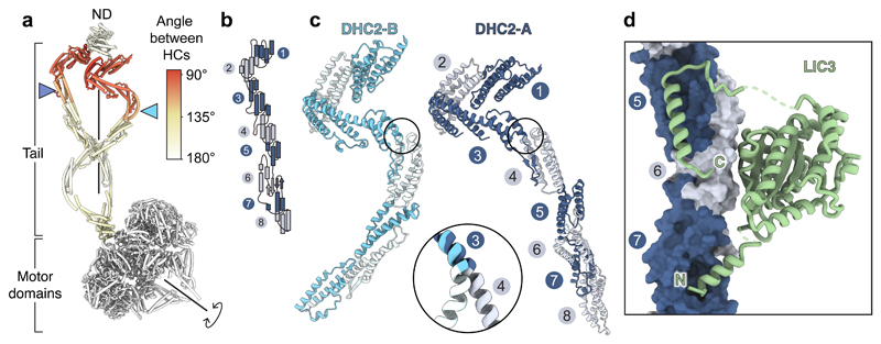
Figure 2
DHC2 asymmetry and LIC3 binding.
( a ) Cylinder representation of the two copies of the DHC2, coloured by the rotation angle relating them. Other subunits omitted for clarity. Blue arrowheads indicate how equivalent helical bundles i...
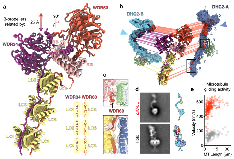
Figure 3
A block of intermediate and light chains controls dynein-2 asymmetry, oligomerization, and activity.
( a ) Ribbon diagram of the intermediate chains WDR34 and WDR60, whose N-proximal extensions are held together by a block of RB and LC8 light chains. Other subunits are omitted for clarity. The transl...
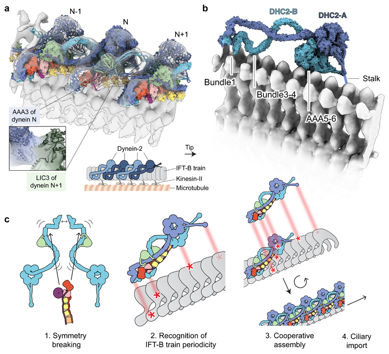
Figure 4
Dynein-2u2019s asymmetric structure matches the periodicity of the IFT train.
( a ) Docking of dynein-2 complexes into a tomogram of an anterograde IFT-B train (EMDB-4303; transparent isosurface) 29 . Individual dynein-2 complexes are shown in alternating surface and cylinder r...
Figure images are served from the NIH/NLM PubMed Central Open Access Subset or Europe PMC; copyright remains with the publishers and authors.
💬 Discussion
0 commentsNo comments yet. Be the first to start a discussion!
Leave a Comment