Abstract
AbstractPolycomb repressive complex 2 (PRC2) is a histone methyltransferase critical for maintaining gene silencing during eukaryotic development. In mammals, PRC2 activity is regulated in part by the selective incorporation of one of two paralogs of the catalytic subunit, EZH1 or EZH2. Each of these enzymes has specialized biological functions that may be partially explained by differences in the multivalent interactions they mediate with chromatin. Here, we present two cryo-EM structures of PRC2:EZH1, one as a monomer and a second one as a dimer bound to a nucleosome. When bound to nucleosome substrate, the PRC2:EZH1 dimer undergoes a dramatic conformational change. We demonstrate that mutation of a divergent EZH1/2 loop abrogates the nucleosome-binding and methyltransferase activities of PRC2:EZH1. Finally, we show that PRC2:EZH1 dimers are more effective than monomers at promoting chromatin compaction, and the divergent EZH1/2 loop is essential for this function, thereby tying together the methyltransferase, nucleosome-binding, and chromatin-compaction activities of PRC2:EZH1. We speculate that the conformational flexibility and the ability to dimerize enable PRC2 to act on the varied chromatin substrates it encounters in the cell.
🔬 Techniques
🔭 Microscopes
🧬 Organisms
💻 Software
✨ Fluorophores
🧪 Sample Preparation
🏭 Microscope Brands
🧪 Reagent Suppliers
📷 Detectors
💻 Software Details
💾 Data Repositories
🏛️ Research Organizations (ROR)
Affiliated research institutions:
📋 Methods
Protein expression and purification
PRC2 core complex Codon-optimized versions of human Strep-EZH1, Flag-SUZ12, His-EED, and His-RBAP48 were cloned into the pBIG1a baculovirus expression plasmid, and the sequence was verified 46 . Bacmids were generated in DH10Bac cells and used to transfect Sf9 cells using Bac-to-Bac protocols (Life Technologies). Baculovirus was used to infect Sf9 or Hi5 cells for expression and harvested 60 h post infection. Cells were lysed in Buffer A (20 mM Tris-HCl, pH 7.5, 300 mM NaCl, 10% v/v glycerol, 1 mM DTT) plus Roche cOmplete protease inhibitors using an Emulsiflex C3 homogenizer (Avestin). Lysates were clarified by centrifugation at 40,000 × g for 20 min at 4 °C, then frozen or further processed. Lysates were bound to Streptavidin beads (Strep-Tactin Macroprep, IBA) for 2 h, then washed extensively in Buffer A without protease inhibitors. Bound PRC2 was cleaved overnight with HRV3C and thrombin in Buffer A at 4 °C. Eluted protein was dialyzed into Q-low buffer (20 mM Tris-HCl, pH 7.5, 150 mM NaCl, 1 mM DTT), bound to a HiTrap Q FF column (GE Healthcare), and eluted with a linear gradient of Q-high buffer (20 mM Tris-HCl, pH 7.5, 1 M NaCl, 1 mM DTT). Fractions containing PRC2 were pooled, concentrated, and loaded onto a Superose 6 Increase column (GE Healthcare) equilibrated with Buffer A. Fractions containing PRC2 were pooled, concentrated, and flash-frozen. EZH1 and SUZ12 variants Mutant versions of EZH1 and SUZ12 were cloned into the pBIG1a expression vector containing the other core subunits of PRC2 and sequence verified. Expressions and purifications were followed according to the protocol for wild-type PRC2. AEBP2 The short isoform of AEBP2 (aa 209–503; UniProt: Q6ZN18 ) was cloned into pGEX6P1 plasmid and used to transform E. coli BL21 cells. Cultures were grown until OD600 reached 0.6, then induced with 0.4 mM isopropyl β-D-1-thiogalactopyranoside (IPTG). Cells were grown for 2 h at 37 °C, then harvested by centrifugation at 3500 × g for 20 min. Cells were resuspended in Buffer A plus protease inhibitors, lysed using an Emulsiflex C3, and clarified by centrifugation at 40,000 × g for 20 min. Lysates were bound to Protino glutathione agarose 4B resin (Macherey-Nagel) for 2 h at 4 °C, then extensively washed with Buffer A. Beads were resuspended in 10 ml of HiTrap S buffer (20 mM HEPES, pH 7.5, 300 mM NaCl, 1 mM DTT) and cleaved overnight with HRV3C protease at 4 °C. Eluted protein was bound to a HiTrap S column (GE Healthcare) and eluted with a linear gradient of HiTrap S buffer containing 1 M NaCl. Fractions containing AEBP2 were pooled, concentrated, and flash-frozen. JARID2 The JARID2 1-367 and JARID2 96-367 fragments were cloned into pGEX6P1 plasmid using standard cloning techniques. Plasmids were transformed into E. coli BL21 RIL cells and overnight cultures were grown. Large-scale cultures were inoculated and grown to OD 600 of 0.4–0.6 at 37 °C, then shifted to 18 °C prior to induction with 250 μM IPTG. Cultures were grown for ~18 h and harvested by centrifugation at 3500 × g for 10 min. Lysates were processed the same as AEBP2. After binding to Protino glutathione agarose 4B resin, protein was eluted using GST-elution buffer (40 mM reduced glutathione, 50 mM Tris-HCl, pH 8.0, 1 mM MgCl 2 , 500 mM NaCl, 20% v/v glycerol, and 5 mM DTT). Fractions were combined and dialyzed overnight at 4 °C into HIC buffer A (1 M ammonium sulfate, 10 mM Tris-HCl, pH 7.5, 1 mM DTT) and loaded onto a HiTrap butyl sepharose column. Protein was eluted with a linear gradient of HIC buffer B (10 mM Tris-HCl, pH 7.5, 1 mM DTT). Fractions containing JARID2 were simultaneously cleaved with HRV3C and dialyzed overnight at 4 °C into Mono Q buffer A (20 mM HEPES, pH 7.6, 50 mM KCl, 5% v/v glycerol, 1 mM DTT). Free GST and uncut protein were removed by passing the sample over glutathione sepharose resin. The flow-through was loaded onto a Mono Q column and eluted with a linear gradient of Mono Q buffer containing 1 M KCl. Fractions containing JARID2 were pooled and flash-frozen. PRC2 plus AEBP2 and JARID2 To purify core PRC2 with accessory factors, 1.5 molar excess of either AEBP2 alone or AEBP2 and JARID2 (either aa 1–367 or 96–367) were mixed with core PRC2 and incubated on ice for 30 min, followed by purification over a Superose 6 Increase column equilibrated with Buffer A. Fractions containing monomeric or dimeric versions of the complexes were pooled and flash-frozen. Histone proteins Individual Xenopus laevis wild-type and mutant histones were expressed and purified based on Luger et al. 47 . Individual histone genes in pET3a vectors were transformed into E. coli BL21 DE3 pLysS. Fresh colonies were used to inoculate 5-mL cultures and were grown overnight at 37 °C. The next day 3-L cultures were inoculated from the overnight cultures and grown until OD 600 of 0.4 at 37 °C. Cultures were induced by adding IPTG to 400 μM and shaken at 37 °C for 2 h. Cells were harvested by centrifugation at 3500 × g for 10 min and resuspended in inclusion body wash buffer (50 mM Tris-HCl, pH 7.5, 100 mM NaCl, 1 mM benzamidine, 1 mM β-mercaptoethanol), and flash-frozen. Cell pellets were thawed and lysed with a Tissumizer (Tekmar). Lysed cells were centrifuged 15,000 × g for 20 min at 4 °C. The pellets were resuspended with wash buffer plus 1% Triton X-100 and then centrifuged again as before. The Triton X-100 wash was repeated twice more, and then the pellets were washed twice with wash buffer without Triton X-100. After the final wash, 1 mL of DMSO was added to the pelleted inclusion bodies and they were incubated for 30 min. In all, 20 mL of unfolding buffer was added to each pellet (7 M guanidine hydrochloride, 20 mM sodium acetate, pH 5.2, 10 mM DTT) and gently stirred for 1 hr then centrifuged 25,000 × g for 10 min. The resulting supernatants were purified on a 2-L column packed with Superdex 200 and equilibrated with SAUDE200 (7 M Urea, 20 mM Sodium Acetate, pH 5.2, 200 mM NaCl, 1 mM EDTA, 5 mM β-mercaptoethanol). The fractions containing histones were pooled and loaded onto a 500-mL SP Sepharose column equilibrated into SAUDE200 and eluted with a linear gradient of SAUDE1000 (7 M urea, 20 mM sodium acetate, pH 5.2, 1000 mM NaCl, 1 mM EDTA, 5 mM β-mercaptoethanol). Fractions containing pure histones were dialyzed extensively into water containing 1 mM β-mercaptoethanol and lyophilized until further use. Ubiquitinated H2A was prepared as described for histone H2B 35 . Histone H2A K119C was expressed and purified according to the protocol for the wild-type histones. To generate ubiquitin, His-tagged ubiquitin G76C in pET was transformed into SoluBL21, and cultures were grown to an OD 600 of 0.4 at 37 °C and induced with 400 μM IPTG. Cultures were grown for an additional 4 h. Bacterial pellets were resuspended in ubiquitin lysis buffer (50 mM Tris-HCl, pH 8.0, 300 mM NaCl, 10 mM imidazole, 5 mM β-mercaptoethanol, plus Roche cOmplete protease inhibitors) and lysed using an Emulsiflex C3 homogenizer. The lysate was bound to Ni-NTA agarose (QIAGEN) and eluted with lysis buffer plus 300 mM imidazole. Fractions containing ubiquitin were dialyzed into 20 mM Tris-HCl, pH 8.0, 50 mM NaCl, 0.2 mM EDTA, 10 mM β-mercaptoethanol, and then loaded onto a HiTrap Q HP (GE Healthcare) column and eluted with a linear gradient of loading buffer supplemented with 1 M NaCl. Fractions containing ubiquitin were extensively dialyzed into deionized water supplemented with 1 mM acetic acid followed by lyophilization. To prepare cross-linked H2AUb, lyophilized pellets of His-tagged ubiquitin G76C and H2A K119C were dissolved in resuspension buffer (10 mM acetic acid, 7 M urea) to 10 mg/ml. Proteins were mixed to 2:1 molar ratio of Ub to H2A. Sodium tetraborate and TCEP were added to final concentrations of 50 mM and 5 mM, incubated for 30 min on ice. Next, 0.1 M 1,3, dichloroacetone (Sigma) in dimethylformamide (Sigma) was added to 0.5 molar equivalents of sulfhydryl groups and incubated on ice for 1 h. Reactions were quenched by adding β-mercaptoethanol to 5 mM. Reactions were purified over Ni-NTA agarose (QIAGEN), lyophilized extensively against deionized water supplemented with 1 mM β-mercaptoethanol, and stored at −80 °C until used. To generate histone octamers, lyophilized aliquots of the desired individual histones were resuspended in unfolding buffer (7 M guanidine hydrochloride, 20 mM sodium acetate, pH 5.2, 10 mM DTT) for 30 min, then mixed in equimolar ratios. The mixture of histones was dialyzed extensively into refolding buffer (2 M NaCl, 10 mM Tris-HCl, pH 7.5, 1 mM EDTA, 5 mM β-mercaptoethanol). The refolded histone octamers were purified on a Superdex 200 column equilibrated into refolding buffer. The fractions containing octamers were pooled, concentrated, and stored at 4 °C until use.
Show full methods section
Protein expression and purification
PRC2 core complex Codon-optimized versions of human Strep-EZH1, Flag-SUZ12, His-EED, and His-RBAP48 were cloned into the pBIG1a baculovirus expression plasmid, and the sequence was verified 46 . Bacmids were generated in DH10Bac cells and used to transfect Sf9 cells using Bac-to-Bac protocols (Life Technologies). Baculovirus was used to infect Sf9 or Hi5 cells for expression and harvested 60 h post infection. Cells were lysed in Buffer A (20 mM Tris-HCl, pH 7.5, 300 mM NaCl, 10% v/v glycerol, 1 mM DTT) plus Roche cOmplete protease inhibitors using an Emulsiflex C3 homogenizer (Avestin). Lysates were clarified by centrifugation at 40,000 × g for 20 min at 4 °C, then frozen or further processed. Lysates were bound to Streptavidin beads (Strep-Tactin Macroprep, IBA) for 2 h, then washed extensively in Buffer A without protease inhibitors. Bound PRC2 was cleaved overnight with HRV3C and thrombin in Buffer A at 4 °C. Eluted protein was dialyzed into Q-low buffer (20 mM Tris-HCl, pH 7.5, 150 mM NaCl, 1 mM DTT), bound to a HiTrap Q FF column (GE Healthcare), and eluted with a linear gradient of Q-high buffer (20 mM Tris-HCl, pH 7.5, 1 M NaCl, 1 mM DTT). Fractions containing PRC2 were pooled, concentrated, and loaded onto a Superose 6 Increase column (GE Healthcare) equilibrated with Buffer A. Fractions containing PRC2 were pooled, concentrated, and flash-frozen. EZH1 and SUZ12 variants Mutant versions of EZH1 and SUZ12 were cloned into the pBIG1a expression vector containing the other core subunits of PRC2 and sequence verified. Expressions and purifications were followed according to the protocol for wild-type PRC2. AEBP2 The short isoform of AEBP2 (aa 209–503; UniProt: Q6ZN18 ) was cloned into pGEX6P1 plasmid and used to transform E. coli BL21 cells. Cultures were grown until OD600 reached 0.6, then induced with 0.4 mM isopropyl β-D-1-thiogalactopyranoside (IPTG). Cells were grown for 2 h at 37 °C, then harvested by centrifugation at 3500 × g for 20 min. Cells were resuspended in Buffer A plus protease inhibitors, lysed using an Emulsiflex C3, and clarified by centrifugation at 40,000 × g for 20 min. Lysates were bound to Protino glutathione agarose 4B resin (Macherey-Nagel) for 2 h at 4 °C, then extensively washed with Buffer A. Beads were resuspended in 10 ml of HiTrap S buffer (20 mM HEPES, pH 7.5, 300 mM NaCl, 1 mM DTT) and cleaved overnight with HRV3C protease at 4 °C. Eluted protein was bound to a HiTrap S column (GE Healthcare) and eluted with a linear gradient of HiTrap S buffer containing 1 M NaCl. Fractions containing AEBP2 were pooled, concentrated, and flash-frozen. JARID2 The JARID2 1-367 and JARID2 96-367 fragments were cloned into pGEX6P1 plasmid using standard cloning techniques. Plasmids were transformed into E. coli BL21 RIL cells and overnight cultures were grown. Large-scale cultures were inoculated and grown to OD 600 of 0.4–0.6 at 37 °C, then shifted to 18 °C prior to induction with 250 μM IPTG. Cultures were grown for ~18 h and harvested by centrifugation at 3500 × g for 10 min. Lysates were processed the same as AEBP2. After binding to Protino glutathione agarose 4B resin, protein was eluted using GST-elution buffer (40 mM reduced glutathione, 50 mM Tris-HCl, pH 8.0, 1 mM MgCl 2 , 500 mM NaCl, 20% v/v glycerol, and 5 mM DTT). Fractions were combined and dialyzed overnight at 4 °C into HIC buffer A (1 M ammonium sulfate, 10 mM Tris-HCl, pH 7.5, 1 mM DTT) and loaded onto a HiTrap butyl sepharose column. Protein was eluted with a linear gradient of HIC buffer B (10 mM Tris-HCl, pH 7.5, 1 mM DTT). Fractions containing JARID2 were simultaneously cleaved with HRV3C and dialyzed overnight at 4 °C into Mono Q buffer A (20 mM HEPES, pH 7.6, 50 mM KCl, 5% v/v glycerol, 1 mM DTT). Free GST and uncut protein were removed by passing the sample over glutathione sepharose resin. The flow-through was loaded onto a Mono Q column and eluted with a linear gradient of Mono Q buffer containing 1 M KCl. Fractions containing JARID2 were pooled and flash-frozen. PRC2 plus AEBP2 and JARID2 To purify core PRC2 with accessory factors, 1.5 molar excess of either AEBP2 alone or AEBP2 and JARID2 (either aa 1–367 or 96–367) were mixed with core PRC2 and incubated on ice for 30 min, followed by purification over a Superose 6 Increase column equilibrated with Buffer A. Fractions containing monomeric or dimeric versions of the complexes were pooled and flash-frozen. Histone proteins Individual Xenopus laevis wild-type and mutant histones were expressed and purified based on Luger et al. 47 . Individual histone genes in pET3a vectors were transformed into E. coli BL21 DE3 pLysS. Fresh colonies were used to inoculate 5-mL cultures and were grown overnight at 37 °C. The next day 3-L cultures were inoculated from the overnight cultures and grown until OD 600 of 0.4 at 37 °C. Cultures were induced by adding IPTG to 400 μM and shaken at 37 °C for 2 h. Cells were harvested by centrifugation at 3500 × g for 10 min and resuspended in inclusion body wash buffer (50 mM Tris-HCl, pH 7.5, 100 mM NaCl, 1 mM benzamidine, 1 mM β-mercaptoethanol), and flash-frozen. Cell pellets were thawed and lysed with a Tissumizer (Tekmar). Lysed cells were centrifuged 15,000 × g for 20 min at 4 °C. The pellets were resuspended with wash buffer plus 1% Triton X-100 and then centrifuged again as before. The Triton X-100 wash was repeated twice more, and then the pellets were washed twice with wash buffer without Triton X-100. After the final wash, 1 mL of DMSO was added to the pelleted inclusion bodies and they were incubated for 30 min. In all, 20 mL of unfolding buffer was added to each pellet (7 M guanidine hydrochloride, 20 mM sodium acetate, pH 5.2, 10 mM DTT) and gently stirred for 1 hr then centrifuged 25,000 × g for 10 min. The resulting supernatants were purified on a 2-L column packed with Superdex 200 and equilibrated with SAUDE200 (7 M Urea, 20 mM Sodium Acetate, pH 5.2, 200 mM NaCl, 1 mM EDTA, 5 mM β-mercaptoethanol). The fractions containing histones were pooled and loaded onto a 500-mL SP Sepharose column equilibrated into SAUDE200 and eluted with a linear gradient of SAUDE1000 (7 M urea, 20 mM sodium acetate, pH 5.2, 1000 mM NaCl, 1 mM EDTA, 5 mM β-mercaptoethanol). Fractions containing pure histones were dialyzed extensively into water containing 1 mM β-mercaptoethanol and lyophilized until further use. Ubiquitinated H2A was prepared as described for histone H2B 35 . Histone H2A K119C was expressed and purified according to the protocol for the wild-type histones. To generate ubiquitin, His-tagged ubiquitin G76C in pET was transformed into SoluBL21, and cultures were grown to an OD 600 of 0.4 at 37 °C and induced with 400 μM IPTG. Cultures were grown for an additional 4 h. Bacterial pellets were resuspended in ubiquitin lysis buffer (50 mM Tris-HCl, pH 8.0, 300 mM NaCl, 10 mM imidazole, 5 mM β-mercaptoethanol, plus Roche cOmplete protease inhibitors) and lysed using an Emulsiflex C3 homogenizer. The lysate was bound to Ni-NTA agarose (QIAGEN) and eluted with lysis buffer plus 300 mM imidazole. Fractions containing ubiquitin were dialyzed into 20 mM Tris-HCl, pH 8.0, 50 mM NaCl, 0.2 mM EDTA, 10 mM β-mercaptoethanol, and then loaded onto a HiTrap Q HP (GE Healthcare) column and eluted with a linear gradient of loading buffer supplemented with 1 M NaCl. Fractions containing ubiquitin were extensively dialyzed into deionized water supplemented with 1 mM acetic acid followed by lyophilization. To prepare cross-linked H2AUb, lyophilized pellets of His-tagged ubiquitin G76C and H2A K119C were dissolved in resuspension buffer (10 mM acetic acid, 7 M urea) to 10 mg/ml. Proteins were mixed to 2:1 molar ratio of Ub to H2A. Sodium tetraborate and TCEP were added to final concentrations of 50 mM and 5 mM, incubated for 30 min on ice. Next, 0.1 M 1,3, dichloroacetone (Sigma) in dimethylformamide (Sigma) was added to 0.5 molar equivalents of sulfhydryl groups and incubated on ice for 1 h. Reactions were quenched by adding β-mercaptoethanol to 5 mM. Reactions were purified over Ni-NTA agarose (QIAGEN), lyophilized extensively against deionized water supplemented with 1 mM β-mercaptoethanol, and stored at −80 °C until used. To generate histone octamers, lyophilized aliquots of the desired individual histones were resuspended in unfolding buffer (7 M guanidine hydrochloride, 20 mM sodium acetate, pH 5.2, 10 mM DTT) for 30 min, then mixed in equimolar ratios. The mixture of histones was dialyzed extensively into refolding buffer (2 M NaCl, 10 mM Tris-HCl, pH 7.5, 1 mM EDTA, 5 mM β-mercaptoethanol). The refolded histone octamers were purified on a Superdex 200 column equilibrated into refolding buffer. The fractions containing octamers were pooled, concentrated, and stored at 4 °C until use.
Nucleosomal DNA generation
Plasmids containing 601 DNA fragments for assemblies were prepared using large-scale alkaline lysis 48 . Large-scale cultures (12 L) of E. coli containing plasmid DNA were grown overnight at 37 °C. Cells were harvested by centrifugation at 3500 × g for 10 min, then resuspended with 80 mL of P1 (10 mM EDTA, pH 8) per liter of culture and combined. In total, 160 mL of P2 (0.2 M NaOH, 1% sodium dodecyl sulfate) per liter of culture was added by shaking vigorously and then incubated on ice for 20 min. In all, 160 mL of P3 (4 M potassium acetate, 2 M acetic acid) per liter of culture was added and mixed by gently inverting followed by incubation on ice for 20 min. Lysates were centrifuged at 3500 × g for 20 min, and then the supernatants were filtered through miracloth. In all, 0.5 volumes of isopropyl alcohol were added, and the mixture was stirred for 60 min at 4 °C. The precipitated plasmids were centrifuged 3500 × g for 30 min, air dried, and resuspended in 50 mL TE buffer. In total, 0.5 mg of RNAse A (Thermo) was added and incubated at 37 °C overnight. Solid potassium chloride was added to the plasmid to adjust the final concentration to 2 M. The mixture was purified over a 500-mL Sepharose 6 column equilibrated in plasmid buffer (50 mM Tris-HCl, pH 7.5, 2 M KCl, 1 mM EDTA). Fractions containing plasmid were pooled and precipitated with isopropyl alcohol, centrifuged 3500 × g for 30 min, dried, and resuspended in TE until use. Plasmids were digested with EcoRV to liberate the fragment containing nucleosome-positioning sequences and purified using fractional PEG precipitation. Overnight digestions were mixed with 5 M NaCl to obtain 0.5 M NaCl. A solution of 40% PEG 6000 in 0.5 M NaCl was added to 4.5%, incubated on ice for 1 hr, and centrifuged 25,000 × g for 20 min. Supernatants and pellets were run on an agarose gel to monitor precipitation. Additional PEG solution was added in 0.5% increments, and the process was repeated until the desired fragment of DNA was separated from the plasmid backbone. The purified fragments were precipitated with 2.5 volumes of ethanol overnight at −20 °C. DNA was resuspended in TE to a concentration of 2 mg/ml and filtered until use. Nucleosome assemblies Nucleosomes were assembled using gradient dialysis, as described elsewhere 49 . Briefly, purified DNA and histone octamers were assembled in a high salt buffer (2 M KCl, 10 mM Tris-HCl, 7.5, 1 mM EDTA, 1 mM DTT) and dialyzed into low-salt buffer (0.25 M KCl, 10 mM Tris-HCl, 7.5, 1 mM EDTA, 1 mM DTT) via a peristaltic pump over 16 h. After addition, the assemblies were dialyzed for 4 h into a low-salt buffer and then overnight into TE (10 mM Tris-HCl, 7.5, 1 mM EDTA, 1 mM DTT). Nucleosome arrays were stored until used. Mononucleosomes were further purified over a ResourceQ column (GE Healthcare) equilibrated with TE and eluted with TE plus 1 M NaCl. Fractions containing nucleosomes were dialyzed into TE and stored at 4 °C until used. Optimized DNA/octamer ratios were determined empirically using small-scale reactions, and reactions were then scaled up. Nucleosome assemblies were verified by running samples on native PAGE gels. Electrophoretic mobility-shift assay (EMSA) Proteins and nucleosome samples were dialyzed into EMSA binding buffer (EBB) (10 mM HEPES, pH 7.9, 50 mM KCl, 5% v/v glycerol, 5 mM DTT). Serial dilutions of PRC2 proteins were made in EBB, and then an equal volume of 25 nM nucleosomes in EBB plus 0.5 mg/ml BSA was added to each PRC2 dilution. Reactions were incubated at room temperature for 30 min, loaded onto 3.5% native polyacrylamide gels, and run at 120 V for 70 min using 0.3× Tris Borate EDTA (TBE) running buffer (30 mM Tris base, 30 mM boric acid, 0.6 mM EDTA). Gels were stained with SYBR gold (Thermo Fisher) for 15 min and scanned with a Typhoon Imager (GE Healthcare). Scans of the gels are included in the accompanying Source Data File. DNA bands were quantified using ImageJ 2 50 and graphed using Prism 8 (GraphPad) with data from three independent replicates.
Methyltransferase assays
Histone methyltransferase (HMT) assays were performed in a total volume of 15 μl containing HMT buffer (50 mM Tris-HCl, pH 8.5, 5 mM MgCl 2 , and 4 mM DTT) with 500 nM of 3 H-labeled S-adenosylmethionine (PerkinElmer), 300 nM of nucleosomes, and recombinant human PRC2 at the indicated concentrations. Reaction mixtures were incubated for 60 min at 30 °C and stopped by adding 4 μl of STOP buffer (0.2 M Tris-HCl, pH 6.8, 20% v/v glycerol, 10% m/v SDS, 10 mM β-mercaptoethanol, and 0.05% bromophenol blue). A titration of PRC2 (from 5 to 60 nM) was performed under these conditions to establish that the HMT reactions were within the linear range. After the addition of STOP buffer, samples were incubated for 5 min at 95 °C and separated on SDS-PAGE gels. The gels were subjected to Coomassie blue staining for protein visualization and subsequently transferred to 0.45-μm PVDF membranes (Millipore) using standard Western blotting techniques and exposed to autoradiography film (Denville Scientific). All reactions were done in triplicate. Uncropped scans of the gels are included in the accompanying Source Data File.
MTase-Glo assays
(Promega) were performed according to the manufacturer’s recommendations. Briefly, reactions were assembled in a total volume of 8 μl in MTase-Glo buffer (50 mM Tris-HCl, pH 8.0, 1 mM MgCl 2 , 50 mM NaCl, 0.1 mg/ml BSA, 4 mM DTT) with 10 μM SAM, 1 μM nucleosomes, and recombinant human PRC2 at the indicated concentrations. Reactions were incubated for 20 min at 25 °C and stopped by adding 2 μl of 0.5% trifluoroacetic acid. 2 μl of 5× MTase-Glo reagent was added, and reactions were incubated for 30 min at 25 °C. In total, 10 μl of detection reagent was added and reactions were incubated for 30 min at 25 °C, followed by measuring luminescence in an EnSpire plate reader (PerkinElmer). Reactions were done in triplicate and graphed using Prism 8.
Cryo-EM sample preparation and data collection
PRC2:EZH1 was cross-linked using the GraFix protocol 51 . Briefly, protein complexes were dialyzed into Buffer GA (20 mM HEPES, pH 7.9, 50 mM KCl, 5% v/v glycerol, 1 mM DTT). A 10–40% glycerol gradient was created in 4 ml of Buffer GB (20 mM HEPES, pH 7.9, 50 mM KCl, 10% v/v glycerol, 1 mM DTT) and Buffer GC (20 mM HEPES, pH 7.9, 50 mM KCl, 40% v/v glycerol, 0.1% v/v glutaraldehyde, 1 mM DTT). PRC2:EZH1 was layered onto the top of the gradient and centrifuged in a SW 60Ti rotor for 14 h at 100,000 × g . Fractions were analyzed by SDS-PAGE, and relevant fractions were pooled, concentrated, and dialyzed into freezing buffer (20 mM HEPES, pH 7.9, 50 mM KCl, 1 mM MgCl 2 , 1 mM DTT). To generate PRC2:EZH1 complexes with nucleosomes, PRC2:EZH1-containing JARID 1-367 was mixed 2:1 with nucleosomes and dialyzed into 20 mM HEPES, pH 7.9, 5% v/v glycerol, 1 mM DTT for 6 h. S-adenosylmethionine was added to a final concentration of 0.1 μM and samples were incubated for 1 h at room temperature before processing using GraFix as described for the monomeric PRC2:EZH1 complexes. Three samples were analyzed (Table 1 ): PRC2 in complex with AEBP2 (a), PRC2 in complex with AEBP2, and JARID2 96-367 in a monomeric form (b) and in a dimeric form bound to nucleosome (c). The homogeneity of samples was first examined by negative-stain EM using 0.7% (v/v) uranyl formate, as described 52 . Protein preparations that showed monodispersed particles of homogeneous size and shape were used to prepare vitrified samples for cryo-EM. Vitrified grids were prepared with a Vitrobot Mark IV (Thermo Fisher Scientific) set at 100% humidity and 4 °C. Table 1 Cryo-EM data collection, refinement, and validation statistics. #1 PRC2:EZH1–AEBP2 #2 PRC2:EZH1–AEBP2–JARID2 #3 PRC2-nuclesome complex, nucleosome #4 PRC2-nucleosome complex, PRC2_A #5 PRC2-nucleosome complex, PRC2_B #6 PRC2-nucleosome complex, composite map (EMD-23022) (EMD-23021) (EMD-23026) (EMD-23024) (EMD-23025) (EMD-23103) (PDB 7KSO) (PDB 7KTQ) (PDB 7KSR) (PDB 7KTP) Data collection and processing Magnification 22,500 22,500 64,000 64,000 64,000 64,000 Voltage (kV) 300 300 300 300 300 300 Electron exposure (e–/Å 2 ) 47 47 56.7 56.7 56.7 56.7 Defocus range (μm) −1.5 to −3.0 −1.5 to −3.0 −1.0 to −2.5 −1.0 to −2.5 −1.0 to −2.5 −1.0 to −2.5 Pixel size (Å) 1.3 1.3 1.35 1.35 1.35 1.35 Symmetry imposed C1 C1 C1 C1 C1 C1 Initial particle images (no.) 3,142,334 1,608,434 3,410,000 3,410,000 3,410,000 3,410,000 Final particle images (no.) 155,809 211,110 56,616 18,151 26,440 * Map resolution (Å) 4.1 3.9 3.3 4.1 4.8 * FSC threshold 0.143 0.143 0.143 0.143 0.143 Map resolution range (Å) 3.9–6.6 3.7–6.4 3.1–5.8 3.7–6.4 3.7–6.4 * Refinement Initial model used (PDB code) 5HYN, 2XU7, 2YB8, 5FXY, 5WAI 1KX5, 5WCU Model #2 Model #2 Model resolution (Å) 4.1 3.5 4.3 6.2 FSC threshold 0.5 0.5 0.5 0.5 Model resolution range (Å) Map sharpening B factor (Å 2 ) −135 −122 −81 −104 −120 Model composition Non-hydrogen atoms 13,075 12,745 11,863 12,008 Protein residues 1690 752 1524 1544 Ligands 8 0 7 7 B factors (Å 2 ) mean Protein 95.32 31.08 124.73 191.01 Ligand 150.31 N/A 136.69 219.75 R.m.s. deviations Bond lengths (Å) 0.005 0.005 0.005 0.005 Bond angles (°) 1.16 0.877 1.135 1.157 Validation MolProbity score 1.5 1.3 1.33 1.3 Clashscore 3.83 5.59 1.91 2.11 Poor rotamers (%) 0.29 0 0 0 Ramachandran plot Favored (%) 95.33 98.51 94.47 95.49 Allowed (%) 4.67 1.49 5.53 4.51 Disallowed (%) 0 0 0 0 * Composite map comprised of #3–5, see individual maps for statistics. For PRC2:EZH1–AEBP2 and monomeric PRC2:EZH1–AEBP2–JARID2, 4-µl aliquots at 0.05 mg/ml were applied to glow-discharged Quantifoil 300 mesh 1.2/1.3 gold grids. The grids were blotted for 3 s with a blot force setting of −2 and then plunged into liquid ethane. Before freezing in liquid ethane, grids without an additional carbon film were blotted for 3 s after a 5-s waiting time, whereas grids with an additional carbon layer were blotted for 1 s after a 20-s waiting time. Grids were screened on a Talos Arctica electron microscope (Thermo Fisher Scientific) operated at an acceleration voltage of 200 kV. Since particles at the center of holes tended to aggregate, images were taken at the edge of the holes. Image stacks were collected with a Titan Krios electron microscope (Thermo Fisher Scientific) in the Cryo-EM Resource Center at the Rockefeller University operated at an acceleration voltage of 300 kV. Data were collected at a nominal magnification of ×22,500 (calibrated pixel size of 1.3 Å on the specimen level) with a K2 Summit camera (Gatan) in super-resolution counting mode. Exposures of 10 s were dose-fractionated into 40 frames (250 ms per frame), with a dose rate of 8 electrons/pixel/s (~1.18 electrons/Å 2 /frame), resulting in a total dose of 47 electrons/Å 2 . The images were recorded with SerialEM 3.8 53 , using defocus values ranging from −1.5 to −3 µm. For the dimeric PRC2:EZH1–AEBP2–JARID2 complex bound to core nucleosomes containing H3K27M and H2AUb modifications, samples at 0.12 mg/ml were applied to glow-discharged Quantifoil 200 mesh 1.2/1.3 gold grids. The grids were blotted for 3 s with a blot force setting of −1 and then plunged into liquid ethane. The image stacks were collected on a Titan Krios electron microscope in the Pacific Northwest Cryo-EM Center at a calibrated pixel size of 1.35 Å on the specimen level with a K3 camera in counting mode. Exposures of 10 s were dose-fractionated into 50 frames (200 ms per frame), with a dose rate of 0.84 electrons/pixel/frame, resulting in a total dose of 56.7 electrons/Å 2 .
Cryo-EM image processing
Image stacks recorded in super-resolution mode were binned over 2 × 2 pixels. All image stacks were motion-corrected, dose-weighted, and summed in Motioncorr2 54 (Supplementary Figs. 1a , 2a ). The CTF parameters were determined with Ctffind4 55 . The particles were autopicked with Gautomatch ( http://www.mrc-lmb.cam.ac.uk/kzhang/Gautomatch/ ) using templates obtained by 2D classification of small datasets (~3000 particles) of manually picked particles. All subsequent image-processing steps, including 2D and 3D classification, refinement, postprocessing, and local resolution estimation, were performed in RELION-2.1 56 . For the PRC2:EZH1–AEBP2 complex, 3,142,334 particles were autopicked from 5527 micrographs. The particles were extracted into 180 × 180-pixel images and subjected to 2D classification. The particles in the classes that showed the most detailed averages were used to calculate an initial map of the PRC2:EZH1–AEBP2 complex in cryoSPARC v2 57 . After removing classes whose averages showed ice contamination or edges of the carbon film, the remaining 2,629,432 particles were subjected to 3D classification into ten classes using the map generated by cryoSPARC as the initial reference map. One class showed secondary structure. This map and four additional maps generated from it (the EED1–EZH1 sub-complex by itself, centered and off-center, and the RBAP48–SUZ12–AEBP2 sub-complex by itself, centered and off-center) were used as reference maps for a supervised 3D classification. The 677,847 particles assigned to the full PRC2:EZH1–AEBP2 complex were then subjected to unsupervised 3D classification into six classes using as reference map the same map that was used to generate the references for the supervised classification (Supplementary Fig. 1c , green map). The 245,208 particles in the two classes showing the most detail were combined and subjected to another round of 3D classification into six classes. The 155,809 particles in the four classes showing the most detailed structure were combined and refined, yielding a map at 4.1-Å resolution after postprocessing (Supplementary Fig. 3d ). For the monomeric PRC2:EZH1–AEBP2–JARID2 complex, a total of 1,608,434 particles were autopicked from 4831 micrographs. The particles were extracted into 180 × 180-pixel images and subjected to 2D classification. Compared with the PRC2:EZH1–AEBP2 sample, a larger number of averages showed the intact complex, suggesting that JARID2 stabilizes the complex. After removing classes representing ice contamination and carbon edges as well as classes whose averages showed no features, the remaining 1,025,945 particles were subjected to 3D classification into eight classes. The 236,094 particles in the class showing the most structural detail were subjected to the second round of 3D classification into six classes. The 211,110 particles in the five classes showing the most detailed structure were combined and refined, yielding a map at 3.9-Å resolution after postprocessing. To further improve the quality of the map, the 236,094 particles from the best class resulting from the first 3D classification were combined with the 245,208 particles from the best two classes resulting from the first unsupervised 3D classification of the PRC2:EZH1–AEBP2 dataset and subjected to 3D classification into six classes. The 329,184 particles from the four classes showing the most detailed structure were combined and refined, yielding a map at 3.9-Å resolution after postprocessing (Supplementary Fig. 2c ). The Bayesian polishing and CTF refinement procedures implemented in RELION-3.1 did not improve map quality. For the dimeric PRC2:EZH1–AEBP2–JARID2 complex bound to core nucleosomes containing H3K27M and H2AUb modifications, a total of 7658 movies were collected at the Pacific Northwest Cryo-EM Center using a Titan Krios and K3 camera (Supplementary Fig. 5a ). Patch motion correction and patch CTF correction were done in cryoSPARC v2 57 . In total, 3,410,00 particles were autopicked from 7384 aligned micrographs using references generated from a subset of ~1000 manually picked particles. The particles were extracted into 300 × 300-pixel images and subjected to multiple rounds of 2D classification in cryoSPARC resulting in 742,000 particles (Supplementary Fig. 5b ). cryoSPARC was used to generate an ab initio map that was subsequently used in multiple rounds of heterogeneous refinement. Classes containing stronger density for two PRC2 complexes bound to a nucleosome were selected and used as input particles for additional heterogenous refinements until no further classification was observed. A stack of 109,858 particles was imported into RELION-3.0 58 for final processing (Supplementary Fig. 5c ). Signal subtraction and refinements using masks were employed to generate initial maps of the nucleosome and each monomer of PRC2 individually. Particles from each component were subjected to 3D classification without alignment, and the best classes were used to refine each component individually (Supplementary Fig. 5d ). In total, 56,616 particles yielded a map of the nucleosome at 3.3-Å resolution after postprocessing, 18,151 particles yielded a map of PRC2:EZH1–AEBP2–JARID2 complex “A” at 4.1-Å resolution, and 26,440 particles yielded a map of PRC2:EZH1–AEBP2–JARID2 complex “B” at 4.8-Å resolution (Supplementary Fig. 6a–c ). Model building and refinement of the monomeric PRC2 Initial fitting of subunits Ezh1, EED, RBAP48, and VEFS domain of SUZ12: PDB: 5HYN was used to model EZH1, EED, and SUZ12 561-686 (VEFS). PDB: 2XU7 was used for modeling RBAP48 27 , 59 . The coordinates of each subunit were individually fit into the density using UCSF Chimera’s v1.14 “Fit in map” function 60 . Coot v0.8 was used for manual adjustment of domains, secondary-structure elements, and side chains into densities 61 . The complete model was refined using real-space refinement in PHENIX v1 62 . The EZH2 model in PDB: 5HYN was manually replaced with a sequence from EZH1 where applicable. Additional portions were added from an in silico EZH1 model prediction (Swiss-Model: Q92800 ) 63 . Zinc atoms were rigid-body fit into the appropriate density based on PDB: 5HYN.
SUZ12 model
The “neck” region (aa 494–562), Zinc finger (aa 362–495), and ZnB/WDB1 (aa 78–146) of SUZ12 were built using models from PDB: 2YB8 and PDB: 5FXY and manual building from scratch in Coot based on the secondary-structure predictions from PSIPRED v3.3 and using aromatic residues as anchor points 31 , 32 . A crystal structure of the lower lobe (PDB: 5WAI) was used as a starting model for the rest of SUZ12, followed by manual building in Coot 29 . AEBP2 and JARID2 models The AEBP2 model was built based on PDB: 5WAI 28 , 29 . Residues 391–418 of AEBP2 contributing to a lower lobe protein–protein interaction helix bundle were manually built in Coot by using a dummy α-helix chain and using the aromatic residues in the sequence as anchor points. Residues 149–177 of JARID2 were modeled based on PDB: 5WAI and manual fitting to the cryo-EM density in Coot. Refinement of PRC2:EZH1 After local adjustments of secondary-structure elements and side chains into densities in Coot, the complete model was refined in real-space (PHENIX) using secondary structure, rotamer, and Ramachandran restraints in 100 iterations. It was then visually inspected, manually adjusted where appropriate, and evaluated for Ramachandran outliers. Figures were prepared using Chimera v1.14 60 , ChimeraX v1.0 64 , and PyMol v2 (Schrödinger). Size-exclusion chromatography and multiangle light scattering A TSKgel G4000SW xl HPLC column (TOSOH) was equilibrated in MALS buffer (20 mM Tris-HCl, pH 7.5, 300 mM NaCl, and 1 mM DTT) at 0.5 ml/min using a Waters HPLC system. PRC2 complexes were diluted to 1.25 mg/ml in MALS buffer and 30 μl samples were injected onto the column. Multiangle light scattering data were collected using a miniDAWN and Optilab rEX, and analyzed using ASTRA (Wyatt).
Chromatin-compaction assay
Chromatin-compaction experiments were based on the phase-separation assays in Gibson et al. 37 . A fragment of DNA containing twelve 601 nucleosome-positioning sequences with 187 base pairs (12_187_601) nucleosome repeat length was prepared as described above for single 601 nucleosome-positioning constructs 47 . Xenopus laevis histone H2B containing a single mutant cysteine residue (120C) was reduced by dissolving the lyophilized protein in Reducing Buffer (RB) (20 mM Tris-HCl, pH 7.5, 6 M guanidine HCl, 5 mM EDTA, 75 mM β-mercaptoethanol). After 90 min at room temperature, the histones were transferred into RB without β-mercaptoethanol using a HiTrap 5-ml desalting column. A 5:1 molar excess of fluorescein-5-maleimide (Molecular Probes) dissolved in dimethylsulfoxide (DMSO) was added to the reduced histones and incubated at room temperature overnight. Free dye was removed by extensive dialysis into unfolding buffer (6 M guanidine HCl, 20 mM Tris-HCl, pH 7.5, 5 mM DTT). Labeled and unlabeled histones were mixed at a 0.5:0.5:1:1:1 ratio of labeled H2B:unlabeled H2B:H2A:H3:H4 in unfolding buffer and assembled into octamers as described above. Labeled octamers were mixed 1:10 with unlabeled octamers and used to assemble nucleosome arrays with 12_187_601. PRC2 samples were extensively dialyzed into phase-separation buffer (PSB) (25 mM Tris-HCl, pH 7.5, 150 mM KCl, 0.1 mM EDTA, 10% v/v glycerol, 5 mM DTT). Labeled chromatin at 12 μM nucleosomes in Tris-EDTA buffer (TE) (10 mM Tris pH 8.0, 0.1 mM EDTA, 1 mM DTT) was diluted 1:1 to 6 μM using 2× chromatin dilution buffer (50 mM Tris-HCl, pH 7.5, 0.2 mM EDTA, 0.4 mg/ml BSA, 10 mM DTT). Serial dilutions of PRC2 complexes were made in PSB, and then an equal volume of chromatin was mixed with PRC2 to induce phase separation. Reactions were incubated at room temperature for 30 min, then imaged on a Zeiss 880 confocal microscope using Zen 2.5 (Zeiss). Reporting summary Further information on research design is available in the Nature Research Reporting Summary linked to this article.
Supplementary information Supplementary Information Reporting Summary Description of Additional Supplementary Files Supplementary Movie 1
📊 Figures
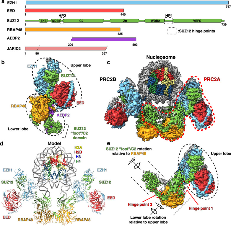
Fig. 1
Structures of monomeric and dimeric PRC2:EZH1.
a Cartoon representation of the proteins used to generate PRC2:EZH1 complexes. Hinge points (HP) 1 (aa 535u2013561) and 2 (aa 146u2013155) are regions in SUZ12 that appear to allow the upper and lower...
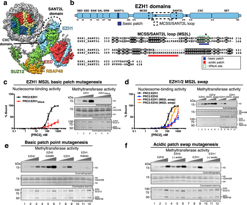
Fig. 2
The MCSS/SANT2L loop is important for PRC2 activity.
a Composite map of one PRC2:EZH1 bound to nucleosome with regions that likely interact with DNA indicated by dashed circles. b Upper panel: Cartoon representation of EZH1 showing basic patches. Lower ...
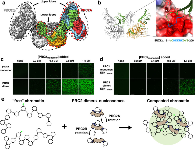
Fig. 3
PRC2 dimers promote chromatin compaction.
a Cryo-EM map showing the PRC2:EZH1 dimer. Subunits are colored as in Fig. 1 . One PRC2 monomer is highlighted in color and by a red dashed line. b , left panel: Isolated view of the dimeric interface...
Figure images are served from the NIH/NLM PubMed Central Open Access Subset or Europe PMC; copyright remains with the publishers and authors.
💬 Discussion
0 commentsNo comments yet. Be the first to start a discussion!
Leave a Comment