Abstract
AbstractWe report a lipid‐based strategy to visualize Golgi structure and dynamics at super‐resolution in live cells. The method is based on two novel reagents: a trans‐cyclooctene‐containing ceramide lipid (Cer‐TCO) and a highly reactive, tetrazine‐tagged near‐IR dye (SiR‐Tz). These reagents assemble via an extremely rapid “tetrazine‐click” reaction into Cer‐SiR, a highly photostable “vital dye” that enables prolonged live‐cell imaging of the Golgi apparatus by 3D confocal and STED microscopy. Cer‐SiR is nontoxic at concentrations as high as 2 μM and does not perturb the mobility of Golgi‐resident enzymes or the traffic of cargo from the endoplasmic reticulum through the Golgi and to the plasma membrane.
🔬 Techniques
💻 Software
✨ Fluorophores
🧪 Sample Preparation
🔬 Cell Lines
🏭 Microscope Brands
🧪 Reagent Suppliers
💻 Software Details
🏛️ Research Organizations (ROR)
Affiliated research institutions:
📊 Figures
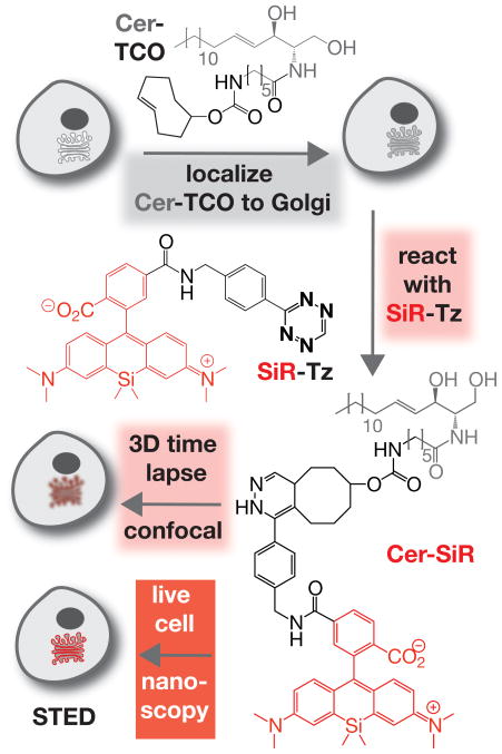
Figure 1
Two-step procedure for high-density labeling of the Golgi in living cells. Cells are treated first with Cer-TCO, a trans -cyclooctene-containing ceramide lipid, and then reacted with SiR-Tz, a tetrazi...
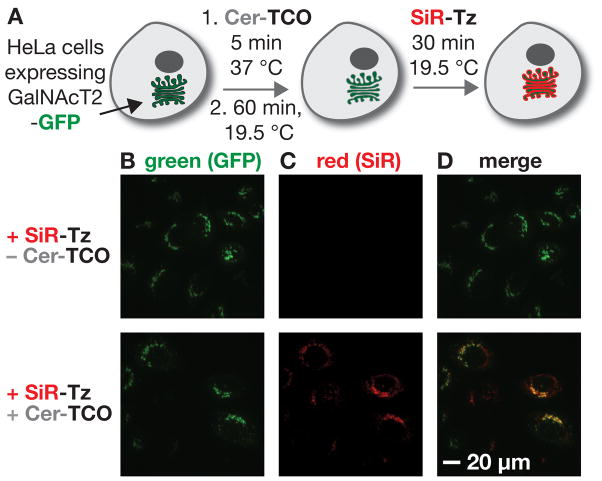
Figure 2
Cer-TCO localizes and reacts with SiR-Tz to visualize the Golgi in live cells. a) HeLa cells expressing the Golgi reporter protein GalNAcT2-GFP were treated with Cer-TCO (2 u03bcM), subjected to a tem...
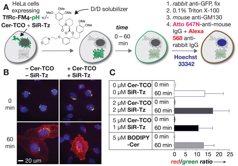
Figure 3
a) Endpoint trafficking assay using TfRc-F M 4-pH and u2018D/Du2019 solubilizer to distinguish between cargo that reached the plasma membrane or remained inside the cell. b) HeLa cells expressing TfRc...
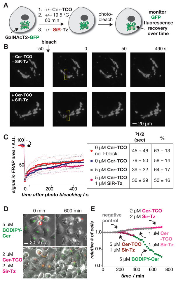
Figure 4
Intra-Golgi trafficking is unaffected by Cer-TCO and SiR-Tz. a) Cells expressing GalNAcT2-GFP (green) are labeled with Cer-TCO and SiR-Tz, photobleached, and the fluorescence recovery after photobleac...
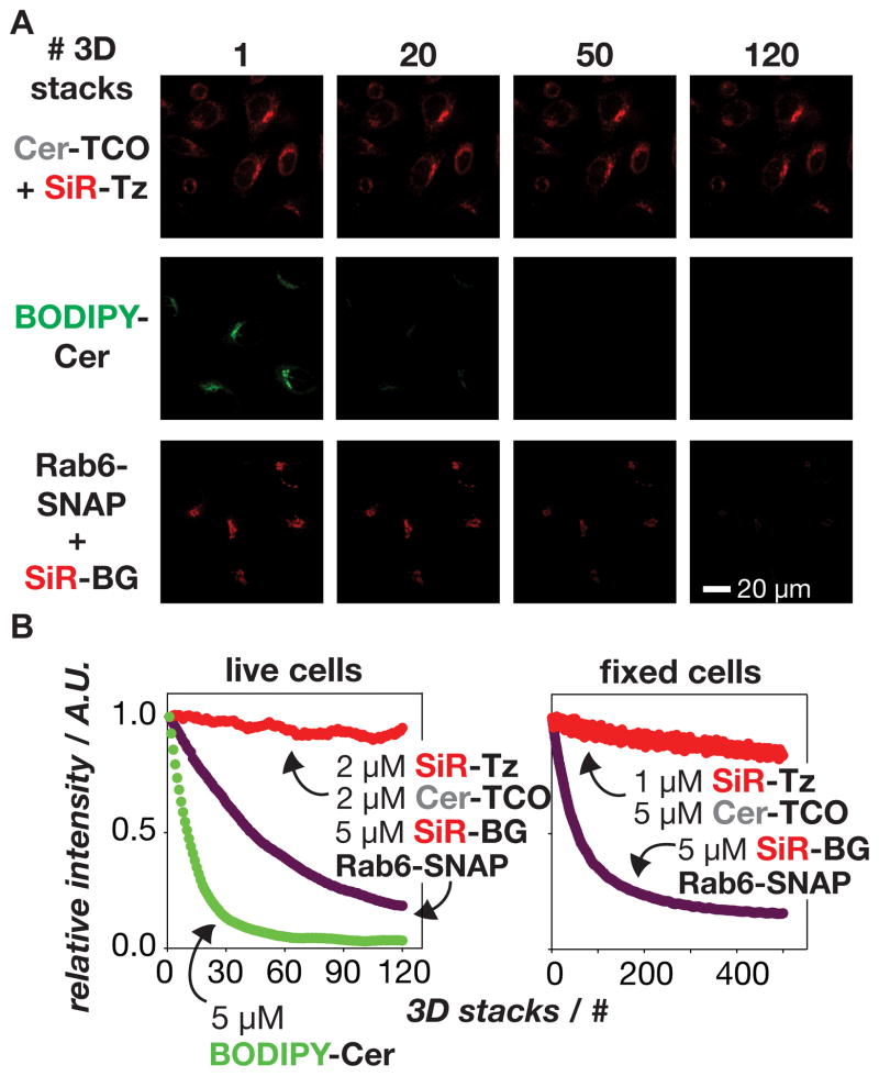
Figure 5
Golgi labeled with the lipid Cer-SiR is extremely stable to prolonged illumination using spinning disk confocal microscopy. a) Images show cells labeled with Cer-SiR (Cer-TCO+SiR-Tz), BODIPY-Cer, or t...
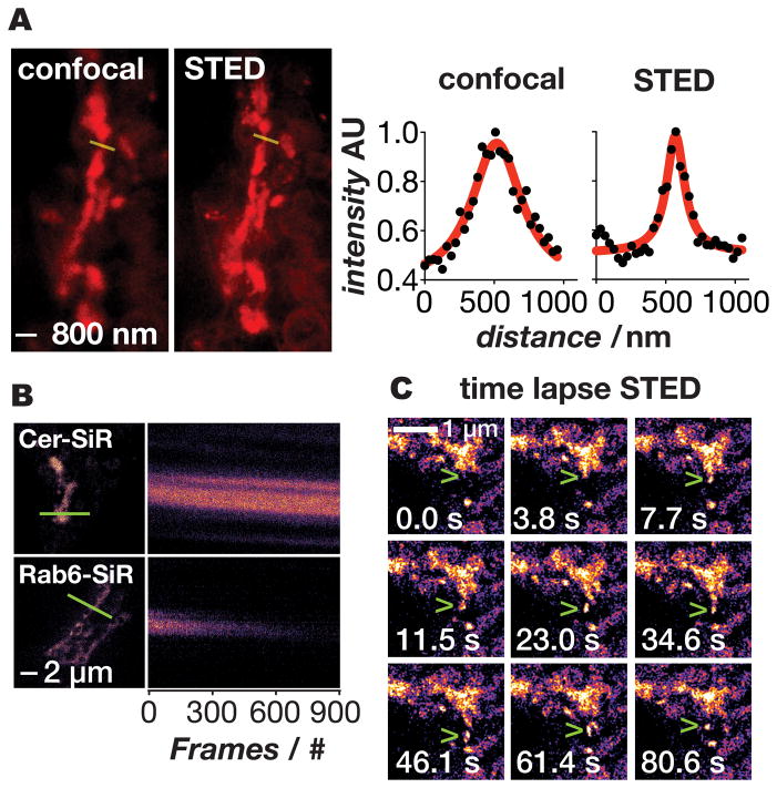
Figure 6
Super-resolution imaging of the Golgi in live cells using Cer-SiR. a) Confocal and STED images of the Golgi in live cells treated with Cer-TCO and SiR-Tz. Line traces through the Golgi (yellow) show t...
Figure images are served from the NIH/NLM PubMed Central Open Access Subset or Europe PMC; copyright remains with the publishers and authors.
💬 Discussion
0 commentsNo comments yet. Be the first to start a discussion!
Leave a Comment