Abstract
AbstractThe rhodamine system is a flexible framework for building small‐molecule fluorescent probes. Changing N‐substitution patterns and replacing the xanthene oxygen with a dimethylsilicon moiety can shift the absorption and fluorescence emission maxima of rhodamine dyes to longer wavelengths. Acylation of the rhodamine nitrogen atoms forces the molecule to adopt a nonfluorescent lactone form, providing a convenient method to make fluorogenic compounds. Herein, we take advantage of all of these structural manipulations and describe a novel photoactivatable fluorophore based on a Si‐containing analogue of Q‐rhodamine. This probe is the first example of a “caged” Si‐rhodamine, exhibits higher photon counts compared to established localization microscopy dyes, and is sufficiently red‐shifted to allow multicolor imaging. The dye is a useful label for super‐resolution imaging and constitutes a new scaffold for far‐red fluorogenic molecules.
🔬 Techniques
✨ Fluorophores
🧪 Sample Preparation
🔬 Cell Lines
🏛️ Research Organizations (ROR)
Affiliated research institutions:
📊 Figures
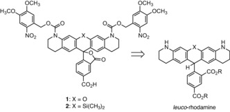
Scheme 1
Structural formula and retrosynthesis of photoactivatable (u201ccagedu201d) Rh Q 1 and SiRh Q 2 .
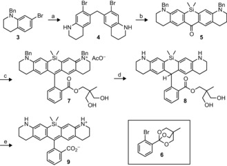
Scheme 2
Synthesis of SiRh Q ( 9 ). a)u2005H 2 CO, AcOH, 60u2009u00b0C, 77u2009%. b)u2005i. sec u2010BuLi, THF, u221278u2009u00b0C, (CH 3 ) 2 SiCl 2 . ii. KMnO 4 , acetone, u221215u2009u00b0C, 37u2009%, (two s...
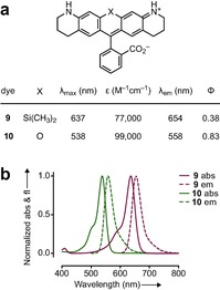
Figure 1
a)u2005Spectral properties for rhodamines 9 and 10 . b)u2005Absorption (abs) and fluorescence emission (em) spectra for rhodamines 9 and 10 . All measurements were taken in aqueous buffer (10u2005m m ...
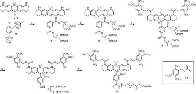
Scheme 3
Synthesis of 5u2010carboxyu2010NVOC 2 u2010SiRh Q ( 2 ) and phalloidin conjugate 17 . a)u2005i. t BuLi, THF, u221278u2009u00b0C, 1 n HCl. ii. MOMCl, DIEA, CH 2 Cl 2 , 82u2009% (two steps). b)u2005NH 4...
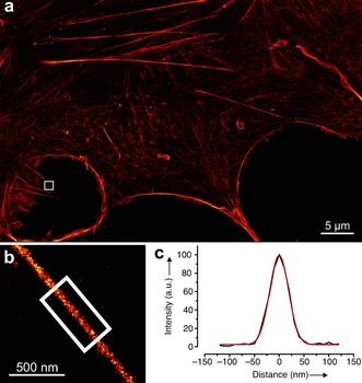
Figure 2
a)u2005Localization microscopy image (HILO) of COSu20107 cell stained with phalloidin conjugate 17 ; scale bar: 5u2005u03bcm. b)u2005Expanded image of boxed region inu2005(a) showing a protruding filo...
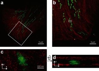
Figure 3
a)u2005Two color iPALM image of a fixed U2OS cell labeled with phalloidin conjugate 17 (actin, red) and mEos2 (mitochondria, green). b)u2005Enlarged image of boxed area inu2005(a). c) x , y projection...
Figure images are served from the NIH/NLM PubMed Central Open Access Subset or Europe PMC; copyright remains with the publishers and authors.
💬 Discussion
0 commentsNo comments yet. Be the first to start a discussion!
Leave a Comment