Abstract
Significance The spike proteins of most current severe acute respiratory syndrome coronavirus 2 isolates contain a D614G substitution, by comparison with the spike protein of initial isolates. In this study we present high-resolution, single-particle cryo-electron microscopy structures of the G614 spike variant showing that it adopts a predominantly open conformation, unlike the D614 spike that is mostly closed. We conclude that the D614G substitution promotes “opening” of the spike, priming it for binding to the receptor ACE2 and possibly for its subsequent role in membrane fusion. The observed open conformation of the G614 spike may be the reason for the current virus’ reported increased infectivity and its current predominance.
🔬 Techniques
🔭 Microscopes
💻 Software
🧪 Sample Preparation
🏭 Microscope Brands
🧪 Reagent Suppliers
📷 Detectors
💻 Software Details
💾 Data Repositories
🏛️ Research Organizations (ROR)
Affiliated research institutions:
📋 Methods
Protein Production. The construct coding for the D614G mutant was based on the furin-uncleavable version of the SARS-CoV-2 spike protein ectodomain with a set of stabilizing mutations (R682S, R685S, K986P, and K987P) that we described before ( 5 ). The G614 spike protein was produced very similarly to the D614 spike we described before ( 5 ). Briefly, it was expressed in Expi293F cells (Gibco) growing in suspension at 37 °C in an 8% CO 2 atmosphere transfected with ExpiFectamine 293 (Gibco) and 1 mg of DNA per liter of culture. The enhancers were added 20 h after the transfection, according to the manufacturer’s instructions (Gibco), and the cells were then moved to 32 °C and the supernatant containing the protein was harvested on the fifth day posttransfection. The collected supernatant was clarified, bound overnight to TALON beads (Takara), briefly washed, eluted with 200 mM imidazole, concentrated, and gel-filtered on a Superdex 200 Increase 10/300 GL column (GE Life Sciences) into 150 mM NaCl and 20 mM Tris, pH 8. Cryo-EM Sample Preparation and Data Collection. The G614 spike glycoprotein was frozen on 200-mesh Quantifoil R2/2 grids glow-discharged for 30 s at 25 mA. Four microliters of spike protein at ∼0.5 mg/mL in 150 mM NaCl and 20 mM Tris, pH 8, supplemented with 0.1% octyl glucoside was applied on a grid at 4 °C, blotted for 4 to 4.5 s using a Vitrobot MkIII, and plunge-frozen into liquid ethane. Data were collected using a Titan Krios operating at 300 kV. Images were recorded using a Falcon III detector operating in electron counting mode. Images were recorded as a 40-s exposure, fractionated into 32 frames, with an accumulated dose of 36.8 e/Å 2 . The calibrated pixel size was 0.85 Å and images were collected at various defoci between 1.0 and 3.0 µm. Cryo-EM Data Processing. The frames of the collected movies were aligned using MotionCor2 ( 17 ) implemented in RELION ( 18 ), and the Contrast Transfer Function (CTF) was fitted using CTFfind4 ( 19 ). Particles were picked using crYOLO ( 20 ) using a model trained on manually picked micrographs. Picked particles were subjected to two rounds of two-dimensional classification in cryoSPARC ( 21 ), retaining classes with clear secondary structure. An ab initio three-dimensional (3D) model was generated using cryoSPARC, which was used as an initial model for 3D classification in RELION, separating into 10 classes. Particles in classes which pertained to the closed form, one erect RBD form, and two erect RBDs form were refined using RELION 3D-Autorefine, followed by Bayesian polishing ( 22 ). The final refinements were carried out in cryoSPARC using the homogeneous refinement protocol, coupled to CTF refinement. C3 symmetry was imposed on the closed form. The final maps had local resolution estimated using blocres ( 23 ) implemented in cryoSPARC, followed by local resolution filtering and global B-factor sharpening ( 24 ) in cryoSPARC. The image processing workflow is summarized in SI Appendix , Fig. 3 . Model Building. Models were built based on our previously published structures for the closed wild-type SARS-CoV-2 spike (Protein Data Bank [PDB] ID code 6ZGE) ( 5 ), one erect RBD (PDB ID code 6ZGG) ( 5 ), and two erect RBDs (PDB ID code 7A93) ( 6 ). Models were fitted into the density and manually adjusted using Coot ( 25 ). The closed structure had an S1 structure with large deviations in the positioning of the S1 subdomains. The model was initially built by rigid body refinement in PHENIX ( 26 ), followed by adjustment in Coot. All models were real-space-refined and validated using PHENIX ( Table 1 ). Accuracy of model building for previously deposited D614 structures was compared by measuring Q-scores using the UCSF Chimera plugin ( 27 , 28 ).
Show full methods section
Protein Production. The construct coding for the D614G mutant was based on the furin-uncleavable version of the SARS-CoV-2 spike protein ectodomain with a set of stabilizing mutations (R682S, R685S, K986P, and K987P) that we described before ( 5 ). The G614 spike protein was produced very similarly to the D614 spike we described before ( 5 ). Briefly, it was expressed in Expi293F cells (Gibco) growing in suspension at 37 °C in an 8% CO 2 atmosphere transfected with ExpiFectamine 293 (Gibco) and 1 mg of DNA per liter of culture. The enhancers were added 20 h after the transfection, according to the manufacturer’s instructions (Gibco), and the cells were then moved to 32 °C and the supernatant containing the protein was harvested on the fifth day posttransfection. The collected supernatant was clarified, bound overnight to TALON beads (Takara), briefly washed, eluted with 200 mM imidazole, concentrated, and gel-filtered on a Superdex 200 Increase 10/300 GL column (GE Life Sciences) into 150 mM NaCl and 20 mM Tris, pH 8. Cryo-EM Sample Preparation and Data Collection. The G614 spike glycoprotein was frozen on 200-mesh Quantifoil R2/2 grids glow-discharged for 30 s at 25 mA. Four microliters of spike protein at ∼0.5 mg/mL in 150 mM NaCl and 20 mM Tris, pH 8, supplemented with 0.1% octyl glucoside was applied on a grid at 4 °C, blotted for 4 to 4.5 s using a Vitrobot MkIII, and plunge-frozen into liquid ethane. Data were collected using a Titan Krios operating at 300 kV. Images were recorded using a Falcon III detector operating in electron counting mode. Images were recorded as a 40-s exposure, fractionated into 32 frames, with an accumulated dose of 36.8 e/Å 2 . The calibrated pixel size was 0.85 Å and images were collected at various defoci between 1.0 and 3.0 µm. Cryo-EM Data Processing. The frames of the collected movies were aligned using MotionCor2 ( 17 ) implemented in RELION ( 18 ), and the Contrast Transfer Function (CTF) was fitted using CTFfind4 ( 19 ). Particles were picked using crYOLO ( 20 ) using a model trained on manually picked micrographs. Picked particles were subjected to two rounds of two-dimensional classification in cryoSPARC ( 21 ), retaining classes with clear secondary structure. An ab initio three-dimensional (3D) model was generated using cryoSPARC, which was used as an initial model for 3D classification in RELION, separating into 10 classes. Particles in classes which pertained to the closed form, one erect RBD form, and two erect RBDs form were refined using RELION 3D-Autorefine, followed by Bayesian polishing ( 22 ). The final refinements were carried out in cryoSPARC using the homogeneous refinement protocol, coupled to CTF refinement. C3 symmetry was imposed on the closed form. The final maps had local resolution estimated using blocres ( 23 ) implemented in cryoSPARC, followed by local resolution filtering and global B-factor sharpening ( 24 ) in cryoSPARC. The image processing workflow is summarized in SI Appendix , Fig. 3 . Model Building. Models were built based on our previously published structures for the closed wild-type SARS-CoV-2 spike (Protein Data Bank [PDB] ID code 6ZGE) ( 5 ), one erect RBD (PDB ID code 6ZGG) ( 5 ), and two erect RBDs (PDB ID code 7A93) ( 6 ). Models were fitted into the density and manually adjusted using Coot ( 25 ). The closed structure had an S1 structure with large deviations in the positioning of the S1 subdomains. The model was initially built by rigid body refinement in PHENIX ( 26 ), followed by adjustment in Coot. All models were real-space-refined and validated using PHENIX ( Table 1 ). Accuracy of model building for previously deposited D614 structures was compared by measuring Q-scores using the UCSF Chimera plugin ( 27 , 28 ).
📊 Figures
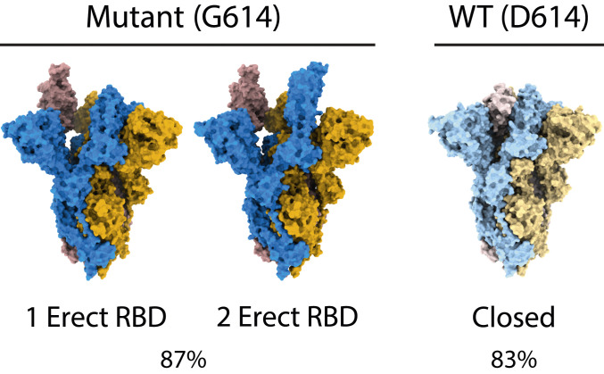
Fig. 1.
Space-filling representation of the two predominant open forms of G614 spike and the predominant closed form of D614 spike. The one- and two-RBD-erect forms of G614 spike make up 87% of particles, whi...
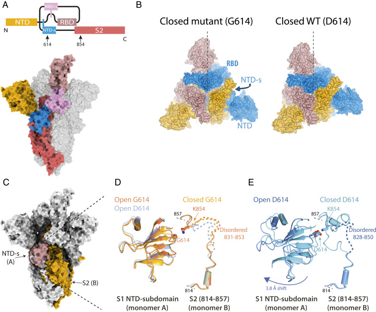
Fig. 2.
Structural differences between the G614 and D614 spike. ( A ) Bar diagram, showing domain connectivity of spike and locations of residues 614 and 854 ( Top ), with domains colored on single monomer of...
Figure images are served from the NIH/NLM PubMed Central Open Access Subset or Europe PMC; copyright remains with the publishers and authors.
💬 Discussion
0 commentsNo comments yet. Be the first to start a discussion!
Leave a Comment