Abstract
Interaction between the nascent polypeptide chain and the ribosomal exit tunnel can modulate the rate of translation and induce translational arrest to regulate expression of downstream genes. The ribosomal tunnel also provides a protected environment for initial protein folding events. Here, we present a 2.9 Å cryo-electron microscopy structure of a ribosome stalled during translation of the extremely compacted VemP nascent chain. The nascent chain forms two α-helices connected by an α-turn and a loop, enabling a total of 37 amino acids to be observed within the first 50-55 Å of the exit tunnel. The structure reveals how α-helix formation directly within the peptidyltransferase center of the ribosome interferes with aminoacyl-tRNA accommodation, suggesting that during canonical translation, a major role of the exit tunnel is to prevent excessive secondary structure formation that can interfere with the peptidyltransferase activity of the ribosome.
🔬 Techniques
🧬 Organisms
🧪 Sample Preparation
🏭 Microscope Brands
🧪 Reagent Suppliers
💻 Software Details
📋 Protocols
💾 Data Repositories
🏛️ Research Organizations (ROR)
Affiliated research institutions:
📋 Methods
DNA manipulations To test the effect of adding the extra C-terminal VemP sequence to the identified 19aa stalling window ( Ishii et al., 2015 ), the VemP ‘short’ plasmid was constructed from a previously designed pING1 plasmid carrying a lepB -based construct harboring a SecM arrest peptide ( Ismail et al., 2012 ). In order to produce a soluble, non-membrane targeted variant, transmembrane helix 1 and 2 of Lep were deleted using PCR. To replace the SecM arrest peptide with the VemP arrest sequence, the plasmid was amplified using a primer pair producing a linear plasmid lacking the SecM arrest peptide. Gibson assembly ( Gibson et al., 2009 ) was then used to ligate synthesized oligonucleotides corresponding to the VemP arrest peptide (in bold) and its following three residues, HRISGWKETNAMYVALNSQ FSA, into the plasmid. Overlap PCR ( Liu and Naismith, 2008 ) was used to replace the seven Lep residues just upstream of the VemP ‘short’ arrest peptide with the corresponding seven residues from the native VemP and by GSGSGSG to generate VemP ‘long’, FYHFTSDHRISGWKETNAMYVALNSQFSA, and VemP ‘GS’, GSGSGSGHRISGWKETNAMYVALNSQFSA, respectively. Overlap PCR was also used to generate other variants of VemP used in pulse-labelling analysis. For in vitro translation and cryo-grid sample preparation, the full VemP gene without the signal sequence (∆SS-VemP, A26-A159) was cloned from synthesized V. alginolyticus genome by KOD XtremeTM Hot-Start DNA Polymerase (Novagen, MA, USA), and subsequently inserted to modified plasmid p7XNH3 by FX cloning method ( Geertsma, 2014 ). The complete construct contains an N-terminal His-tag for purification, a FLAG-tag for detection and a TEV-protease cleavage site as a linker sequence. The C-terminus following the ∆SS-VemP comprises a spacer containing a 3C-protease cleavage site and an HA-tag. MPWIYLRKLILLLFAMVLLPVHVSAAQIDHKAHVPHFSKLQPFVAVSVSPNSSVDFSEASEESSQSPVSEGHASLDSVALFNSQRWTSYLREGLDDEHVDFVGDLTTPFYADAGYAYSLMDINWRHNQSTFYHFTSDHRISGWKETNAMYVALNSQFSA(TAA-Stop x2) is the total amino acid sequence of the construct. In vivo pulse-labelling E. coli MC1061 carrying plasmids with the different lepB -based constructs described in the paper were cultured overnight at 37°C in M9 minimal medium supplemented with 19 natural amino acids (1 μg/ml; no methionine, 100 μg/ml thiamine, 0.1 mM CaCl 2 , 2 mM MgSO 4 , 0.4% (w/v) fructose, and 100 μg/ml ampicillin). Overnight cultures were back-diluted to OD 600 ~0.05–0.1 and grown 2.5 to 3.5 hr to an OD 600 of 0.2–0.35. Protein expression was induced with 0.2% (w/v) arabinose for 5 min, whereafter expressed proteins were radiolabelled with [ 35 S]methionine for 2 min at 37°C. Ice-cold trichloroacetic acid (TCA) was added to a final concentration of 10% and the samples were incubated on ice for 30 min. The samples were centrifuged for 5 min at 20,800 x g at 4°C and the precipitates were washed with cold acetone, and spun again for 5 min at 20,800 x g at 4°C. The precipitates were resolubilized in Tris-SDS solution (10 mM Tris-HCl, pH 7.5, and 2% SDS) at 95°C for 10 min and spun for 5 min at room temperature. The protein of interest was then immunoprecipitated using anti-LepB antibody and the resulting samples were prepared for SDS-PAGE analysis with Laemmli sample buffer supplemented with 400 μg/ml RNase A. Gels were analyzed with a Fuji FLA-3000 phosphorimager and ImageGauge V4.23 software. Quantification of protein bands was performed using EasyQuant (in-house developed software). The fraction of full-length values (fFL) was calculated using the formula fFL = IFL / (IFL+IA), where IFL is the quantified intensity of the full-length protein band and IA is the quantified intensity of the arrested protein band. Experiments were repeated in triplicate and standard errors of the mean were calculated. In vitro transcription and translation For the RNase treatment assay and the final large scale purification for cryo-grid, RTS 100 E.coli HY Kit (5 PRIME) was used (transcription and translation coupled). 500 μL reaction was incubated at 30°C for 35 min with 12.6 pmol PCR-product template generated upon the ∆SS-VemP construct. RNase was added to the reaction where indicated, and incubated at 30°C for an additional 10 min. For the time course, the same amount of PCR-product template was used with the PURExpress In Vitro Protein Synthesis Kit (New England Biolabs #E6800S, transcription and translation coupled). Reactions were incubated at 37°C for various times (25/40/55/70/85/100 min). For western blotting, reaction products were separated by either home-made or commercial NuPAGE 12% Bis-Tris gels (Invitrogen, CA, USA) with 1x MOPS buffer. Proteins were blotted to nitrocellulose membrane (Carl Roth, Germany), incubated with mouse anti FLAG M2 HRP (Sigma, Germany) and visualized by ChemiDoc MP System (Bio-Rad). Purification of the VemP-SRC 500 μL in vitro translation reaction was loaded onto 10–50% sucrose gradient prepared with Buffer C (25 mM pH 7.2 HEPES-KOH, 100 mM KOAc, 10 mM Mg(OAc) 2 , 0.01% DDM, 1/1,000 complete protease inhibitor (Roche, Germany), 0.2 U/mL RNase, 2 mM 2-mercaptoethanol) and centrifuged for 3 hr in a Beckman coulter SW40 Ti swinging bucket rotor with 35,000 r.p.m. at 4°C. Gradients were separated on a Biocomp Gradient Station and fractions containing 70S ribosomal particles were collected and loaded onto a Talon metal affinity chromatography column (1.5 ml resin) pre-equilibrated in 10 mL buffer C containing 10 μg/mL bulk tRNA. The column was washed with 25 ml buffer C until no significant absorption (OD 260 ) could be detected in the wash fractions. The VemP-SRC, bound to the Talon matrix by the VemP N-terminal His-tag, was eluted in 750 μL buffer C containing 150 mM imidazole. The elution was pelleted for 4 hr 20 min in a Beckman Coulter TLA 120.2 fixed-angle rotor with 51,000 r.p.m. at 4°C. 15.6 pmol VemP-SRC pellet was resuspended in ice-cold buffer C without DDM, aliquoted and snap-frozen.
Show full methods section
DNA manipulations To test the effect of adding the extra C-terminal VemP sequence to the identified 19aa stalling window ( Ishii et al., 2015 ), the VemP ‘short’ plasmid was constructed from a previously designed pING1 plasmid carrying a lepB -based construct harboring a SecM arrest peptide ( Ismail et al., 2012 ). In order to produce a soluble, non-membrane targeted variant, transmembrane helix 1 and 2 of Lep were deleted using PCR. To replace the SecM arrest peptide with the VemP arrest sequence, the plasmid was amplified using a primer pair producing a linear plasmid lacking the SecM arrest peptide. Gibson assembly ( Gibson et al., 2009 ) was then used to ligate synthesized oligonucleotides corresponding to the VemP arrest peptide (in bold) and its following three residues, HRISGWKETNAMYVALNSQ FSA, into the plasmid. Overlap PCR ( Liu and Naismith, 2008 ) was used to replace the seven Lep residues just upstream of the VemP ‘short’ arrest peptide with the corresponding seven residues from the native VemP and by GSGSGSG to generate VemP ‘long’, FYHFTSDHRISGWKETNAMYVALNSQFSA, and VemP ‘GS’, GSGSGSGHRISGWKETNAMYVALNSQFSA, respectively. Overlap PCR was also used to generate other variants of VemP used in pulse-labelling analysis. For in vitro translation and cryo-grid sample preparation, the full VemP gene without the signal sequence (∆SS-VemP, A26-A159) was cloned from synthesized V. alginolyticus genome by KOD XtremeTM Hot-Start DNA Polymerase (Novagen, MA, USA), and subsequently inserted to modified plasmid p7XNH3 by FX cloning method ( Geertsma, 2014 ). The complete construct contains an N-terminal His-tag for purification, a FLAG-tag for detection and a TEV-protease cleavage site as a linker sequence. The C-terminus following the ∆SS-VemP comprises a spacer containing a 3C-protease cleavage site and an HA-tag. MPWIYLRKLILLLFAMVLLPVHVSAAQIDHKAHVPHFSKLQPFVAVSVSPNSSVDFSEASEESSQSPVSEGHASLDSVALFNSQRWTSYLREGLDDEHVDFVGDLTTPFYADAGYAYSLMDINWRHNQSTFYHFTSDHRISGWKETNAMYVALNSQFSA(TAA-Stop x2) is the total amino acid sequence of the construct. In vivo pulse-labelling E. coli MC1061 carrying plasmids with the different lepB -based constructs described in the paper were cultured overnight at 37°C in M9 minimal medium supplemented with 19 natural amino acids (1 μg/ml; no methionine, 100 μg/ml thiamine, 0.1 mM CaCl 2 , 2 mM MgSO 4 , 0.4% (w/v) fructose, and 100 μg/ml ampicillin). Overnight cultures were back-diluted to OD 600 ~0.05–0.1 and grown 2.5 to 3.5 hr to an OD 600 of 0.2–0.35. Protein expression was induced with 0.2% (w/v) arabinose for 5 min, whereafter expressed proteins were radiolabelled with [ 35 S]methionine for 2 min at 37°C. Ice-cold trichloroacetic acid (TCA) was added to a final concentration of 10% and the samples were incubated on ice for 30 min. The samples were centrifuged for 5 min at 20,800 x g at 4°C and the precipitates were washed with cold acetone, and spun again for 5 min at 20,800 x g at 4°C. The precipitates were resolubilized in Tris-SDS solution (10 mM Tris-HCl, pH 7.5, and 2% SDS) at 95°C for 10 min and spun for 5 min at room temperature. The protein of interest was then immunoprecipitated using anti-LepB antibody and the resulting samples were prepared for SDS-PAGE analysis with Laemmli sample buffer supplemented with 400 μg/ml RNase A. Gels were analyzed with a Fuji FLA-3000 phosphorimager and ImageGauge V4.23 software. Quantification of protein bands was performed using EasyQuant (in-house developed software). The fraction of full-length values (fFL) was calculated using the formula fFL = IFL / (IFL+IA), where IFL is the quantified intensity of the full-length protein band and IA is the quantified intensity of the arrested protein band. Experiments were repeated in triplicate and standard errors of the mean were calculated. In vitro transcription and translation For the RNase treatment assay and the final large scale purification for cryo-grid, RTS 100 E.coli HY Kit (5 PRIME) was used (transcription and translation coupled). 500 μL reaction was incubated at 30°C for 35 min with 12.6 pmol PCR-product template generated upon the ∆SS-VemP construct. RNase was added to the reaction where indicated, and incubated at 30°C for an additional 10 min. For the time course, the same amount of PCR-product template was used with the PURExpress In Vitro Protein Synthesis Kit (New England Biolabs #E6800S, transcription and translation coupled). Reactions were incubated at 37°C for various times (25/40/55/70/85/100 min). For western blotting, reaction products were separated by either home-made or commercial NuPAGE 12% Bis-Tris gels (Invitrogen, CA, USA) with 1x MOPS buffer. Proteins were blotted to nitrocellulose membrane (Carl Roth, Germany), incubated with mouse anti FLAG M2 HRP (Sigma, Germany) and visualized by ChemiDoc MP System (Bio-Rad). Purification of the VemP-SRC 500 μL in vitro translation reaction was loaded onto 10–50% sucrose gradient prepared with Buffer C (25 mM pH 7.2 HEPES-KOH, 100 mM KOAc, 10 mM Mg(OAc) 2 , 0.01% DDM, 1/1,000 complete protease inhibitor (Roche, Germany), 0.2 U/mL RNase, 2 mM 2-mercaptoethanol) and centrifuged for 3 hr in a Beckman coulter SW40 Ti swinging bucket rotor with 35,000 r.p.m. at 4°C. Gradients were separated on a Biocomp Gradient Station and fractions containing 70S ribosomal particles were collected and loaded onto a Talon metal affinity chromatography column (1.5 ml resin) pre-equilibrated in 10 mL buffer C containing 10 μg/mL bulk tRNA. The column was washed with 25 ml buffer C until no significant absorption (OD 260 ) could be detected in the wash fractions. The VemP-SRC, bound to the Talon matrix by the VemP N-terminal His-tag, was eluted in 750 μL buffer C containing 150 mM imidazole. The elution was pelleted for 4 hr 20 min in a Beckman Coulter TLA 120.2 fixed-angle rotor with 51,000 r.p.m. at 4°C. 15.6 pmol VemP-SRC pellet was resuspended in ice-cold buffer C without DDM, aliquoted and snap-frozen.
Cryo-electron microscopy and single particle reconstruction
E. coli VemP-SRC (6 OD 260 /mL) was applied to 2 nm pre-coated Quantifoil R3/3 holey carbon supported grids and vitrified using Vitrobot Mark IV (FEI). Data collection was performed using EM-TOOLS (TVIPS GmbH) on a Titan Krios transmission electron microscope equipped with a Falcon II direct electron detector (FEI) at 300kV at a pixel size of 1.084 Å and a defocus range of 1–2.5 μm. 10 frames (dose per frame of 2.5 e - /Å 2 ) were aligned using MotionCor2 ( Li et al., 2013 ). Power-spectra, defocus values and astigmatism were determined with CTFFIND4 software ( Rohou and Grigorieff, 2015 ). 5735 micrographs were manually inspected in real space and in the meantime filtered by threshold of resolution at 4.5 Å and astigmatism at 5% resulting in 4849 micrographs. 850,433 particles were automatically picked by Gautomatch ( http://www.mrc-lmb.cam.ac.uk/kzhang/ ) and single particles were processed using RELION 1.4 ( Scheres, 2012 ). After 2D classification, 789,006 particles were subjected to 3D refinement using E. coli 70S ribosome as reference structure, followed by several further rounds of 3D classifications, including tRNA-focused sorting (64 rounds) and sorting (58 rounds) with a ribosome mask ( Figure 1—figure supplement 2 ). One major class containing 400,024 particles (62%) was further refined, resulting in a final reconstruction with an average resolution of 2.9 Å (0.143 FSC) ( Figure 1—figure supplement 3 ). The map was subsequently B-factored by EMBfactor ( Fernández et al., 2008 ) with the FSC. Finally, the local resolution was calculated using ResMap ( Kucukelbir et al., 2014 ). Molecular modeling and refinement of the VemP-SRC The molecular model for the ribosomal proteins and rRNA of the 70S ribosome of the VemP-SRC was based on the molecular model from cryo-EM reconstruction of the E. coli 70S ribosome (PDB ID 5JU8) ( Arenz et al., 2016 ), except that the bL31 was based on the (PDB ID 5LZD) ( Fischer et al., 2016 ). The molecular model for Gln-tRNA was based on a crystal structure (PDB ID 1GSG) ( Rould et al., 1989 ). The molecular models were initially fitted as a rigid body into the cryo-EM density map of the corresponding stalled complex using UCSF Chimera ( Pettersen et al., 2004 ). The complete atomic model of the VemP-SRC was manually adjusted using Coot ( Emsley and Cowtan, 2004 ) and refined using phenix.real_space_refine with restraints obtained by phenix.secondary_structure_restraints ( Adams et al., 2010 ). The Phenix refined model was further refined using REFMAC ( Vagin et al., 2004 ) to validate the overfitting as previously described ( Brown et al., 2015 ) ( Figure 1—figure supplement 3 ). The statistics of the refined model were calculated using Molprobity ( Chen et al., 2010 ) are presented in Table 1 .
Figure preparation
The protein secondary structure prediction of the native VemP sequence was performed using the MPI bioinformatics Toolkit ( Alva et al., 2016 ) with the prediction method written by B. Rost ( Rost, 2001 ). Figures showing electron densities and atomic models were generated using either UCSF Chimera ( Pettersen et al., 2004 ) or PyMol Molecular Graphics Systems (Version 1.8 Schrödinger, LLC,). Accession codes The cryo-electron microscopy map for the VemP-SRC has been deposited in the EMDataBank with the accession code EMD-3713. The respective coordinates for electron-microscopy-based model of the VemP-SRC are deposited in the ProteinDataBank (5NWY).
Additional files Major datasets
The following dataset was generated: Su T , Cheng J , Sohmen D , Hedman R , Berninghausen O , von Heijne G , Wilson DN , Beckmann R , 2017 , 2.9 A cryo-EM structure of VemP-stalled ribosome-nascent chain complex , http://www.rcsb.org/pdb/explore/explore.do?structureId=5NWY , Publicly available at the RCSBProtein Data Bank (accession no: 5NWY)
📊 Figures
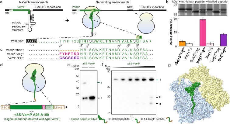
Figure 1.
Biochemical and structural analysis of VemP stalling.
( a ) Schematic representation of the VemP-SecDF2 mRNA encoding VemP leader peptide with N-terminal signal sequence (SS) and C-terminal stalling region (green), followed by a stem-loop structure that ...
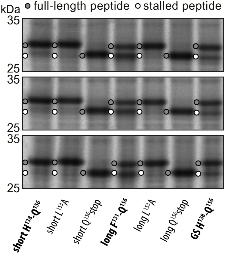
Figure 1u2014figure supplement 1.
Triplicates of the pulse-chase analysis.
In vivo pulse-chase analysis with different VemP constructs; VemP u2018shortu2019 (H138u2013Q156), VemP u2018longu2019 (F131u2013Q156), VemP u2018GSu2019 and VemP mutants L153A and Q156*. The triplica...
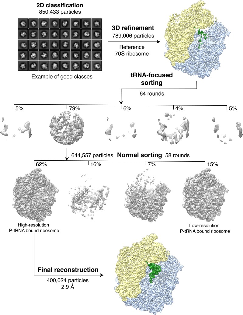
Figure 1u2014figure supplement 2.
Classification of the VemP-SRC.
A total of 850,433 extracted particles were subjected to 2D classification with 8 x binned images, 200 initial classes and 100 rounds in order to filter away non-particle contaminants. A subset of 789...
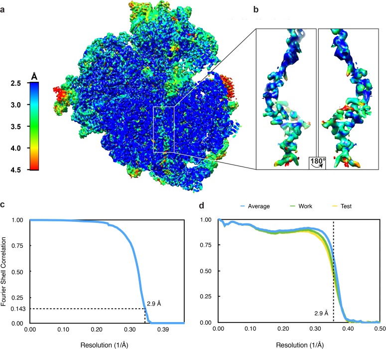
Figure 1u2014figure supplement 3.
Resolution of the VemP-SRC.
( a ) Transverse section of the VemP-SRC showing local resolution of the VemP nascent chain within the ribosomal tunnel. ( b ) Zoomed-in view of the extracted VemP nascent chain coloured according to ...
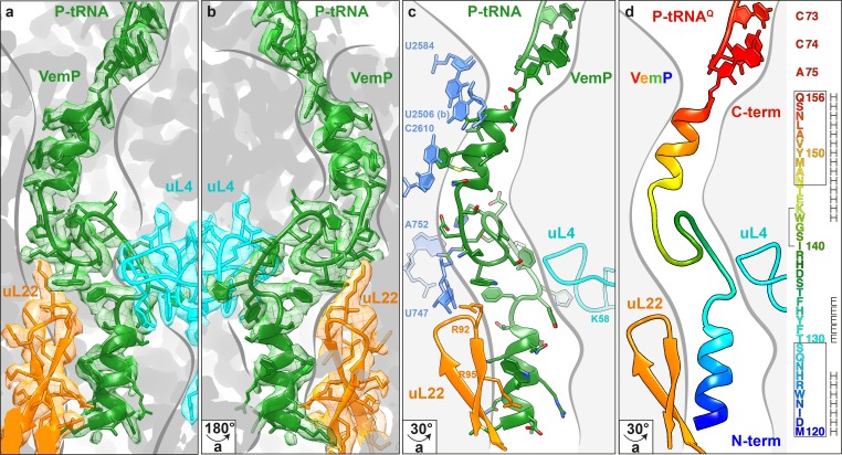
Figure 2.
Overview of the VemP nascent chain in the ribosomal tunnel.
( a u2013 b ) Isolated electron density and molecular model for the VemP nascent chain connected to the P-tRNA (green) in the ribosomal tunnel (grey) with ribosomal proteins uL4 (cyan) and uL22 (orang...
Video 1.
Cryo-EM density and model for the VemP nascent chain.
Video showing the quality of the cryo-EM density (green mesh) and fit of the molecular model (green ribbon) of the VemP nascent chain and CCA-end of the P-tRNA. DOI: http://dx.doi.org/10.7554/eLife.25...
Video 2.
Cryo-EM density and model for u03b1-turn and loop of the VemP nascent chain.
Video showing a zoom of the cryo-EM density (green mesh) and fit of the molecular model (stick representation) of the u03b1-turn and loop of the VemP nascent chain. DOI: http://dx.doi.org/10.7554/eLif...
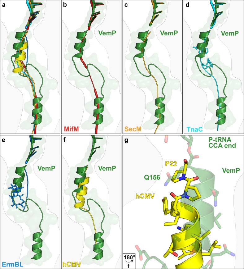
Figure 3.
Comparison of the VemP nascent chain in the ribosomal tunnel with other stalling peptides.
( a ) Overall superposition of VemP (model in green, surface in light green) with MifM (red, PDB ID 3J9W) ( Sohmen et al., 2015 ), SecM (orange, PDB ID 3JBU) ( Zhang et al., 2015 ), hCMV (yellow, PDB ...
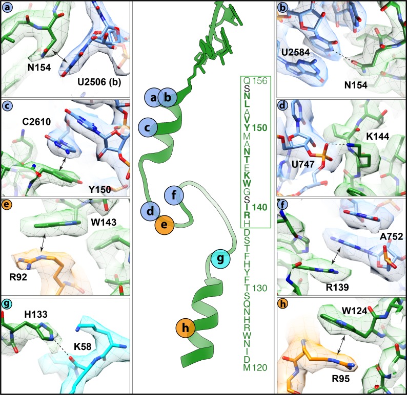
Figure 4.
Interactions of the VemP nascent chain with the ribosomal tunnel (with density shown).
( a ) N154 of VemP (green) stacks upon U2506(b) of the 23S rRNA (blue). ( b ) N154 of VemP is within hydrogen bond distance of nucleotide U2584. ( c ) Y150 of VemP stacks upon C2610. ( d ) K144 of Vem...
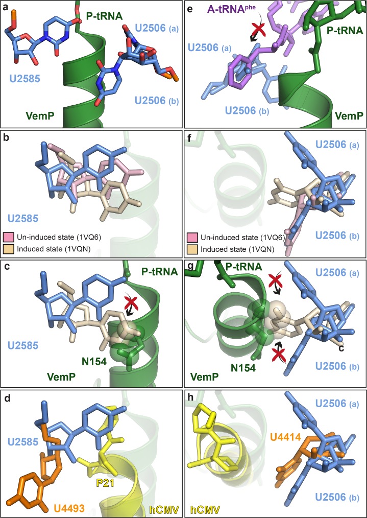
Figure 5.
VemP stabilizes the uninduced state of the PTC to inhibit A-tRNA accommodation.
( a ) Conformation of U2585 and U2506 relative to VemP (green) at the PTC of the VemP-SRC. ( b ) Conformation of U2585 (blue) in the VemP-SRC compared with the uninduced (pink, PDB ID 1VQ6) and induce...
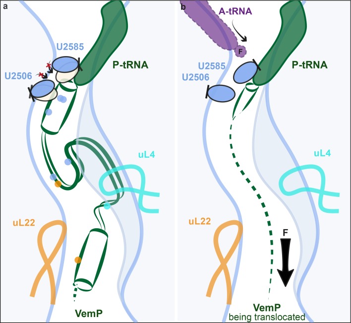
Figure 6.
Model for VemP-mediated translation stalling and relief.
( a u2013 b ) Schematic summarizing the molecular basis for ( a ) VemP-dependent translation arrest and ( b ) relief of arrest via the pulling-force (indicated by arrow) on VemP protein translocation....
Figure images are served from the NIH/NLM PubMed Central Open Access Subset or Europe PMC; copyright remains with the publishers and authors.
💬 Discussion
0 commentsNo comments yet. Be the first to start a discussion!
Leave a Comment