Abstract
The conversion of multinucleate postmitotic muscle fibers to dividing mononucleate progeny cells (cellularisation) occurs during limb regeneration in salamanders, but the cellular events and molecular regulation underlying this remarkable process are not understood. The homeobox gene Msx1 has been studied as an antagonist of muscle differentiation, and its expression in cultured mouse myotubes induces about 5% of the cells to undergo cellularisation and viable fragmentation, but its relevance for the endogenous programme of salamander regeneration is unknown. We dissociated muscle fibers from the limb of larval salamanders and plated them in culture. Most of the fibers were activated by dissociation to mobilise their nuclei and undergo cellularisation or breakage into viable multinucleate fragments. This was followed by microinjection of a lineage tracer into single fibers and analysis of the labelled progeny cells, as well as by time-lapse microscopy. The fibers showing morphological plasticity selectively expressed Msx1 mRNA and protein. The uptake of morpholino antisense oligonucleotides directed to Msx1 led to a specific decrease in expression of Msx1 protein in myonuclei and marked inhibition of cellularisation and fragmentation. Myofibers of the salamander respond to dissociation by activation of an endogenous programme of cellularisation and fragmentation. Lineage tracing demonstrates that cycling mononucleate progeny cells are derived from a single myofiber. The induction of Msx1 expression is required to activate this programme. Our understanding of the regulation of plasticity in postmitotic salamander cells should inform strategies to promote regeneration in other contexts.
🔬 Techniques
🔭 Microscopes
💻 Software
✨ Fluorophores
🧪 Sample Preparation
🏭 Microscope Brands
🧪 Reagent Suppliers
📷 Detectors
💻 Software Details
💾 Data Repositories
🏛️ Research Organizations (ROR)
Affiliated research institutions:
📋 Methods
Tissue dissociation and culture of myofibers
The forelimbs and hind limbs of the larval spotted salamander ( A. maculatum ) or axolotl ( A. mexicanum ) (3–5 cm size) were removed, the epidermis was peeled off, and the tissue was rinsed in serum-free amphibian MEM (AMEM) ( Ferretti and Brockes 1988 ) prior to dissociation for 3 h at 26 °C in PBS containing 0.15% collagenase (Type 1A, Sigma, St. Louis, Missouri, United States), 0.8% Dispase II (Roche, Basel, Switzerland), 0.15% crystalline bovine serum albumin, 0.3% D-glucose, and 0.15 mg/ml DNase I (Roche). After 30 min of incubation, the tissues were gently triturated through a fire-polished glass pasteur pipette to aid the detachment of myofibers from the bone. After incubation, the suspension was triturated several times, centrifuged at 400 rpm for 10 min, resuspended in AMEM, and filtered through a 35 μm sieve (VWR International, Poole, United Kingdom) to remove most of the mononucleate cells. The retentate was rinsed with AMEM and plated on 35 mm Falcon Primaria (Becton-Dickinson, Palo Alto, California, United States) tissue culture dishes. Cultures were maintained at 25 °C with 2.5% CO 2 in a humidified incubator as described elsewhere ( Ferretti and Brockes 1988 ). After attachment of the myofibers to the culture dish by overnight incubation, the culture media was replaced with serum-free AMEM or AMEM supplemented with 10% foetal bovine serum.
Labelling and implantation of myofibers
Myofibers were dissociated as above, retained in suspension in a sterile bacteriological dish (Bibby Sterilin, Stone, United Kingdom), and incubated with 10 μM CellTracker Orange CMTMR (Molecular Probes, Eugene, Oregon, United States) for 30 min at 25 °C. The labelling was terminated by addition of 10% AMEM, and the cells were incubated for 45 min at 25 °C to permit enzymatic activation of the dye. The cell suspension was diluted several fold to allow observation of myofibers at low density. The forelimbs of axolotl larvae (7–10 cm size) were amputated at mid humerus level under tricaine (0.1%) anaesthesia 48 h before injection of labelled myofibers (see Figure 2 A). The animals were anaesthetized, and the forelimbs were positioned under a stereo zoom microscope. The myofiber suspension was placed under inverted microscope, and the myofibers were drawn into a glass micropipette (30 μm tip diameter) using an oil-driven manual microinjector (Sutter Instruments, Novato, California, United States) mounted on a Narishige (Tokyo, Japan) MMO-1 micromanipulator. The skin was punctured with a tungsten needle in order to introduce the blunt end of the micropipette. Three to eight myofibers were picked, examined carefully to verify the absence of any adherent mononucleate cells, and injected into each limb regenerate. Contralateral limbs were mock injected with medium from the suspension. The regenerates were removed at 48 h and 96 h after injection, fixed in 4% paraformaldehyde (PFA), and processed ( Kumar et al. 2000 ). Serial longitudinal sections of 60 μm were cut on a cryostat (Leica, Solms, Germany), air dried, dehydrated in PBS, and counterstained with 2.5 μM Sytox Green (Molecular Probes). The sections were observed under epifluorescence on an Axiophot microscope (Zeiss, Jena, Germany). Microinjection of cultured myofibers with conjugated dextran Myofibers were incubated in AMEM containing 2,3-butanedionne monoxime (BDM) (4 mM) for 30 min to prevent contraction of the myofibers ( Bettencourt-Dias et al. 2003 ) and maintained in the same medium during microinjection. The culture dishes were placed under a Zeiss Axiovert microscope and microinjected with TR-conjugated dextran (TR–dextran, 70 kDa; Molecular Probes). The medium was changed immediately after injection and the cultures were returned to the incubator. To identify the labelled myofibers and their mononucleate progeny, cultures were counterstained in Syto13 (200 nM; Molecular Probes) live nucleic acid stain for 30 min and observed under fluorescence microscope with a dual band pass (FITC/TRITC) filter. Live imaging of myofiber plasticity To record the coordinates of the myofibers in culture, the dish was scored underneath with a scalpel, and cells in each grid square were observed daily and images were acquired with a CCD camera (Sony, Tokyo, Japan). For time-lapse microscopy, myofiber cultures were placed under an Axiovert microscope fitted with an incubation chamber maintained at 26 °C and 3% CO 2 , and phase contrast or variable relief contrast (VAREL) (Zeiss) images were acquired using a digital camera controlled through Image-Pro Plus software (Media Cybernetics, Silver Spring, Maryland, United States). A sequence gallery was created using Image Pro-Plus and images of interest were selected, digitally enhanced, and processed in Adobe Photoshop 6.0 (Adobe, San Jose, California, United States). [ 3 H]thymidine labelling. Myofibers were incubated in 1 μCi/ml [ 3 H] thymidine (Amersham Biosciences, Little Chalfont, United Kingdom) for 24 h, fixed in 1% glutaraldehyde, and processed for autoradiography ( Velloso et al. 2000 ).
Show full methods section
Tissue dissociation and culture of myofibers
The forelimbs and hind limbs of the larval spotted salamander ( A. maculatum ) or axolotl ( A. mexicanum ) (3–5 cm size) were removed, the epidermis was peeled off, and the tissue was rinsed in serum-free amphibian MEM (AMEM) ( Ferretti and Brockes 1988 ) prior to dissociation for 3 h at 26 °C in PBS containing 0.15% collagenase (Type 1A, Sigma, St. Louis, Missouri, United States), 0.8% Dispase II (Roche, Basel, Switzerland), 0.15% crystalline bovine serum albumin, 0.3% D-glucose, and 0.15 mg/ml DNase I (Roche). After 30 min of incubation, the tissues were gently triturated through a fire-polished glass pasteur pipette to aid the detachment of myofibers from the bone. After incubation, the suspension was triturated several times, centrifuged at 400 rpm for 10 min, resuspended in AMEM, and filtered through a 35 μm sieve (VWR International, Poole, United Kingdom) to remove most of the mononucleate cells. The retentate was rinsed with AMEM and plated on 35 mm Falcon Primaria (Becton-Dickinson, Palo Alto, California, United States) tissue culture dishes. Cultures were maintained at 25 °C with 2.5% CO 2 in a humidified incubator as described elsewhere ( Ferretti and Brockes 1988 ). After attachment of the myofibers to the culture dish by overnight incubation, the culture media was replaced with serum-free AMEM or AMEM supplemented with 10% foetal bovine serum.
Labelling and implantation of myofibers
Myofibers were dissociated as above, retained in suspension in a sterile bacteriological dish (Bibby Sterilin, Stone, United Kingdom), and incubated with 10 μM CellTracker Orange CMTMR (Molecular Probes, Eugene, Oregon, United States) for 30 min at 25 °C. The labelling was terminated by addition of 10% AMEM, and the cells were incubated for 45 min at 25 °C to permit enzymatic activation of the dye. The cell suspension was diluted several fold to allow observation of myofibers at low density. The forelimbs of axolotl larvae (7–10 cm size) were amputated at mid humerus level under tricaine (0.1%) anaesthesia 48 h before injection of labelled myofibers (see Figure 2 A). The animals were anaesthetized, and the forelimbs were positioned under a stereo zoom microscope. The myofiber suspension was placed under inverted microscope, and the myofibers were drawn into a glass micropipette (30 μm tip diameter) using an oil-driven manual microinjector (Sutter Instruments, Novato, California, United States) mounted on a Narishige (Tokyo, Japan) MMO-1 micromanipulator. The skin was punctured with a tungsten needle in order to introduce the blunt end of the micropipette. Three to eight myofibers were picked, examined carefully to verify the absence of any adherent mononucleate cells, and injected into each limb regenerate. Contralateral limbs were mock injected with medium from the suspension. The regenerates were removed at 48 h and 96 h after injection, fixed in 4% paraformaldehyde (PFA), and processed ( Kumar et al. 2000 ). Serial longitudinal sections of 60 μm were cut on a cryostat (Leica, Solms, Germany), air dried, dehydrated in PBS, and counterstained with 2.5 μM Sytox Green (Molecular Probes). The sections were observed under epifluorescence on an Axiophot microscope (Zeiss, Jena, Germany). Microinjection of cultured myofibers with conjugated dextran Myofibers were incubated in AMEM containing 2,3-butanedionne monoxime (BDM) (4 mM) for 30 min to prevent contraction of the myofibers ( Bettencourt-Dias et al. 2003 ) and maintained in the same medium during microinjection. The culture dishes were placed under a Zeiss Axiovert microscope and microinjected with TR-conjugated dextran (TR–dextran, 70 kDa; Molecular Probes). The medium was changed immediately after injection and the cultures were returned to the incubator. To identify the labelled myofibers and their mononucleate progeny, cultures were counterstained in Syto13 (200 nM; Molecular Probes) live nucleic acid stain for 30 min and observed under fluorescence microscope with a dual band pass (FITC/TRITC) filter. Live imaging of myofiber plasticity To record the coordinates of the myofibers in culture, the dish was scored underneath with a scalpel, and cells in each grid square were observed daily and images were acquired with a CCD camera (Sony, Tokyo, Japan). For time-lapse microscopy, myofiber cultures were placed under an Axiovert microscope fitted with an incubation chamber maintained at 26 °C and 3% CO 2 , and phase contrast or variable relief contrast (VAREL) (Zeiss) images were acquired using a digital camera controlled through Image-Pro Plus software (Media Cybernetics, Silver Spring, Maryland, United States). A sequence gallery was created using Image Pro-Plus and images of interest were selected, digitally enhanced, and processed in Adobe Photoshop 6.0 (Adobe, San Jose, California, United States). [ 3 H]thymidine labelling. Myofibers were incubated in 1 μCi/ml [ 3 H] thymidine (Amersham Biosciences, Little Chalfont, United Kingdom) for 24 h, fixed in 1% glutaraldehyde, and processed for autoradiography ( Velloso et al. 2000 ).
DNA cytometry
DNA content in myofiber nuclei and TR–dextran-labelled mononucleate progeny was measured quantitatively after fixation and staining of the nuclei with Hoechst 33258 (2 μg/ml; Sigma). Baseline values for nuclear DNA content in cycling axolotl mononucleate cells were measured in parallel after incorporation of 5-deoxy-2′-bromouridine (BrdU) (1 μM; see Tanaka et al. 1997 ). The BrdU-labelled cells were processed for double immunofluorescence with monoclonal antibody against BrdU and rabbit antibody to anti-phosphohistone H3 ( Velloso et al. 2000 ; Bettencourt-Dias et al. 2003 ). The nuclei were counterstained with Hoechst dye as above. All images were acquired using 12-bit cooled CCD camera (Photonic Sciences, Robertsbridge, United Kingdom), maintaining camera and microscope settings identical between various samples, corrected for uneven illumination and background using software functions, and processed using classification and measuring routines in Image-Pro Plus software. Scoring of plasticity in myofibers A viable nucleic acid stain such as Syto 13 or Hoechst 33342 (Molecular Probes) was routinely used in cultures to visualize and score the myofibers. The quiescent or inactive myofiber nuclei were aligned along the fiber (see Figures 1 and 7 D), and the cell did not show any cytoplasmic extensions from the axis. The nuclei in active fibers moved along the axis of the fiber to form aggregations that were localized either in the middle or towards the end of the cell (see Figure 3 A, 3 B, and 3 G). In most cases, this resulted in formation of localised cytoplasmic protrusions in the vicinity of the nuclei. Myofibers were classified and counted based on the above criteria. Taxol inhibition assay. Dissociated myofibers were plated in medium containing taxol (2 μM; Sigma). Parallel control cultures were incubated in vehicle (DMSO) in a similar way. The cultures were fixed at 48 h after treatment and processed for tubulin immunofluorescence or in situ hybridisation. Functional assay for Msx-1 using morpholino antisense oligonucleotides Morpholino-based antisense oligonucleotides of 25 oligomere were prepared to target the translation initiation site of axolotl Msx1 gene (5′-CGGTCTGCATCCTCTGCTTGCTTAG-3′) by Gene Tools Inc. (Corvallis, Oregon, United States). Invert control oligos of Msx1 (5′-GATTGCTTCGTCTCCTAGCTCTGGC-3′) and standard control oligos (5′-CCTCTTACCTCAGTTACAATTTATA-3′) from the supplier were used as controls. 3′-Biotin-end-labelled standard control oligos were used for evaluating the uptake of morpholino oligonucleotides by myofibers. Oligo stock solutions were prepared according to guidelines from the manufacturer and stored at 4 °C. The morpholino oligos were added at a concentration of 10 μM to the dissociation cocktail and the myofibers were dissociated as described. After purification of myofibers, fresh morpholino oligos were added to the culture medium. After sequential washes to remove any adherent morpholino ( McKeon et al. 2001 ), cultures were fixed at 48 h after treatment, prior to analysis. Detection of morpholino uptake by immunofluorescence. Tyramide signal amplification (PerkinElmer Life Sciences Inc., Wellesley, Massachusetts, United States) coupled with enzyme-linked immunofluorescence (ELF97, Molecular Probes) was employed to localize the uptake of morpholino oligos in cultured myofibers. Myofiber cultures were fixed at 48 h in 0.5% PFA containing 0.05% glutaraldehyde for 15 min on ice. The fixative was replaced with freshly made 0.1% NaBH 4 solution and incubated for 5 min. The manufacturer's protocol was employed for TSA amplification with the ELF97 modification. The samples were developed in ELF reaction buffer under fluorescence microscope for 10–20 s and images were acquired using a cooled digital camera. In situ hybridisation The axolotl Msx1 cDNA (a kind gift from H. Ide, Tohoku University, Sendai, Japan) was cloned into Bluescribe vector (Stratagene, La Jolla, California, United States), and probes were generated as described elsewhere ( Koshiba et al. 1998 ). A 0.7 kb axolotl EF-1α fragment (kindly provided by D. Gardiner and S. Bryant, University of California, Irvine, United States) was cloned into PCR II vector (Invitrogen, Carlsbad, California, United States) and linearised with XhoI (antisense), and a riboprobe was generated with SP6 RNA polymerase. Newt Rad ( NRad , a gift from K. Yoshizato, Hiroshima University, Hiroshima, Japan) probe was generated from a fragment of approximately 400 bp from Bluescribe vector after linearising with either HindIII (antisense) or EcoRI (sense), and riboprobes were synthesized using T3 and T7 RNA polymerase respectively ( Shimizu-Nishikawa et al. 2001 ). Axolotl EF1α and NRad probes were used as positive controls, while neomycin ( Cash et al. 1998 ), NRad sense, and Msx1 sense probes served as negative controls. For in situ hybridisation, the myofiber cultures were incubated in BDM (4 mM), fixed in chilled 1% glutaraldehyde for 15 min, postfixed in 4% PFA, and washed in 0.3% PBT. In situ hybridisation was essentially as described elsewhere ( Kumar et al. 2000 ), with minor modifications in the hybridisation temperature.
Antibodies and immunofluorescence
Myofiber cultures were routinely fixed in ice-cold 0.5% PFA containing 0.05% glutaraldehyde for 10 min on ice. For β-tubulin staining, 5 μM Taxol (Sigma) was incorporated into the fixative. After fixation, the culture was treated with freshly prepared 0.1% NaBH 4 for 5 min to reduce nonspecific fluorescence. The samples were post-fixed in ice-cold methanol at −20 °C for 10 min, washed three to four times in 0.3% PBT, and blocked in PBT containing 10% goat serum. The primary antibodies were to MHC and titin, and BrdU monoclonal antibody and rabbit polyclonal antibodies to phosphohistone H3, were all as described elsewhere ( Tanaka et al. 1997 ; Kumar et al. 2000 ; Velloso et al. 2000 ; Bettencourt-Dias et al. 2003 ). For localisation of β-tubulin, the culture was fixed and washed overnight in 0.3% PBT and incubated with mouse monoclonal β-tubulin antibody (1:100; clone TUB 2.1; Sigma) overnight at 4 °C. The samples were washed extensively in GS/PBT and incubated in TR-conjugated goat anti-mouse antibody (1 μg/ml; Molecular Probes). The nuclei were counterstained with Hoechst 33258 (2 μg/ml). A rabbit polyclonal antibody raised against the full-length mouse Msx1 homeoprotein was used to detect expression of Msx1 protein (BAbCO, Richmond, California, United States). When a full-length expression construct of axolotl Msx1 was expressed in mouse cells by transient transfection, the antibody gave strong and specific staining of nuclei in transfected cells ( Figure S2 ). The samples were fixed and processed as before and incubated with Msx1 antibody (1:1000) overnight at 4 °C. After several washes, the cultures were incubated with FITC-conjugated goat anti-rabbit antibodies (1:100; DakoCytomation, Cambridgeshire, United Kingdom), and the nuclei were counterstained with Hoechst. A control rabbit polyclonal antibody was processed in parallel to obtain a baseline value for quantitative fluorescence measurements on immunostained nuclei. The myofiber cultures stained for β-tubulin, MHC, or titin, or cultures injected with TR–dextran were observed under confocal laser scanning microscope (Leica). The images were acquired as z -stacks, and composite maximum projection images were generated through Leica software. Samples stained for Msx1 protein were observed under a Zeiss Axioplan microscope and images were acquired with an Axiocam digital camera. The fluorescence intensity in myofiber nuclei was measured in Axiovision software (Zeiss), and the data were analysed by one-way analysis of variance (ANOVA) followed by multiple range test using Instat (Graphpad Software Inc., San Diego, California, United States).
Supporting Information Figure S1 Live Striated Myofiber Dissociated from the Limb of a Larval Salamander The myonuclei incorporate Syto13 live nuclear stain. The myofiber was observed with VAREL optics at 24 h after plating. Scale bar, 100 μm. (4.1 MB TIF). Click here for additional data file. Figure S2 Expression of Newt Msx1 in Mouse PS Cells by Transient Transfection Nuclear localisation of Msx1 protein (green) was detected with a rabbit polyclonal antibody generated against the full-length mouse Msx1 homeoprotein. (5.6 MB TIF). Click here for additional data file. Video S1 Time-Lapse Video Analysis of Nuclear Migration in a Myofiber Time-lapse sequence was begun 6 h after plating of the myofiber on to a culture dish. The images were taken at 6 min intervals under 32× VAREL objective magnification. (110 KB AVI). Click here for additional data file.
📊 Figures
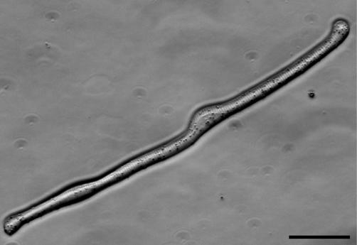
Figure 1
A Live Striated Myofiber from the Larval Salamander
Photomicrograph of a live striated myofiber dissociated from the larval limb musculature and adhering to the culture dish in serum-free medium. This cell has the appearance of a normal quiescent fiber...
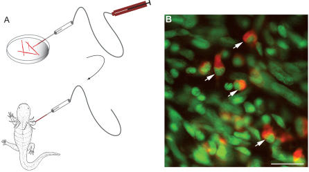
Figure 2
Cellularisation of Striated Myofibers after Implantation into a Larval Limb Blastema
(A) Schematic diagram of procedure. After dissociation of larval limb musculature, the cells were loaded with a cell tracker dye and single myofibers taken up into a suction micropipette, prior to inj...
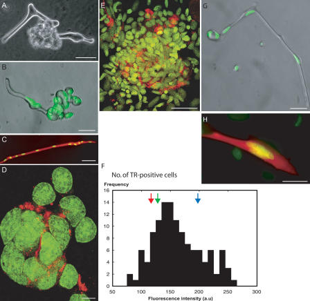
Figure 3
Plasticity of Isolated Myofibers
(A) Phase-contrast micrograph of a live cell at 3 d after plating, showing a lobulated structure in the middle of the fiber. (B) Micrograph of a live fiber at 2 d after plating, showing budding of nuc...
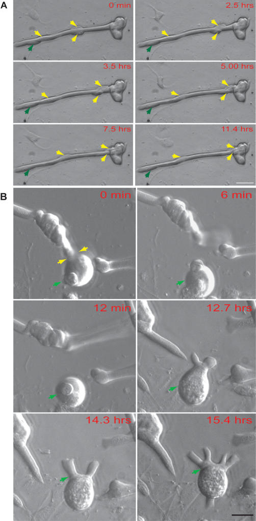
Figure 4
Analysis of Nuclear Migration and Fragmentation by Time-Lapse Microscopy
(A) Single frames illustrating the migration of three nuclei (yellow arrows) along a myofiber, of which two are incorporated into a terminal aggregate by 11.4 h. One nucleus (green arrow) remained sta...
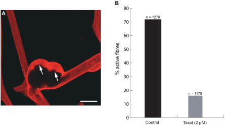
Figure 5
Plasticity and Microtubule Depolymerization
(A) The distribution of microtubules surrounding a multinucleate aggregate on a myofiber, as analysed by staining with anti-u03b2-tubulin. Note the relatively disordered state of the tubulin (arrowed)...
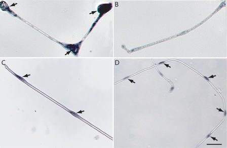
Figure 6
Analysis of mRNA Expression in Myofibers at 48 h by In Situ Hybridisation
(A) Expression of Msx1 mRNA in active myofiber. Note the accumulation of reaction product around nuclear aggregates (arrowed). (B) Absence of significant Msx1 mRNA expression in a quiescent fiber. Thi...
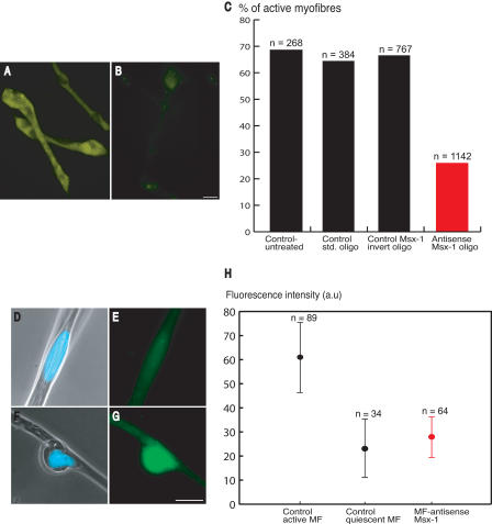
Figure 7
Analysis of the Functional Role of Msx1 Expression by Exposure to Morpholino Antisense Oligonucleotides
(A and B) Uptake of morpholino by myofibers. Myofibers were dissociated in the presence of biotinylated (A) or control (B) morpholinos and analysed by tyramide signal amplification at 24 h after plati...
Figure images are served from the NIH/NLM PubMed Central Open Access Subset or Europe PMC; copyright remains with the publishers and authors.
💬 Discussion
0 commentsNo comments yet. Be the first to start a discussion!
Leave a Comment