Abstract
Correlated network activity is important in the development of many neural circuits. Purkinje cells are among the first neurons to populate the cerebellar cortex, where they sprout exuberant axon collaterals. We used multiple patch-clamp recordings targeted with two-photon microscopy to characterize monosynaptic connections between the Purkinje cells of juvenile mice. We found that Purkinje cell axon collaterals projected asymmetrically in the sagittal plane, directed away from the lobule apex. On the basis of our anatomical and physiological characterization of this connection, we constructed a network model that robustly generated waves of activity that traveled along chains of connected Purkinje cells. Consistent with the model, we observed traveling waves of activity in Purkinje cells in sagittal slices from young mice that require GABA(A) receptor-mediated transmission and intact Purkinje cell axon collaterals. These traveling waves are absent in adult mice, suggesting they have a developmental role in wiring the cerebellar cortical microcircuit.
🔬 Techniques
💻 Software
✨ Fluorophores
🧪 Sample Preparation
💻 Software Details
💾 Data Repositories
🏛️ Research Organizations (ROR)
Affiliated research institutions:
📋 Methods
Experiments All experiments were carried out in accordance with the animal care and handling guidelines approved by the U.K. Home Office. Acute cerebellar slices were prepared from P3 – P25 L7-tau-GFP mice 15 (or in a few cases, from GAD65-GFP mice 16 , 17 ) using standard techniques 19 . All electrophysiological experiments were carried out at 33-35°C. For confocal and electron microscopy, L7-tau-GFP mouse pups (P8 or P18) were transcardially perfused with fixative, and immunohistochemical and immmunogold labelling was performed. For further details see Supplementary Methods .
Network modeling
A network simulation consisting of 50 synaptically connected Purkinje cells was implemented in NEURON using a biophysical model of the Purkinje cell based on an existing model for spontaneously firing Purkinje cells 23 . The anatomical and physiological parameters of the model were tuned to replicate our experimental data. Further details are available in Supplementary Methods .
Data analysis and statistics
Data are reported as means ± s.e.m. unless otherwise indicated. Data analysis was performed using Igor Pro and Matlab. Comparisons were made using either paired, two-tailed Student’s t-tests or unpaired two-tailed Student’s t-tests assuming unequal variances. Further details are available in Supplementary Methods .
Supplementary Material Supplementary Figures, Methods, References
📊 Figures
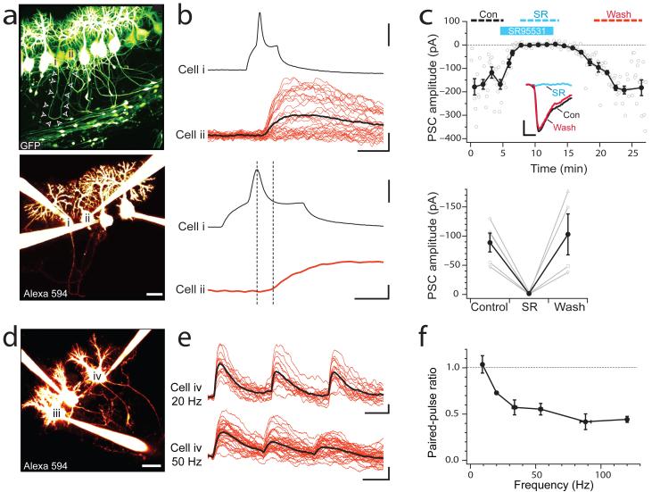
Figure 1
Unitary synaptic connections between neighboring Purkinje cells
( a ) Top: Two-photon image of Purkinje cells from a P9 L7-tau-GFP mouse. Bottom: Quadruple whole-cell recordings of Purkinje cells selected from the GFP image above, imaged with Alexa 594 in the inte...
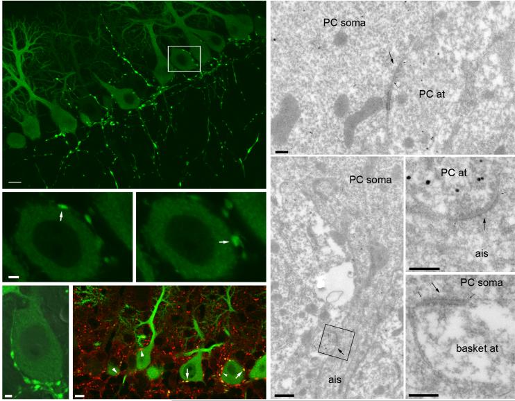
Figure 2
Purkinje cell local axon collaterals establish synapses on other Purkinje cells
( a ) Confocal laser scanning microscopic image of the cerebellar cortex of an L7-tau-GFP mouse. Scale bar, 10 u03bcm. ( b, c ) High magnification single optical section images from the area boxed in ...
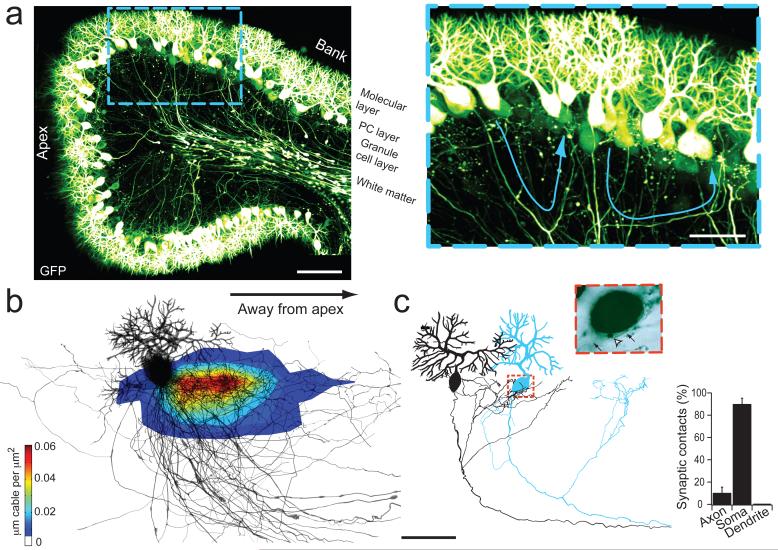
Figure 3
Anatomical distribution of Purkinje cell axon collaterals and Purkinjeu2013Purkinje synapses
( a ) Image of a lobule from a P9 mouse (left) and high-magnification image of the region indicated by the blue dashed box (right), with two axon collaterals highlighted with blue arrows. Scale bar, 1...
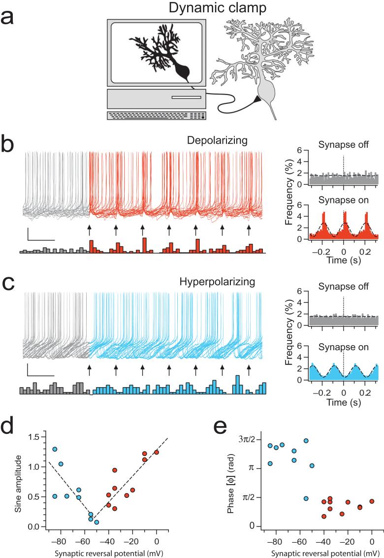
Figure 4
Purkinje cells synchronize in different phases depending on synaptic reversal potential
( a ) Schematic illustration of recording configuration where a computer-generated dynamic clamp synaptic conductance replaces the input from a presynaptic Purkinje cell. ( b ) After a four-second-lon...
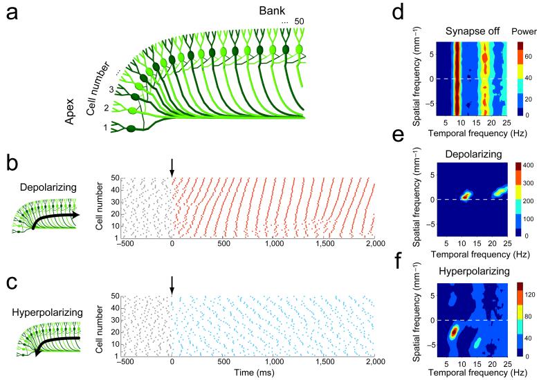
Figure 5
Waves of activity in a network model of Purkinje cells
( a ) Schematic illustration of the Purkinje cell axon collateral network model (cells numbered starting at the apex of the folium). Each Purkinje cell was connected to the basally-located nearest nei...
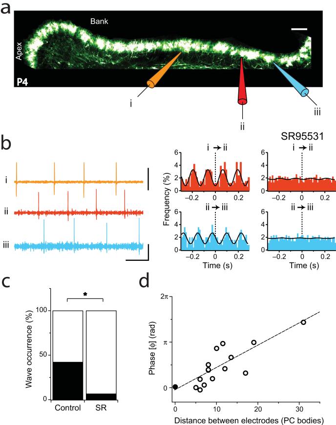
Figure 6
Traveling waves in sagittal cerebellar slices
( a ) Cerebellar folium of a P4 L7-tau-GFP mouse illustrating the recording configuration in ( b ). Scale bar, 50 u03bcm. ( b ) Left: sample extracellular recording traces from electrodes in ( a ), sh...
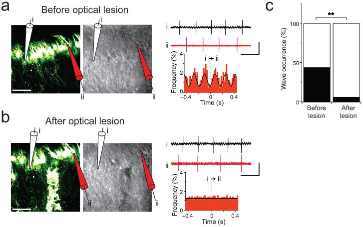
Figure 7
Optical lesion of Purkinje axon collaterals abolishes traveling waves
( a ) Left: Two-photon image (left) and corresponding laser-scanning Dodt contrast image (middle) of L7-tau-GFP Purkinje cells from a P6 mouse illustrating the recording configuration. Scale bar, 50 u...
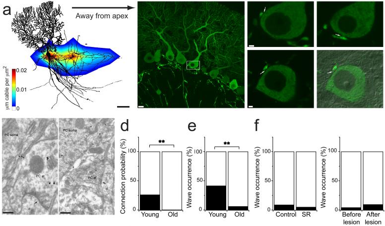
Figure 8
PC-PC connectivity and traveling waves are absent in older animals
( a ) Density plot of Purkinje cell axon collaterals from older mice reveals collaterals project asymmetrically away from lobule apex (n = 14; P17-22; cf. Fig 3b , Methods). Scale bar, 50 u03bcm. ( b ...
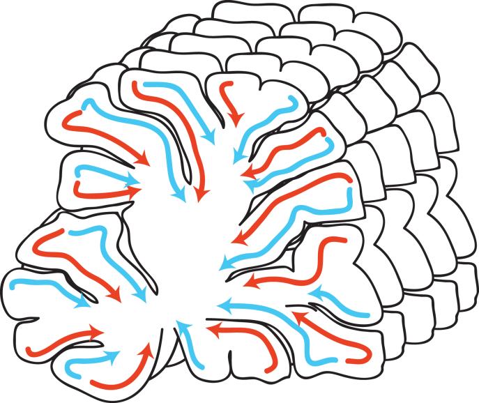
Figure 9
Traveling waves define a novel functional unit in the developing cerebellar cortex
A schematic illustration of the functional organization of the juvenile cerebellar cortex described in this study. Purkinje cells form functional units in the sagittal plane defined by their asymmetri...
Figure images are served from the NIH/NLM PubMed Central Open Access Subset or Europe PMC; copyright remains with the publishers and authors.
💬 Discussion
0 commentsNo comments yet. Be the first to start a discussion!
Leave a Comment