Abstract
Nicotinic acid adenine dinucleotide phosphate (NAADP) is a potent intracellular Ca(2+) signalling second messenger, but the mechanism of NAADP-induced Ca(2+) release is still poorly understood. The present study tested the hypothesis that NAADP induces Ca(2+) release from the lysosomal store via a TRP-ML1 (transient receptor potential-mucolipin 1)-mediated Ca(2+) release channel in coronary arterial myocytes (CAMs). RT-PCR and Western blot analyses demonstrated that TRP-ML1 was present in CAMs, and fluorescence resonance energy transfer (FRET) detection revealed that the TRP-ML1 was closely associated with some lysosomal proteins in these CAMs. ET-1, a well-known NAADP stimulator, was found to induce a local Ca(2+) burst from lysosomes followed by a global Ca(2+) release. This lysosome-associated Ca(2+) release was significantly inhibited in the TRP-ML1 siRNA pre-treated CAMs by 46.8 +/- 12.6% in the local Ca(2+) burst and 73.3 +/- 14.9% in the global Ca(2+) wave. In the reconstituted lysosomal channels from CAMs, NAADP activated Ca(2+) release channels at concentrations of 1-1000 nM, but neither activators (1 microM IP(3), 5 microM Rya) nor blockers (100 microM 2-APB, 50 microM Rya) of sarcoplasmic reticulum (SR) Ca(2+) release channels had effect on the channel activity. Moreover, TRP-ML1 gene silencing reduced this NAADP-sensitive Ca(2+) release channel activity in lysosomes by 71.5 +/- 18.5%. Immunoprecipitation or blockade of TRP-ML1 by anti-TRP-ML1 antibodies almost abolished NAADP-induced activation of lysosomal Ca(2+) channels (to 14.0 +/- 4.4% of control). These results for the first time provide direct evidence that an NAADP-sensitive Ca(2+) release channel is characteristic of TRP-ML1 channels.
🔬 Techniques
✨ Fluorophores
🧪 Sample Preparation
🏭 Microscope Brands
🧪 Reagent Suppliers
🏛️ Research Organizations (ROR)
Affiliated research institutions:
📋 Methods
Culture of CAMs
The bovine CAMs were cultured as described previously [ 20 , 21 ]. Briefly, the vessels were first rinsed with 5% FBS in medium 199 containing 25 mM HEPES with 1% penicillin, 0.3% gentamycin and 0.3% nystatin and then cut into segments, and the lumen was filled with 0.4% collagenase in medium 199. After 30 min. of incubation at 37°C, the vessels were flushed with medium 199. The strips of denuded arteries were placed into gelatin-coated flasks with medium 199 containing 10% FBS with 1% L-glutamine, 0.1% tylosin and 1% penicillin-streptomycin. CAMs migrated to the flasks within 3–5 days. Once growth was established, the vessels were removed and cells were grown in medium 199 containing 20% FBS. The identification of CAMs was based on positive staining by an anti-β-actin antibody. All studies were performed with cells of 2–4 passages except where specified. Demonstration of TRP-ML1 expression in coronary arterial myocytes (CAMs) For reverse transcriptase PCR (RT-PCR) analysis, total RNA was isolated from primary cultured CAMs by Trizole (Invitrogen, CA, USA), and 25, 50, 100 and 200 ng of different amount of total RNA was transcribed to cDNA by iScripTM cDNA synthesis kit (Invitrogen) in 20 μl reaction mixture. These synthesized cDNAs were used for TRP-ML1 (Accession number: BC118374 ) PCR reaction by PCR Supermix (Invitrogen) with primers of 5′-GCCAGTTACAGGAACCTCACG-3′ and 5′-CCAGAAGGATGTACCAGCCATT-3′ at a final concentration of 200 nM. Thirty cycles of PCR were performed in a thermal cycler with a denaturing phase of 30 sec. at 94°C, annealing phase of 45 sec. at 58°C and extension phase of 1 min. at 72°C. Glyceraldehyde-3-phosphate dehydrogenase (GAPDH) (Accession number: NM 001034034) was used as control with primers of 5′-CCACGAGAAGTATAACAACACCC-3′ and 5′- TGAAGTCGCAGGAGACAACC-3′. Meanwhile, the levels of TRP-ML subfamily were also determined with primers of 5′-GTTTCATCGGCTAAGGAACT-3′ and 5′-TGCCACTGTGAGCTTTATTG-3′ for TRP-ML2 (Accession number: XM_611818 ), and 5′-AATCCTGAGGCTGCTATAAGT-3′ and 5′-TAGGAAGAGGTGCTTGAATG-3′ for TRP-ML3 (Accession number: XM_592179 ). A negative control was performed to verify the PCR condition, which contained all the components of the PCR except the template DNA. The PCR products were separated by 1.2% agarose gel for confirmation of product size. Cell homogenates of 10, 20, 40 and 60 μg from primary cultures of CAMs were used for Western blot analysis with the methods as we described previously [ 11 ]. Anti-TRP-ML1 antibody from Abcam (ab28508, Abcam Inc., MA, USA) was used to probe TRP-ML1 protein according to the manufacturer’s instructions and β-actin was used as loading control. The existence of two other TRP-ML subfamily members of TRP-ML2 and TRP-ML3 were examined with corresponding antibodies from Sigma (St. Louis, MO, USA). Meanwhile, the presence of TRPC (canonical transient receptor potential) channels was also determined with anti-TRPC1 and anti-TRPC3/6/7 antibodies (Santa Cruz Biotechnology, Inc., CA, USA).
Show full methods section
Culture of CAMs
The bovine CAMs were cultured as described previously [ 20 , 21 ]. Briefly, the vessels were first rinsed with 5% FBS in medium 199 containing 25 mM HEPES with 1% penicillin, 0.3% gentamycin and 0.3% nystatin and then cut into segments, and the lumen was filled with 0.4% collagenase in medium 199. After 30 min. of incubation at 37°C, the vessels were flushed with medium 199. The strips of denuded arteries were placed into gelatin-coated flasks with medium 199 containing 10% FBS with 1% L-glutamine, 0.1% tylosin and 1% penicillin-streptomycin. CAMs migrated to the flasks within 3–5 days. Once growth was established, the vessels were removed and cells were grown in medium 199 containing 20% FBS. The identification of CAMs was based on positive staining by an anti-β-actin antibody. All studies were performed with cells of 2–4 passages except where specified. Demonstration of TRP-ML1 expression in coronary arterial myocytes (CAMs) For reverse transcriptase PCR (RT-PCR) analysis, total RNA was isolated from primary cultured CAMs by Trizole (Invitrogen, CA, USA), and 25, 50, 100 and 200 ng of different amount of total RNA was transcribed to cDNA by iScripTM cDNA synthesis kit (Invitrogen) in 20 μl reaction mixture. These synthesized cDNAs were used for TRP-ML1 (Accession number: BC118374 ) PCR reaction by PCR Supermix (Invitrogen) with primers of 5′-GCCAGTTACAGGAACCTCACG-3′ and 5′-CCAGAAGGATGTACCAGCCATT-3′ at a final concentration of 200 nM. Thirty cycles of PCR were performed in a thermal cycler with a denaturing phase of 30 sec. at 94°C, annealing phase of 45 sec. at 58°C and extension phase of 1 min. at 72°C. Glyceraldehyde-3-phosphate dehydrogenase (GAPDH) (Accession number: NM 001034034) was used as control with primers of 5′-CCACGAGAAGTATAACAACACCC-3′ and 5′- TGAAGTCGCAGGAGACAACC-3′. Meanwhile, the levels of TRP-ML subfamily were also determined with primers of 5′-GTTTCATCGGCTAAGGAACT-3′ and 5′-TGCCACTGTGAGCTTTATTG-3′ for TRP-ML2 (Accession number: XM_611818 ), and 5′-AATCCTGAGGCTGCTATAAGT-3′ and 5′-TAGGAAGAGGTGCTTGAATG-3′ for TRP-ML3 (Accession number: XM_592179 ). A negative control was performed to verify the PCR condition, which contained all the components of the PCR except the template DNA. The PCR products were separated by 1.2% agarose gel for confirmation of product size. Cell homogenates of 10, 20, 40 and 60 μg from primary cultures of CAMs were used for Western blot analysis with the methods as we described previously [ 11 ]. Anti-TRP-ML1 antibody from Abcam (ab28508, Abcam Inc., MA, USA) was used to probe TRP-ML1 protein according to the manufacturer’s instructions and β-actin was used as loading control. The existence of two other TRP-ML subfamily members of TRP-ML2 and TRP-ML3 were examined with corresponding antibodies from Sigma (St. Louis, MO, USA). Meanwhile, the presence of TRPC (canonical transient receptor potential) channels was also determined with anti-TRPC1 and anti-TRPC3/6/7 antibodies (Santa Cruz Biotechnology, Inc., CA, USA).
Fluorescence resonance energy transfer
(FRET) determining the presence of TRP-ML1 on lysosomes SubconfluenceCAMs were stained with FITC-conjugated anti-Lamp-1 antibody (553793, BD Pharmingen™, NJ, USA) (FITC/Lamp-1) and TRITC-conjugated F(ab′) 2 (sc-3841, Santa Cruz Biotechnology, Inc.) plus TRP-ML1 primary antibody (ab 28508, Abcam Inc.) (TRITC/TRP-ML1) with a method as described previously [ 22 , 23 ] and then visualized under confocal microscope with excitation/emission wavelength of 494/518 nm and 555/580 nm for FITC and TRITC, respectively. An acceptor bleaching protocol was employed to measure the FRET efficiency [ 22 , 23 ] between FITC/Lamp-1 and TRITC/TRP-ML1, which was calculated through the following formula: E = (FITCpost – FITCpre)/FITCpost × 100%[ 22 ]. TRITC-CTXB (TRITC conjugated with cholera toxin subunit B) was paired with FITC-Lamp-1 to act as negative control, because TRITC-CTXB selectively reacts with ganglioside and is widely used for detection of the cell plasma membrane (PM).
RNA interference of TRP-ML1 in CAMs
RNA silence was achieved by double-stranded siRNA of targeting TRP-ML1 (Accession number: BC118374 ) consisted of 5′-CAGCUUCCGGCUCCUG-3′. A scrambled RNA or Xeragon library scrambled RNA was synthesized for negative control. siRNA transfection was performed according to the manufacturer’s instruction in Qiagen TransMessenger kit (Qiagen, Gaithersburg, MD, USA) as we described previously [ 24 ]. The final concentration of siRNA was 15 nM and the efficiency of TRP-ML1 silencing was assessed by Western blotting analysis. At 36 hrs post-transfection, the TRP-ML1-knocked down CAMs were used to measure intracellular Ca 2+ levels by fluorescent assay or to isolate lysosomes for the channel reconstitution study. Fluorescent microscopic measurement of [Ca 2+ ] i in CAMs Normal CAMs or TRP-ML1 siRNA-treated CAMs were loaded with 10 μM fura-2 at room temperature for 30 min. and washed three times with Ca 2+ -free Hanks’ buffer, which was supplemented with 2 μM EGTA. Endothelin-1 (ET-1, 100 nM)-induced Ca 2+ release was performed with a method described previously [ 9 ]. A fluorescence ratio of excitation at 340 nm to that at 380 nm (F340/F380) was determined after background subtraction, and [Ca 2+ ] i was calculated by using the following equation: [Ca 2+ ] i = K d β[(R- R min )/(R max - R)], where K d for the fura-2-Ca 2+ complex is 224 nM; R is the fluorescence ratio (F340/F380); R max and R min are the maximal and minimal fluorescence ratios measured by addition of 10 μM of Ca 2+ ionophore of ionomycin to Ca 2+ -replete solution (2.5 mM CaCl 2 ) and Ca 2+ -free solution (5 mM EGTA), respectively; and β is the fluorescence ratio at 380-nm excitation determined at R min and R max , respectively. Before and after a 100 nM endothelin-1 (ET-1) treatment, the ratio of fura-2 emissions, when excited at the wavelengths of 340 and 380 nm, was recorded with an inverted microscope (Diaphot 200, Nikon, Tokyo, Japan) and a digital camera (SPOT RT Monochrome, Diagnostic Instruments, Sterling Heights, MI, USA). Metafluor imaging and analysis software was used to acquire, digitize and store the images for off-line processing and statistical analysis.
Preparation of lysosomes from bovine coronary arterial muscle
CAMs were dissociated from circumflex and left anterior descending arteries of bovine hearts, and CAMs homogenates subsequently were processed to lysosomal isolation and purification with our published methods [ 11 , 25 , 26 ]. The purified lysosomal fraction was suspended in sucrose buffer (0.9% NaCl, 0.3 M sucrose and 0.1 μM phenylmethylsulfonyl fluoride) and biochemically confirmed [ 11 ]. PM and SR components were also prepared from bovine coronary arteries as described previously [ 20 , 26 ], and used as controls. The purity of the lysosomal preparation was biochemically identified [ 11 ] and further determined by Western blot analysis using an antibody of LAMP 1 (lysosome-associated membrane protein 1), a lysosomal specific marker, as described previously [ 11 ]. The existence of TRP-ML1 and other two TRP-ML subfamily member of TRP-ML2 and TRP-ML3 were also examined. To clarify whether L-type Ca 2+ channel was expressed on the lysosome, the 1,4-dihydropyridine (DHP) receptor alpha-1 subunit was probed. During Western blot assay, the concentrations of antibodies used were according to the manufacturer’s instructions. LAMP1, TRP-ML1 and DHPR alpha 1 antibodies were purchased from Abcam. TRP-ML2, and 3 antibodies were obtained from Sigma and caveolin antibody was purchased from BD Transduction Laboratories, respectively. Characterization and identity of lysosomal Ca 2+ release channels Purified lysosomes were reconstituted into planar lipid bilayers and biophysical characterization of lysosomal Ca 2+ release channels was performed with a method as we described previously [ 11 , 27 , 28 ]. Pharmacologically, we first investigated the concentration-dependent effects of NAADP on the activity of reconstituted lysosomal Ca 2+ release channels and then examined the effects of a TRP-ML1 blocker, amiloride (1 mM)[ 11 ], an NAADP antagonist of PPADS (50 μM) [ 29 ], a common inhibitor of lysosome function of bafilomycin A1 (100 nM), and voltage-dependent Ca 2+ channel blockers of nifedipine (100 μM) and verapamil (100 μM) on the NAADP-sensitive Ca 2+ release channel activity [ 9 , 11 , 29 , 30 ]. Second, we compared this lysosomal NAADP-sensitive Ca 2+ release channel with IP 3 R and ryanodine receptor/Ca 2+ (RyR/Ca 2+ ) release channels on the SR by applying IP 3 R or RyR agonists and antagonists. Third, we used lysosomes from TRP-ML1 siRNA-treated CAMs or applied immunoprecipitation to deprive TRP-ML1 from the lysosomes to determine whether the channel response to NAADP can be altered. TRP-ML1-deprived lysosome preparations were made as follows: 200 μg lysosome protein in 100 μl resuspension solution was incubated with 10 μg rabbit polyclonal anti-TRP-ML1 antibody (ab28508, Abcam Inc.) at 4°C overnight; then 20 μg agarose-conjugated goat polyclonal secondary antibody to rabbit IgG H&L was added and further incubated at room temperature for another 2 hrs, followed by centrifugation at 200 g for 1 min. to collect supernatant as TRP-ML1-free lysosomal preparations, so that the immunoprecipitation-blocked channel activity would be due to the deprivation of the TRP-ML1 from the lysosomal preparation. Normal rabbit serum was used as a substitute of TRP-ML1 antibody for control preparation. Fourth, we used another anti-TRP-ML1 polyclonal antibody (sc-26269, Santa Cruz Biotechnology, Inc.), which was raised in goat against a peptide mapping at the C terminus of TRP-ML1 of mouse origin, to test channel-blocking effects. A serial diluted anti-TRP-ML1 antibody was added to the bath solution at a final concentration of 1:5000, 1:500 and 1:50 for 5 min., respectively, followed by 1 μM NAADP. Before and after addition of NAADP, the channel currents were recorded at a holding potential of + 40 mV. Normal goat serum (NGS) was used as a substitute for goat polyclonal TRP-ML1 antibody for control experiments.
Statistics
Data are presented as means ± S.E.; the significance of the differences in mean values between multiple groups was examined using an analysis of variance for repeated measures followed by a Duncan’s multiple range test. P < 0.05 was considered statistically significant.
📊 Figures
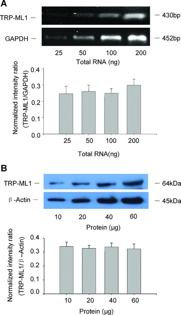
Figure 1
TRP-ML1 mRNA and protein expression in CAMs. (A) Representative gel document of RT-PCR products of TRP-ML1 and GAPDH at different initial RNA concentrations (upper panel). Summarized results in lower ...
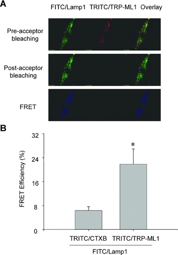
Figure 2
FRET detection of FITC-labelled Lamp-1 and TRITC-labelled TRP-ML1 in CAMs. In Figure 2A, the upper panel shows fluorescent images of a FITC/Lamp1 and TRITC/TRP-ML1 before acceptor bleaching (TRITC ble...
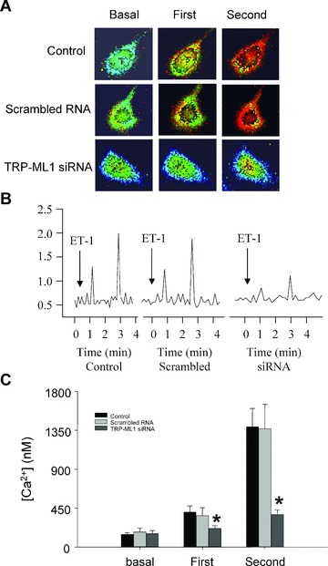
Figure 3
ET-1-induced Ca 2+ release response in TRP-ML1 siRNA treated CAMs. (A) Serial images of fura-2 fluorescence ratio F340/F380 recorded in different treated CAMs. Spatially localized Ca 2+ burst (first p...
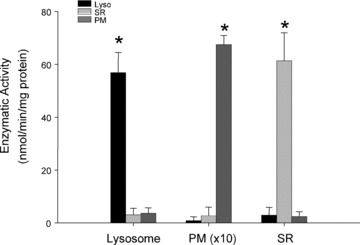
Figure 4
Purity confirmation of lysosome preparations. Summarized results show the conversion rate of 4-nitrophenyl phosphate to 4-nitrophenyl by a lysosome marker enzyme, acid phosphatase in lysosomes (Lyso),...
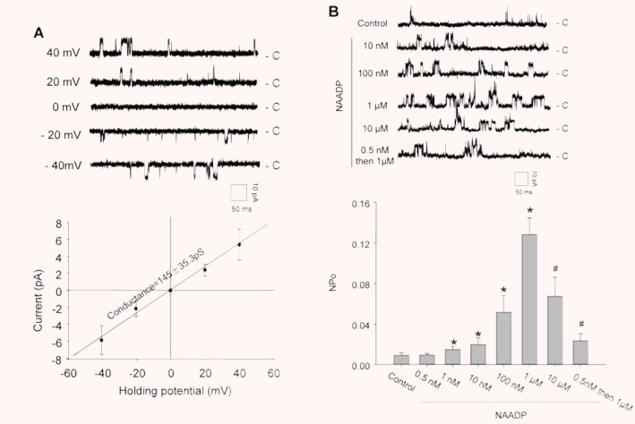
Figure 5
Electrophysiological and pharmacological characterization of reconstituted lysosomal Ca 2+ release channels. (A) Representative recording of NAADP-sensitive Ca 2+ channel currents in the upper panel a...
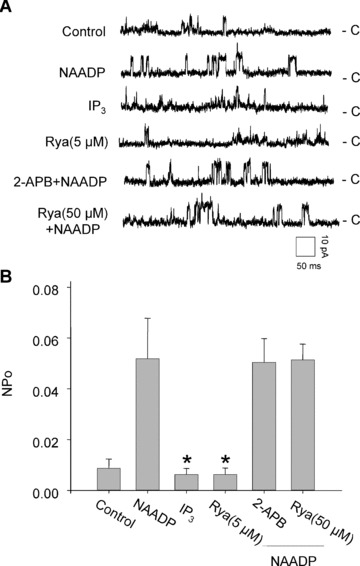
Figure 6
Effects of IP 3 R or RyR agonist and antagonist on the activity of reconstituted lysosomal Ca 2+ release channels. (A) Representative recordings of channel currents under control condition and after a...
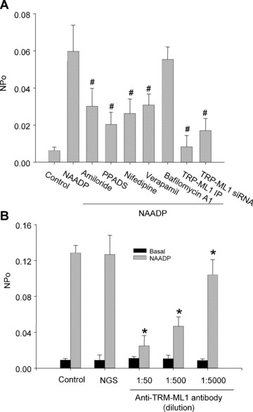
Figure 7
Identification of lysosomal NAADP-sensitive Ca 2+ release channels as TRP-ML1 function by reconstituted lipid bilayer assay. (A) Summarized results show that the channel open probability (NPo) is unde...
Figure images are served from the NIH/NLM PubMed Central Open Access Subset or Europe PMC; copyright remains with the publishers and authors.
💬 Discussion
0 commentsNo comments yet. Be the first to start a discussion!
Leave a Comment