Abstract
The cold and menthol receptor TRPM8 is highly expressed in prostate and prostate cancer (PC). Recently, we identified that TRPM8 is as an ionotropic testosterone receptor. The TRPM8 mRNA is expressed in early prostate tumors with high androgen levels, while anti-androgen therapy greatly reduces its expression. Here, from the chromatin-immunoprecipitation (ChIP) analysis, we found that an androgen response element (ARE) mediates androgen regulation of trpm8. Furthermore, using immunofluorescence, calcium-imaging and planar lipid bilayers, we identified that TRPM8 channel is functionally regulated by androgens in the prostate. Although TRPM8 mRNA is expressed at high levels, we found that the TRPM8 protein undergoes ubiquitination and degradation in PC cells. The mass-spectrometry analysis of TRPM8, immunoprecipitated from LNCaP cells identified ubiquitin-like modifier-activating enzyme 1 (UBA1). PYR-41, a potent inhibitor of initial enzyme in the ubiquitination cascade, UBA1, increased TRPM8 activity on the plasma membrane (PM) of LNCaP cells. Furthermore, PYR-41-mediated PMTRPM8 activity was accompanied by enhanced activation of p53 and Caspase-9. Interestingly, we found that the trpm8 promoter possesses putative binding sites for p53 and that the overexpression of p53 increased the TRPM8 mRNA levels. In addition to the genomic regulation of TRPM8 by AR and p53, our findings indicate that the testosterone-induced PMTRPM8 activity elicits Ca2+ uptake, subsequently causing apoptotic cell death. These findings support the strategy of rescuing PMTRPM8 expression as a new therapeutic application through the regulation of PC cell growth and proliferation.
🔬 Techniques
💻 Software
✨ Fluorophores
🧪 Sample Preparation
🔬 Cell Lines
🏭 Microscope Brands
🧪 Reagent Suppliers
💻 Software Details
🏛️ Research Organizations (ROR)
Affiliated research institutions:
📋 Methods
Ethics statement Human high-density
PC tissue microarray was purchased from US Biomax (Rockville, MD). All the prostate tissue samples were obtained from patients (Age: 21 yrs to 83 yrs) undergoing therapeutic surgery at the US Biomax certified hospitals (see Sdata). Cell lines, transfection The cell lines RWPE1, RWPE2, LNCaP, DU145 and PC3 were obtained from the American Type Culture Collection (Manassas, VA) and were maintained under standard conditions as per ATCC guidelines. LNCaP, PC3 and DU145 cells were cultured in RPMI, F-12K and DMEM/F12K (1:1) medium, respectively supplemented with 10% fetal bovine serum (GIBCO BRL, Lewisville, TX) and 1% penicillin/streptomycin. RWPE1 and RWPE2 cells were grown in keratinocyte-serum free media (K-SFM) containing 50 μg/ml bovine pituitary extract, 5 ng/ml epidermal growth factor and 1% penicillin/streptomycin solution. All cells were cultured in a 37°C incubator with 5% CO 2 humidified atmosphere. HEK-293 stable cells overexpressing TRPM8 (HEK-TRPM8) were established and maintained following standard protocol [ 48 ]. All transfection experiments were performed using FuGENE HD transfection reagent (Roche Diagnostics, Indianapolis, IN) (see Sdata). Plasmids, shRNA construct, antibodies and chemical inhibitors TRPM8 rat cDNA cloned in N-terminus myc-tagged pCDNA3 vector for TRPM8 overexpression (TRPM8OE). TRPM8 trafficking investigated by GFP-TRPM8. The siAR and shTRPM8 purchased from Santa Cruz Biotechnology (Santa Cruz, CA). pCDNA3-empty vector used as control. All antibodies purchased from Santa Cruz Biotechnology; anti-TRPM8/CMR-1 (656-680) from Phoenix Pharmaceuticals (Burlingame, CA); anti- DHT/testosterone from Thermo Scientific Pierce (Rockford, IL); Alexa Flour 488 and 594 purchased from Invitrogen (Invitrogen, Carlsbad, CA). Testosterone, DHT (5α-dihydrotestosterone), PYR-41 (UBA1 inhibitor) and hydroxyflutamide (HF; AR inhibitor) was purchased from Sigma (St. Louis, MO).
Show full methods section
Ethics statement Human high-density
PC tissue microarray was purchased from US Biomax (Rockville, MD). All the prostate tissue samples were obtained from patients (Age: 21 yrs to 83 yrs) undergoing therapeutic surgery at the US Biomax certified hospitals (see Sdata). Cell lines, transfection The cell lines RWPE1, RWPE2, LNCaP, DU145 and PC3 were obtained from the American Type Culture Collection (Manassas, VA) and were maintained under standard conditions as per ATCC guidelines. LNCaP, PC3 and DU145 cells were cultured in RPMI, F-12K and DMEM/F12K (1:1) medium, respectively supplemented with 10% fetal bovine serum (GIBCO BRL, Lewisville, TX) and 1% penicillin/streptomycin. RWPE1 and RWPE2 cells were grown in keratinocyte-serum free media (K-SFM) containing 50 μg/ml bovine pituitary extract, 5 ng/ml epidermal growth factor and 1% penicillin/streptomycin solution. All cells were cultured in a 37°C incubator with 5% CO 2 humidified atmosphere. HEK-293 stable cells overexpressing TRPM8 (HEK-TRPM8) were established and maintained following standard protocol [ 48 ]. All transfection experiments were performed using FuGENE HD transfection reagent (Roche Diagnostics, Indianapolis, IN) (see Sdata). Plasmids, shRNA construct, antibodies and chemical inhibitors TRPM8 rat cDNA cloned in N-terminus myc-tagged pCDNA3 vector for TRPM8 overexpression (TRPM8OE). TRPM8 trafficking investigated by GFP-TRPM8. The siAR and shTRPM8 purchased from Santa Cruz Biotechnology (Santa Cruz, CA). pCDNA3-empty vector used as control. All antibodies purchased from Santa Cruz Biotechnology; anti-TRPM8/CMR-1 (656-680) from Phoenix Pharmaceuticals (Burlingame, CA); anti- DHT/testosterone from Thermo Scientific Pierce (Rockford, IL); Alexa Flour 488 and 594 purchased from Invitrogen (Invitrogen, Carlsbad, CA). Testosterone, DHT (5α-dihydrotestosterone), PYR-41 (UBA1 inhibitor) and hydroxyflutamide (HF; AR inhibitor) was purchased from Sigma (St. Louis, MO).
Chromatin immunoprecipitation
(ChIP) and real time-PCR (RT-PCR) ChIP assay was performed using ChIP-IT TM Express kit from Active Motif (Carlsbad, CA) following supplied protocol. (see SMethods). Sub-cellular protein fractionation, immunoprecipitation, immunoblotting and mass spectrometry Detergent soluble membrane fraction preparation and analysis was done according to standard protocol [ 25 , 49 ].
Ubiquitination and biotinylation assay
For the ubiquitination assay, whole cell lysates (500 μg) were incubated with UbiCapture-Q Matrix (VWR International, Batavia, IL). Biotinylation assays were performed using cell surface protein isolation kit (Thermo Scientific Pierce) (see SMethods). Transmission electron microscopic studies The cells were fixed using fixative solution (2.5% glutaraldehyde in 0.1 M phosphate buffer, pH 7.4). After fixation, samples were buffer rinsed and postfixed with 1% osmium tetroxide, dehydrated (35%, 70%, 95%, 100% ethanol dehydration), and flat embedded in propylene oxide and Epon 812 epoxy resin (Tousimis) in 1:1 ratio at 60 °C for 4 h. A Reichert OMU3 ultramicrotome (Austria) was used to prepare 700A° thin sections that were mounted on 200 copper mesh grids, stained with uranyl acetate and lead citrate. The sections were viewed under a JEOL JEM 100C transmission electron microscope (60 kV). FACS, TUNEL and clonogenic assay The effects of androgen / AR inhibition / TRPM8 overexpression on cancer cell proliferation were examined by FACS, TUNEL and clonogenic assay following standard protocol [ 47 ] (See SMethods).
EMSA and p53 transcription factor assay
The p53 activation in nuclear extracts was monitored using p53 (3) - EMSA (Affymetrix, Santa Clara, CA) and ELISA-based TransAM™ p53 kit (Active Motif) according to the manufacturer's protocol (see SMethods).
Calcium imaging and planar lipid bilayers
Fluorescence measurements of intracellular Ca 2+ concentration of single cells were measured using standard protocol [ 19 , 25 , 48 ]. Planar lipid bilayers measurement and purification of TRPM8 protein from HEK-TRPM8 cells done using standard protocol [ 50 ].
📊 Figures
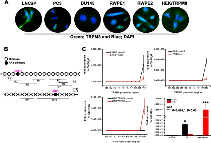
Figure 1
Role of ARE elements in the regulation of trpm8 gene expression
A. Immunocytochemistry analysis for TRPM8 expression. B. Analysis of 2064 bp 5u2032-flanking region of human trpm8 gene (ref| NW_004929306.1 ) identified 2 putative ARE I and ARE II sites. Quantitativ...
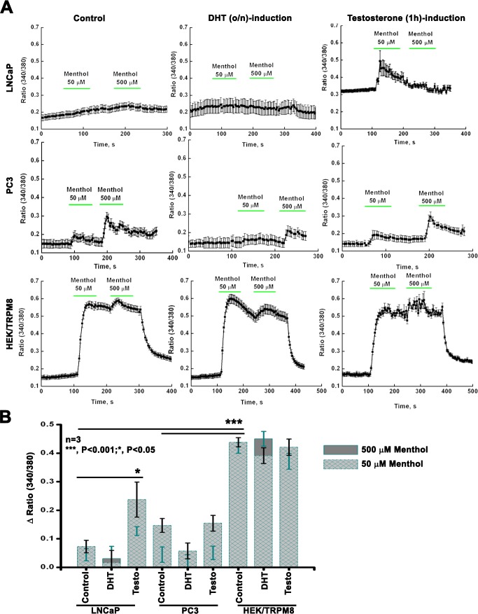
Figure 2
TRPM8 activity by intracellular Ca 2+ -measurements
A. Fluorescence measurements of intracellular Ca 2+ concentration were performed on PC cells upon DHT (o/n) and testosterone (3 h) induction by calcium-imaging. B. The summary of 50 u03bcM and 500 u03...
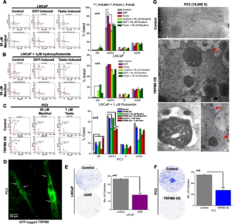
Figure 3
TRPM8 expression suppresses cell viability through the regulation of AR in PC cells
A. - B. FACS analysis of cell cycle progression in DHT and testosterone-induced LNCaP cells both in the A. absence and B. presence of hydroxyflutamide (HF). The cells were then treated with menthol fo...
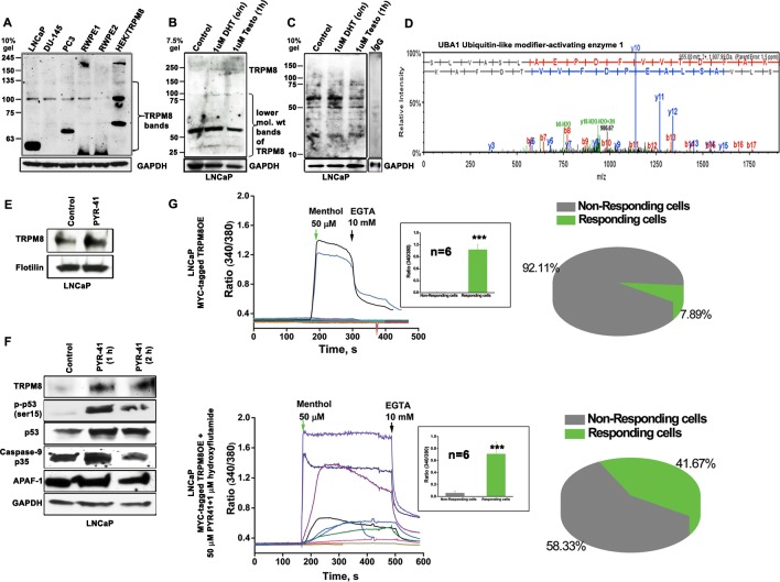
Figure 4
Ubiquitin-mediated proteolytic degradation of TRPM8 in an androgen-responsive LNCaP cells
A. Immunoblot analysis of cell lysate proteins (160 u03bcg) from 5 PC cell lines and HEK-TRPM8 using anti-TRPM8 antibody. B. To determine the lower mol. wt (LMW) product of TRPM8 upon DHT and testoste...
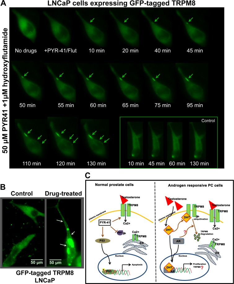
Figure 5
Live-cell imaging for distribution of GFP-TRPM8
A. LNCaP cells were transiently transfected with GFP-tagged TRPM8 construct (1.5 u03bcg) and incubated for 48 h. Cells treated with 50 u03bcM PYR-41/1 u03bcM HF and the distribution of the GFP fused T...
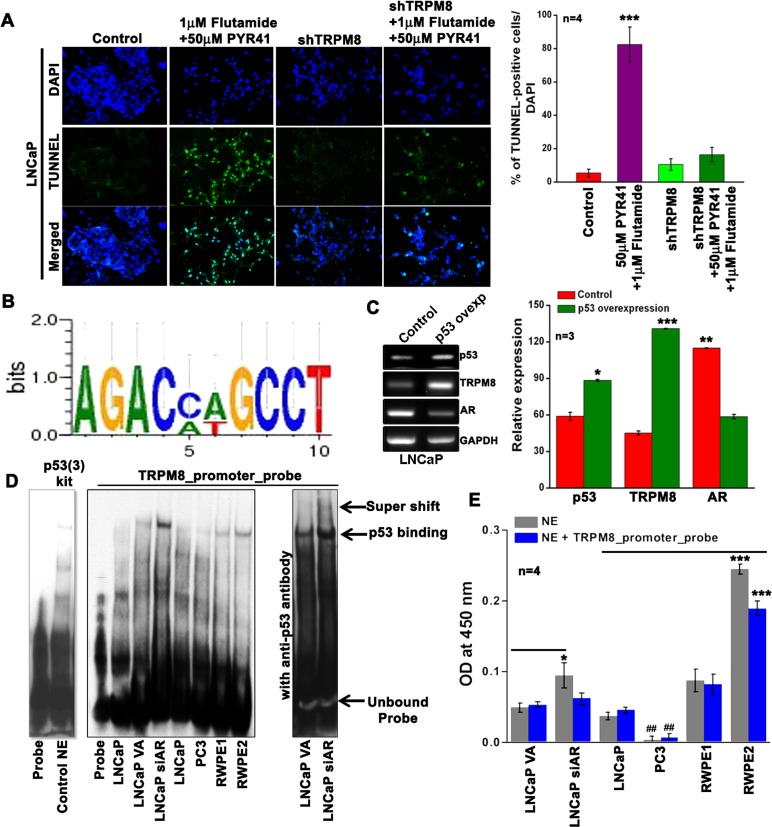
Figure 6
Inverse regulation of TRPM8 by AR and p53
A. TUNEL staining on LNCaP cells. Green fluorescence represents apoptotic cells and Blue fluorescence represents DAPI. B. The 2064 bp 5u2032-flanking region of the human trpm8 gene with ARE I and ARE ...
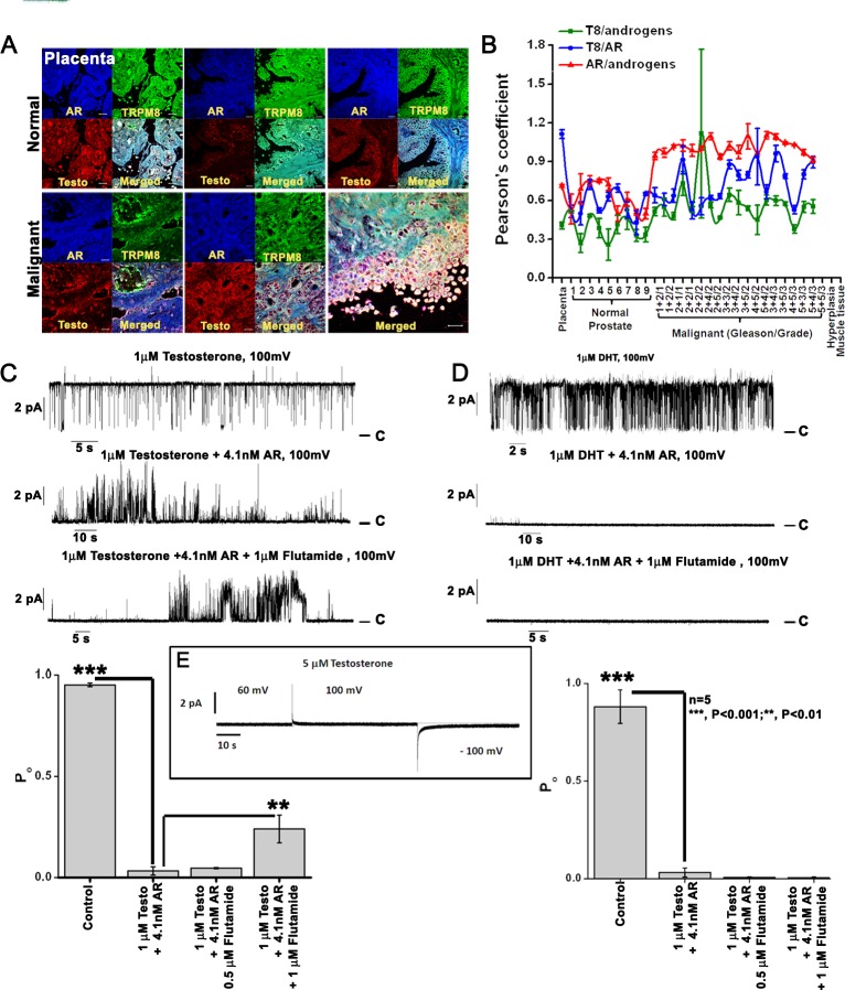
Figure 7
TRPM8 protein is physically associated with androgen-AR proteins
A. Immunohistochemistry analysis of PC tissue microarray (US Biomax) using Alexa fluor secondary antibodies. Images are at 40X magnification. B. The Pearson's coefficient was calculated in ImageJ with...
Figure images are served from the NIH/NLM PubMed Central Open Access Subset or Europe PMC; copyright remains with the publishers and authors.
💬 Discussion
0 commentsNo comments yet. Be the first to start a discussion!
Leave a Comment