Abstract
The progression of a natural bacterial infection is a dynamic process influenced by the physiological characteristics of the target organ. Recent developments in live animal imaging allow for the study of the dynamic microbe-host interplay in real-time as the infection progresses within an organ of a live host. Here we used multiphoton microscopy-based live animal imaging, combined with advanced surgical procedures, to investigate the role of uropathogenic Escherichia coli (UPEC) attachment organelles P and Type 1 fimbriae in renal bacterial infection. A GFP+ expressing variant of UPEC strain CFT073 and genetically well-defined isogenic mutants were microinfused into rat glomerulus or proximal tubules. Within 2 h bacteria colonized along the flat squamous epithelium of the Bowman's capsule despite being exposed to the primary filtrate. When facing the challenge of the filtrate flow in the proximal tubule, the P and Type 1 fimbriae appeared to act in synergy to promote colonization. P fimbriae enhanced early colonization of the tubular epithelium, while Type 1 fimbriae mediated colonization of the center of the tubule via a mechanism believed to involve inter-bacterial binding and biofilm formation. The heterogeneous bacterial community within the tubule subsequently affected renal filtration leading to total obstruction of the nephron within 8 h. Our results reveal the importance of physiological factors such as filtration in determining bacterial colonization patterns, and demonstrate that the spatial resolution of an infectious niche can be as small as the center, or periphery, of a tubule lumen. Furthermore, our data show how secondary physiological injuries such as obstruction contribute to the full pathophysiology of pyelonephritis.
🔬 Techniques
🧬 Organisms
💻 Software
✨ Fluorophores
🧪 Sample Preparation
🔬 Cell Lines
🏭 Microscope Brands
🧪 Reagent Suppliers
🔴 Lasers
💻 Software Details
🏛️ Research Organizations (ROR)
Affiliated research institutions:
📋 Methods
Ethics statement
All studies were performed in accordance with the National Institutes of Health's Guide for the Care and Use of Laboratory Animals and have been approved by the Institutional Animal Care and Use Committee at Indiana University School of Medicine Indianapolis, Indiana, USA (Study number 2453 Amendments 4, 5 and 16), Uppsala Djurfösöksetiska Nämnd, Uppsala, Sweden (Permit number: C14/6) or Stockholm's Norra Djurförsöksetiska Nämnd, Stockholm, Sweden (Permit numbers: N190/05, N347/09, N402/07).
Bacterial strains and plasmids
Bacterial strains used in this study are listed in Table 1 .
Strains
ARD41 and ARD42 were constructed by inserting the gfp + gene into UPEC76 and W3110 as previously described [19] . Briefly, the one-step allelic recombination method was used to achieve site-specific integration of gfp + , under the control of a constitutively active tetracycline promoter P LtetO-1, into the cobS gene [54] . To generate strain ARD43, plasmid pKTH3020, carrying the pap operon, was inserted into strain ARD42 by electroporation. ARD40 was created by inserting the kanamycin resistance cassette from pKD4 into the fimH gene of LT004 deleting 321 bp between nt 5143780- 5144101, using the one step allelic knockout method [54] . Oligonucleotide sequences are listed in Table 1 . All insertions were confirmed by PCR and sequencing (ABI3100, Applied Biosystems). For cloning purposes, bacteria were cultivated in aerated Luria-Bertani broth at 37°C in the presence of ampicillin (Amp, 100 µg/ml), chloramphenicol (Cm, 20 µg/ml) and kanamycin (Km, 50 µg/ml) as required. No alterations in growth rates, capsule morphology or expression of α-hemolysin were observed (data not shown). To prepare bacteria for microinfusion experiments, bacteria from aerated over-night cultures were re-inoculated (1∶100) into fresh LB with antibiotics, cultivated under shaking conditions to OD 600 = 0.6, then washed and concentrated to 10 9 CFU/ml in PBS. Bacteria were maintained on ice (maximum 2 h) until microinfusion. mRNA analysis The renal infection site was dissected using a 5 mm biopsy punch, medulla tissue was removed, and total RNA extraction was performed on the resulting ∼30 mg tissue using Trizol (Invitrogen, Sweden). Experimental triplicates were performed on three separate preparations for both LT004 infected and PBS samples. cDNA was transcribed from 1 µg RNA using the SuperScript III First Strand Synthesis Supermix kit (Invitrogen, Sweden). qRT-PCR was performed using a 7500 Real Time PCR System (Applied Biosystems, Sweden) and the Power SYBR Green PCR Mastermix (Applied Biosystems, Sweden). In all experiments, gfp + was used as an endogenous reference gene. Primer sequences are listed in Table 1 .
Show full methods section
Ethics statement
All studies were performed in accordance with the National Institutes of Health's Guide for the Care and Use of Laboratory Animals and have been approved by the Institutional Animal Care and Use Committee at Indiana University School of Medicine Indianapolis, Indiana, USA (Study number 2453 Amendments 4, 5 and 16), Uppsala Djurfösöksetiska Nämnd, Uppsala, Sweden (Permit number: C14/6) or Stockholm's Norra Djurförsöksetiska Nämnd, Stockholm, Sweden (Permit numbers: N190/05, N347/09, N402/07).
Bacterial strains and plasmids
Bacterial strains used in this study are listed in Table 1 .
Strains
ARD41 and ARD42 were constructed by inserting the gfp + gene into UPEC76 and W3110 as previously described [19] . Briefly, the one-step allelic recombination method was used to achieve site-specific integration of gfp + , under the control of a constitutively active tetracycline promoter P LtetO-1, into the cobS gene [54] . To generate strain ARD43, plasmid pKTH3020, carrying the pap operon, was inserted into strain ARD42 by electroporation. ARD40 was created by inserting the kanamycin resistance cassette from pKD4 into the fimH gene of LT004 deleting 321 bp between nt 5143780- 5144101, using the one step allelic knockout method [54] . Oligonucleotide sequences are listed in Table 1 . All insertions were confirmed by PCR and sequencing (ABI3100, Applied Biosystems). For cloning purposes, bacteria were cultivated in aerated Luria-Bertani broth at 37°C in the presence of ampicillin (Amp, 100 µg/ml), chloramphenicol (Cm, 20 µg/ml) and kanamycin (Km, 50 µg/ml) as required. No alterations in growth rates, capsule morphology or expression of α-hemolysin were observed (data not shown). To prepare bacteria for microinfusion experiments, bacteria from aerated over-night cultures were re-inoculated (1∶100) into fresh LB with antibiotics, cultivated under shaking conditions to OD 600 = 0.6, then washed and concentrated to 10 9 CFU/ml in PBS. Bacteria were maintained on ice (maximum 2 h) until microinfusion. mRNA analysis The renal infection site was dissected using a 5 mm biopsy punch, medulla tissue was removed, and total RNA extraction was performed on the resulting ∼30 mg tissue using Trizol (Invitrogen, Sweden). Experimental triplicates were performed on three separate preparations for both LT004 infected and PBS samples. cDNA was transcribed from 1 µg RNA using the SuperScript III First Strand Synthesis Supermix kit (Invitrogen, Sweden). qRT-PCR was performed using a 7500 Real Time PCR System (Applied Biosystems, Sweden) and the Power SYBR Green PCR Mastermix (Applied Biosystems, Sweden). In all experiments, gfp + was used as an endogenous reference gene. Primer sequences are listed in Table 1 .
Adhesion assays
The human kidney epithelial cell line A498 was grown on coverslips in 24-well cell culture plates in RPMI-1640 media with 10% FCS and 2 mM L-glutamate. Cells were infected with 10 5 CFU/ml LT004, ARD40, or ARD41 for 30 min at 37°C, 5% CO 2 , 95% humidity. Cells were washed 2×5 min in PBS and fixed in 4% paraformaldehyde before microscopic analysis. Image J (U. S. National Institutes of Health, MD, USA, http://rsb.info.nih.gov/ij/ ) was used to evaluate bacterial attachment per 40 cells. Data is pooled from a minimum of 11 tests from 2 independent experiments. Statistical significance was tested using the Student's T-test.
Agglutination assays
To detect PapG mediated agglutination, bacteria were grown overnight on a LB agar plate at 37°C. Agglutination was performed with human RBCs (O type) (8% vol/vol in PBS). To detect FimH mediated agglutination bacteria were grown overnight in a static LB culture at 37°C. Bacteria were added to a yeast suspension (5% vol/vol PBS) in the absence of mannose. Ascending infections Bladders of isofluran-anesthetized female Sprauge-Dawly rats (200 g) (B and K Universal AB) were catheterized and 10 8 CFU of the respective bacterial strains in 1 ml PBS or control PBS were slowly infused. All strains were introduced into 5 separate animals (n = 5). Four-days post infection animals were sacrificed and kidneys removed. Kidneys were homogenized and CFU counts were obtained by plating on selective LB agar plates containing appropriate antibiotics.
Microinfusion procedures
Microinfusion infection was carried out as previously described [19] . Bacteria (10 9 CFU/ml in PBS) were mixed with 1 mg/ml Fast Green FCF (Fisher, Fair Lawn, NJ, USA) and 0.2 mg/ml Cascade blue-conjugated 10 kDa dextran. Bacteria or PBS control suspensions were aspirated into sharpened micropipettes filled with heavy mineral oil. Male Sprague-Dawley (269±30 g) or Munich-Wistar (240±80 g) rats were anesthetized by intra-peritoneal injection of 40–50 mg/kg sodium pentobarbital or 130–150 mg/kg thiobutabarbital (Inactin) (Sigma, St. Louis, MO). Munich-Wistar rats were used to allow for Bowman's capsule injections, and glomerular imaging, due to their surface glomeruli. Surgical procedures performed included a tracheotomy and cannulation of femoral artery, femoral vein and the jugular vein. The left kidney was exposed via a subcostal flank incision, and gently exteriorized. The kidney was supported by a shaped cup and using a Leitz micromanipulator and mercury leveling bulb under stereoscopic microscope observation (96×), the bacterial suspension was infused over a period of 10 min. To allow localization of injection sites Sudan black-stained castor oil was injected into nearby tubules. Injections were performed into either the proximal tubules (LT004 n = 15, ARD40 n = 7, ARD41 n = 12, ARD42 n = 5, ARD43 n = 4, and PBS n = 20) or Bowman's space (LT004 n = 5 and PBS n = 3). Bacteria were infused at an average rate of 43 nl/min corresponding to delivery of 3–6×10 5 CFU per injection.
Multiphoton microscopy
All multiphoton imaging was performed using the set-up previously optimized and described [19] . Images were collected using a Bio-Rad MRC 1024 confocal/2-photon system (Bio-Rad, Hercules, CA) attached to a Nikon Diaphot inverted microscope (Fryer Co, Huntley, IL) with either a Nikon ×60 1.2-NA water-immersion or a 20x objective. Fluorescence excitation was provided by a Tsunami Lite titanium-sapphire laser (Spectraphysics, Mountain View, CA). Image stacks were collected in 1 µm optical steps into the tissue at a depth of approximately 30-100 µm using an excitation wavelength of 810 nm and neutral density filters set to 25–40%. Fluorescent probes were injected as a single bolus via a jugular vein access line. Tetramethylrhodamine-conjugated 500 kDa dextran (∼2.5 mg/400 µl 0.9% saline, Molecular Probes, Eugene, OR) was used to visualize blood flow and Hoechst 33342 (∼600 µg/0.4 ml of 0.9% saline, Molecular Probes, Eugene, OR) to stain cell nuclei. To image filtrate flow 10,000 kDa texas-red dextran was infused via the jugular vein access line until the concentration in Bowman's space reached saturation. Anesthetized rats were placed on the microscope stage with the exposed kidney positioned in a 50 mm-diameter coverslip-bottomed cell culture dish (Warner Instruments, CT, USA) containing isotonic saline. Body temperature was monitored rectally and maintained using a heating pad covering the rat. Arterial blood pressure was regularly monitored and the rat continuously infused, via a femoral venous line, with normal saline (0.9%, 1.5 ml/h) using a syringe pump (Sage Instruments, Freedom CA). During the experiments control regions of either PBS infusion or naïve cortex were routinely checked to verify both fluorescent signal and healthy renal function.
Image processing
Images and data-volumes were processed using Metamorph Image Processing Software (Universal Imaging-Molecular Devices, PA, USA) and Image J (U. S. National Institutes of Health, MD, USA, http://rsb.info.nih.gov/ij/ ). Final figures were prepared with Adobe Photoshop (Adobe, CA, USA). All figures presented are representative images from each experimental set (n numbers listed above).
Biofilm analysis
The bacterial strains were diluted 1∶10 from an LB overnight culture (37°C) to LB medium without NaCl. 0.2 ml was added into the wells of a 96-well microtitre plate, which was incubated at 28°C for 24 h. Following incubation medium containing the planktonic bacteria was decanted and wells were washed three times with PBS. Bacteria attached to the walls of the wells were stained by adding 250 µl/well of crystal violet and incubated 10 min (room temperature) before decanting and drying. Biofilm was imaged using a digital camera. Quantification was performed by dissolving the attached bacteria with 70% ethanol and measuring the optical density at 600 nm. All samples were analyzed in triplicate from three independent experiments, using Student's t-test. Bacteria were grown on LB agar for Western blot analysis. Bacteria (5 mg wet weight) were harvested, re-suspended in sample buffer (0.5 M Tris-HCl, pH 6.8, 87% glycerol, 4% SDS, 0.2% bromphenol blue) and heated at 95°C, 10 min. To equalize the samples, protein content was adjusted using Coomassie blue staining (20% methanol, 10% acetic acid, 0.1% Coomassie brilliant blue G). Proteins were separated by sodium dodecyl sulphate-polyacrylamide gel electrophoresis (12% resolving gel with 4% stacking gel), and transferred to a polyvinylidene difluoride membrane (Hybond-P, Amersham Biosciences). Detection of RpoS was performed according to the manufacturer instruction using a primary anti-mouse monoclonal antibody (2G10, dilution 1∶10 000, NeoClone Biotechnology, Madison) and anti-mouse immunoglobulin G conjugated with horseradish peroxidase (1∶5000, DAKO A/S Denmark). Peroxidase activity on the Hyperfilm ECL (Amersham Biosciences) was recorded using LAS-1000 system (FUJIFILM).
Motility assay
LT004 and ARD40 grown on LB agar plates overnight were re-suspended in water to OD 600 = 0.6. 7 µl of the suspensions were inoculated into the swimming media (0.3% LB agar) and plates were incubated at 37°C for 16 h. The diameter of the swimming zone was then measured. Three experiments were performed with independent cultures in triplicate, and analyzed using Student's t-test. Accession numbers CFT073 – Genebank AE014075 , ref seq. NC-004431; E. coli K-12 W3110 AC_000091 ; fimH Gene ID1037233; pap operon Gene ID 1039518, CobS Protein ID AE016762_190.
Microinfusion procedures
Microinfusion infection was carried out as previously described [19] . Bacteria (10 9 CFU/ml in PBS) were mixed with 1 mg/ml Fast Green FCF (Fisher, Fair Lawn, NJ, USA) and 0.2 mg/ml Cascade blue-conjugated 10 kDa dextran. Bacteria or PBS control suspensions were aspirated into sharpened micropipettes filled with heavy mineral oil. Male Sprague-Dawley (269±30 g) or Munich-Wistar (240±80 g) rats were anesthetized by intra-peritoneal injection of 40–50 mg/kg sodium pentobarbital or 130–150 mg/kg thiobutabarbital (Inactin) (Sigma, St. Louis, MO). Munich-Wistar rats were used to allow for Bowman's capsule injections, and glomerular imaging, due to their surface glomeruli. Surgical procedures performed included a tracheotomy and cannulation of femoral artery, femoral vein and the jugular vein. The left kidney was exposed via a subcostal flank incision, and gently exteriorized. The kidney was supported by a shaped cup and using a Leitz micromanipulator and mercury leveling bulb under stereoscopic microscope observation (96×), the bacterial suspension was infused over a period of 10 min. To allow localization of injection sites Sudan black-stained castor oil was injected into nearby tubules. Injections were performed into either the proximal tubules (LT004 n = 15, ARD40 n = 7, ARD41 n = 12, ARD42 n = 5, ARD43 n = 4, and PBS n = 20) or Bowman's space (LT004 n = 5 and PBS n = 3). Bacteria were infused at an average rate of 43 nl/min corresponding to delivery of 3–6×10 5 CFU per injection.
Supporting Information Figure S1 Movement of ARD42 through a proximal tubule. (A) Image taken at the beginning of the video showing the position of the bacteria and a trace line showing the path they travel over the 70 s duration. (B) Image taken at the end of the video, 70 s later, showing bacterial position. (0.75 MB TIF) Click here for additional data file. Video S1 Video showing movement of ARD42 (green) through a tubule (outlined blue) at 8 h post infection. Total capture time = 60 sec (8.5fps). (0.94 MB MOV) Click here for additional data file. Video S2 Video shows an infection with LT004 (green), 4 h post-infusion. Infected tubule in centre of frame is outlined with cascade blue (blue). 10 kDa dextran is injected intravenously at start of video. For details see Figure 5 . Total capture time = 80 sec (7.2 fps). (1.10 MB MOV) Click here for additional data file. Video S3 Shows same LT004 infection as Video S2 , 8 h post-infusion. 10 kDa dextran is injected intravenously at start of video. Total capture time = 80 sec (7.2 fps). (1.22 MB MOV) Click here for additional data file. Video S4 An animated summary of our hypothesis. This cartoon shows the bacterial infusion into a kidney tubule. A few of the infused bacteria attach to the tubule wall and begin colonization. During the early stages of infection the bacteria express P fimbriae to facilitate epithelial binding. As the bacteria colonize into the tubule centre Type 1 fimbriae expression becomes important in facilitation inter-bacterial binding. Synergy between these two fimbriae allows the bacteria to colonize the tubule and cause nephron obstruction. (6.32 MB MOV) Click here for additional data file.
📊 Figures
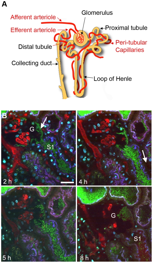
Figure 1
UPEC attachment in Bowman's capsule.
( A ) Schematic showing nephron structure. ( B ) Dynamic imaging of LT004 infused directly into Bowman's capsule. ( 2 h ) Bacteria (green, arrow) conformally lining to Bowman's capsule epithelia of a ...
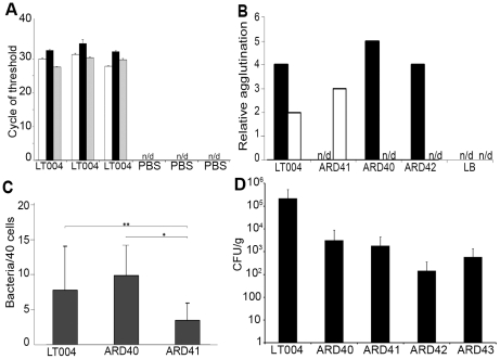
Figure 2
Expression, binding and infection characteristics of P and Type 1 isogenic strains.
( A ) qRT-PCR analysis of micro-dissected tissues 8 h after infusion with LT004 or PBS. Bars show cycle of threshold for detection of gfp + (white), papA_2 (black) and fimA (grey) transcript. n/d u200...
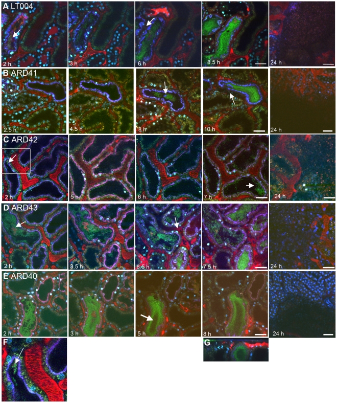
Figure 3
Progression of infection in live animals.
Multiphoton imaging of renal tissue after infusion of indicated strains (green). Injected proximal tubules are outlined by a co-injected 10 kDa cascade-blue dextran (blue); blood plasma is labeled wit...
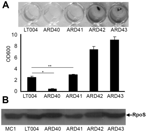
Figure 4
UPEC biofilm formation.
( A ) Crystal violet assay of biofilm formation from indicated strains. Visualization (top) and quantification (bottom) of biofilm at OD 600 . Error bars u200a=u200a standard deviation. * Pu200a=u200a...
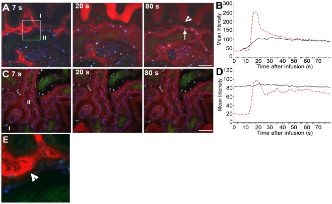
Figure 5
Infection affects renal filtration.
Glomerular filtration in non-infected (I) and infected (II) tubules 4 h (A,B) and 8 h (C, D) after LT004 infusion is visualized 7, 20, and 80 s after iv bolus infusion of 10 kDa dextran (red). These d...
Figure images are served from the NIH/NLM PubMed Central Open Access Subset or Europe PMC; copyright remains with the publishers and authors.
💬 Discussion
0 commentsNo comments yet. Be the first to start a discussion!
Leave a Comment