Abstract
This review provides a practical introduction to superresolution microscopy from the perspective of microbiological research. Because of the small sizes of bacterial cells, superresolution methods are particularly powerful and suitable for revealing details of cellular structures that are not resolvable under conventional fluorescence light microscopy. Here we describe the methodological concepts behind three major categories of superresolution light microscopy: photoactivated localization microscopy (PALM) and stochastic optical reconstruction microscopy (STORM), structured illumination microscopy (SIM) and stimulated emission-depletion (STED) microscopy. We then present recent applications of each of these techniques to microbial systems, which have revealed novel conformations of cellular structures and described new properties of in vivo protein function and interactions. Finally, we discuss the unique issues related to implementing each of these superresolution techniques with bacterial specimens and suggest avenues for future development. The goal of this review is to provide the necessary technical background for interested microbiologists to choose the appropriate superresolution method for their biological systems, and to introduce the practical considerations required for designing and analysing superresolution imaging experiments.
🔬 Techniques
🧬 Organisms
✨ Fluorophores
🔬 Cell Lines
🏛️ Research Organizations (ROR)
Affiliated research institutions:
📋 Methods
Single-molecule localization methods Concept
Photoactivatable and photoswitchable fluorophores are the key to implementing localization-based superresolution techniques. These fluorophores can transition stochastically between bright and dark emission states by exposure to specific wavelengths of light. During super-resolution imaging, activation light is kept at a low level such that within a diffraction-limited area only a single fluorophore is fluorescing at a time ( Fig. 1A ). The positions of these single molecules are then localized with nanometre accuracy by fitting their intensity profiles with a Gaussian function that approximates the microscope’s PSF. Positions collected from thousands of frames are then overlaid to reconstruct a superresolution image. During this long acquisition time, fiducial beads are often added to the sample to track and calibrate stage drift. This principle was first demonstrated in 2006 using photoactivatable fluorescent proteins (PALM) ( Betzig et al ., 2006 ) and cyanine dye pairs (STORM) ( Rust et al ., 2006 ), and resolutions of 10 nm and 20 nm were achieved respectively. Later, this principle was extended to conventional organic dyes, which have been shown to photoswitch robustly under proper buffer conditions ( Folling et al ., 2008 ; Heilemann et al ., 2008 ; Burnette et al ., 2011 ). The general principle of isolating single fluorophores can be further extended to non-photon-driven switching. For example, PAINT (Point Accumulation for Imaging in Nanoscale Topography) utilizes fluorophores that can only be detected when their fluorescence is enhanced ∼ 1000-fold by binding reversibly to a lipid membrane ( Sharonov and Hochstrasser, 2006 ). New observations and insights Single-molecule localization-based superresolution imaging techniques are the simplest to implement instrumentally, and have consequently been most quickly adopted by microbiologists. Using these methods, fine details of a variety of bacterial superstructures that are unresolvable under conventional light microscopy have been revealed. For example, the actin homologue MreB, which is necessary for cell shape maintenance, was shown to adopt helix-like structures in early stages of the Caulobacter crescentus cell cycle and ring-like structures in late stages ( Biteen et al ., 2008 ). The tubulin homologue FtsZ, which is essential for cytokinesis, was found to exist in either a compact helix or ring conformation in non-constricting Escherichia coli cells ( Fig. 2A ) ( Fu et al ., 2010 ). SpoIIIE, the translocase that sequesters DNA into nascent spores in Bacillus subtilis , was found to be confined to the leading edges of constricting spore septa ( Fleming et al ., 2010 ), providing unprecedented structural insight into the protein’s function. In addition to revealing fine details of cellular superstructures, localization-based superresolution techniques have also been used to describe the dynamics and activities of various proteins. By mapping the diffusion and distribution of ribosomes and RNA polymerases in live E. coli cells using PALM, Bakshi et al . determined that the most translation occurs on free mRNA and that co-transcriptional insertion of proteins into the membrane, or ‘transertion’, may exert an expansion force on the nucleoid in the lateral direction ( Bakshi et al ., 2012 ). In support of a chromosome-organizing role for the E. coli nucleoid-associated protein, H-NS, Wang et al . observed distinct clusters of the H-NS in live E. coli cells while other nucleoid-associated proteins (HU, Fis and IHF) were observed to be more scattered ( Wang et al ., 2011 ). Finally, English et al . probed the activity cycle of RelA, a stringent response factor, in E. coli by tracking the diffusion of RelA molecules before and during starvation ( English et al ., 2011 ). They observed that RelA diffuses as a ribosome-bound complex during normal conditions, but becomes cytoplasmic during starvation, indicating that RelA activity occurs off the ribosome. These experiments highlight the ability of superresolution techniques to answer fundamental questions of bacterial cell biology.
Show full methods section
Single-molecule localization methods Concept
Photoactivatable and photoswitchable fluorophores are the key to implementing localization-based superresolution techniques. These fluorophores can transition stochastically between bright and dark emission states by exposure to specific wavelengths of light. During super-resolution imaging, activation light is kept at a low level such that within a diffraction-limited area only a single fluorophore is fluorescing at a time ( Fig. 1A ). The positions of these single molecules are then localized with nanometre accuracy by fitting their intensity profiles with a Gaussian function that approximates the microscope’s PSF. Positions collected from thousands of frames are then overlaid to reconstruct a superresolution image. During this long acquisition time, fiducial beads are often added to the sample to track and calibrate stage drift. This principle was first demonstrated in 2006 using photoactivatable fluorescent proteins (PALM) ( Betzig et al ., 2006 ) and cyanine dye pairs (STORM) ( Rust et al ., 2006 ), and resolutions of 10 nm and 20 nm were achieved respectively. Later, this principle was extended to conventional organic dyes, which have been shown to photoswitch robustly under proper buffer conditions ( Folling et al ., 2008 ; Heilemann et al ., 2008 ; Burnette et al ., 2011 ). The general principle of isolating single fluorophores can be further extended to non-photon-driven switching. For example, PAINT (Point Accumulation for Imaging in Nanoscale Topography) utilizes fluorophores that can only be detected when their fluorescence is enhanced ∼ 1000-fold by binding reversibly to a lipid membrane ( Sharonov and Hochstrasser, 2006 ). New observations and insights Single-molecule localization-based superresolution imaging techniques are the simplest to implement instrumentally, and have consequently been most quickly adopted by microbiologists. Using these methods, fine details of a variety of bacterial superstructures that are unresolvable under conventional light microscopy have been revealed. For example, the actin homologue MreB, which is necessary for cell shape maintenance, was shown to adopt helix-like structures in early stages of the Caulobacter crescentus cell cycle and ring-like structures in late stages ( Biteen et al ., 2008 ). The tubulin homologue FtsZ, which is essential for cytokinesis, was found to exist in either a compact helix or ring conformation in non-constricting Escherichia coli cells ( Fig. 2A ) ( Fu et al ., 2010 ). SpoIIIE, the translocase that sequesters DNA into nascent spores in Bacillus subtilis , was found to be confined to the leading edges of constricting spore septa ( Fleming et al ., 2010 ), providing unprecedented structural insight into the protein’s function. In addition to revealing fine details of cellular superstructures, localization-based superresolution techniques have also been used to describe the dynamics and activities of various proteins. By mapping the diffusion and distribution of ribosomes and RNA polymerases in live E. coli cells using PALM, Bakshi et al . determined that the most translation occurs on free mRNA and that co-transcriptional insertion of proteins into the membrane, or ‘transertion’, may exert an expansion force on the nucleoid in the lateral direction ( Bakshi et al ., 2012 ). In support of a chromosome-organizing role for the E. coli nucleoid-associated protein, H-NS, Wang et al . observed distinct clusters of the H-NS in live E. coli cells while other nucleoid-associated proteins (HU, Fis and IHF) were observed to be more scattered ( Wang et al ., 2011 ). Finally, English et al . probed the activity cycle of RelA, a stringent response factor, in E. coli by tracking the diffusion of RelA molecules before and during starvation ( English et al ., 2011 ). They observed that RelA diffuses as a ribosome-bound complex during normal conditions, but becomes cytoplasmic during starvation, indicating that RelA activity occurs off the ribosome. These experiments highlight the ability of superresolution techniques to answer fundamental questions of bacterial cell biology.
Quantitative measurements
Quantitative measurements such as structural dimensions can also be made from superresolution images. Feature dimensions are often measured as the FWHM that results from fitting feature profiles to Gaussian distributions. From superresolution studies we now know the width of the FtsZ ring in E. coli (110 nm) ( Fu et al ., 2010 ) and C. crescentus (67 nm prior to division; 92 nm during division) ( Biteen et al ., 2012 ), the width of linear ParA bundles that span C. crescentus cells to facilitate chromosome segregation (40 nm) ( Ptacin et al ., 2010 ), and the diameter of crescentin fibres that give C. crescentus its characteristic crescent shape (92 nm, converted to FWHM from reported σ-value) ( Lew et al ., 2011 ). When combined with average protein copy numbers, these measurements can provide valuable information about the arrangement of molecules within structures. For example, because the E. coli FtsZ ring width would be threefold smaller if all FtsZ molecules were packed next to each other, Fu et al . deduced that the FtsZ ring is a loose structure with unoccupied regions that can be occupied by other associated proteins ( Fu et al ., 2010 ). Single-molecule localization-based superresolution imaging also allows molecule density measurements (number of molecules per unit area), which can be analysed for non-uniformity to provide information about molecular interactions. By measuring the distribution of molecule counts within chemotaxis clusters, Greenfield et al . were able to confirm the proposed stochastic assembly mechanism of chemotaxis clusters in E. coli ( Greenfield et al ., 2009 ). By comparison to simulated random distributions, Lee et al . determined that the distribution of HU, a nucleoid-associated protein, is non-uniform, perhaps because HU localization is enhanced in highly transcribed regions ( Lee et al ., 2011 ). Furthermore, using molecule density measurements, three-dimensional information can be extracted from two-dimensional projections. For example, Fu et al . determined that the number of molecules detected per pixel within E. coli FtsZ rings is consistent with a multilayer arrangement of FtsZ protofilaments ( Fu et al ., 2010 ). One caveat (discussed in the section Practical considerations ) to molecule counting, however, is that fluorophores often ‘blink’, yielding multiple observations of the same molecule ( Annibale et al ., 2011a ), so caution must be taken to identify unique molecules.
Multicolour imaging
Multicolour superresolution imaging is an extremely powerful tool for investigating protein–protein or protein–DNA interactions in their native cellular environment. Proteins that appear to colocalize with each other using conventional fluorescence light microscopy may not necessarily be in direct contact because the size of a diffraction-limited spot is much larger than that of a single protein (1–10 nm). Superresolution imaging determines the spatial relationship between two proteins with much higher precision that allows more confident assessment of protein interactions. For example, Ptacin et al . investigated chromosome segregation in C. crescentus by using PALM to simultaneously determine the density of the ParA bundle that spans the cell length and the location of parS loci that serve as chromosomal anchors for ParA ( Fig. 2B ) ( Ptacin et al ., 2010 ). By comparing images of cells at different stages of chromosome segregation (where one parS locus moves across the cell), the authors determined that segregation occurs via retraction of a subset of ParA fibres. Using two-colour imaging in E. coli , Wang et al . found a high degree of colocalization between H-NS and two genes that it regulates ( hdeA and hchA ) but low colocalization between H-NS and lacZ , which it does not regulate, suggesting that H-NS-regulated genes may be organized into close clusters ( Fig. 2C ) ( Wang et al ., 2011 ). As a diffraction-limited spot is similar in size to the E. coli nucleoid, the different H-NS colocalization patterns of these genes would be indistinguishable with conventional microscopy. Multicolour superresolution imaging also has the potential to provide unprecedented detail into host–pathogen interactions. The first demonstration of this was by Lehmann et al ., who performed two-colour superresolution imaging of assembling human immunodeficiency virus (HIV) particles in HeLa cells ( Lehmann et al ., 2011 ). They determined molecule distributions of Gag, the HIV-1 structural protein, and tetherin, a human protein that inhibits release of budding virus particles, and found that most budding HIV-1 particles were colocalized with a single cluster of tetherin, rather than recruiting multiple clusters.
📊 Figures
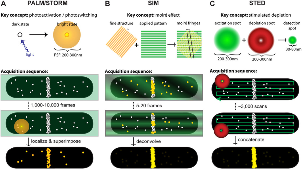
Fig. 1
Key concepts and acquisition schematics for each superresolution technique. In each schematic, molecule positions are shown as white circles, excitation light is represented in green, depletion light ...
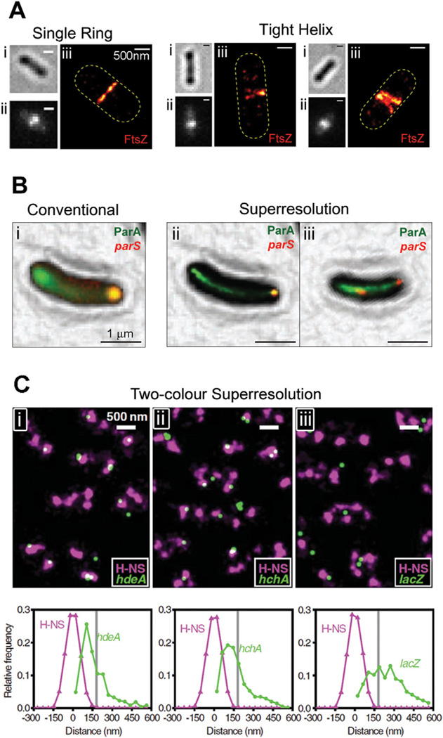
Fig. 2
Single-molecule localization-based superresolution images. A. Superstructures formed by FtsZ-mEos2 in E. coli . For each cell, the brightfield (i), conventional fluorescence (ii) and superresolution (...
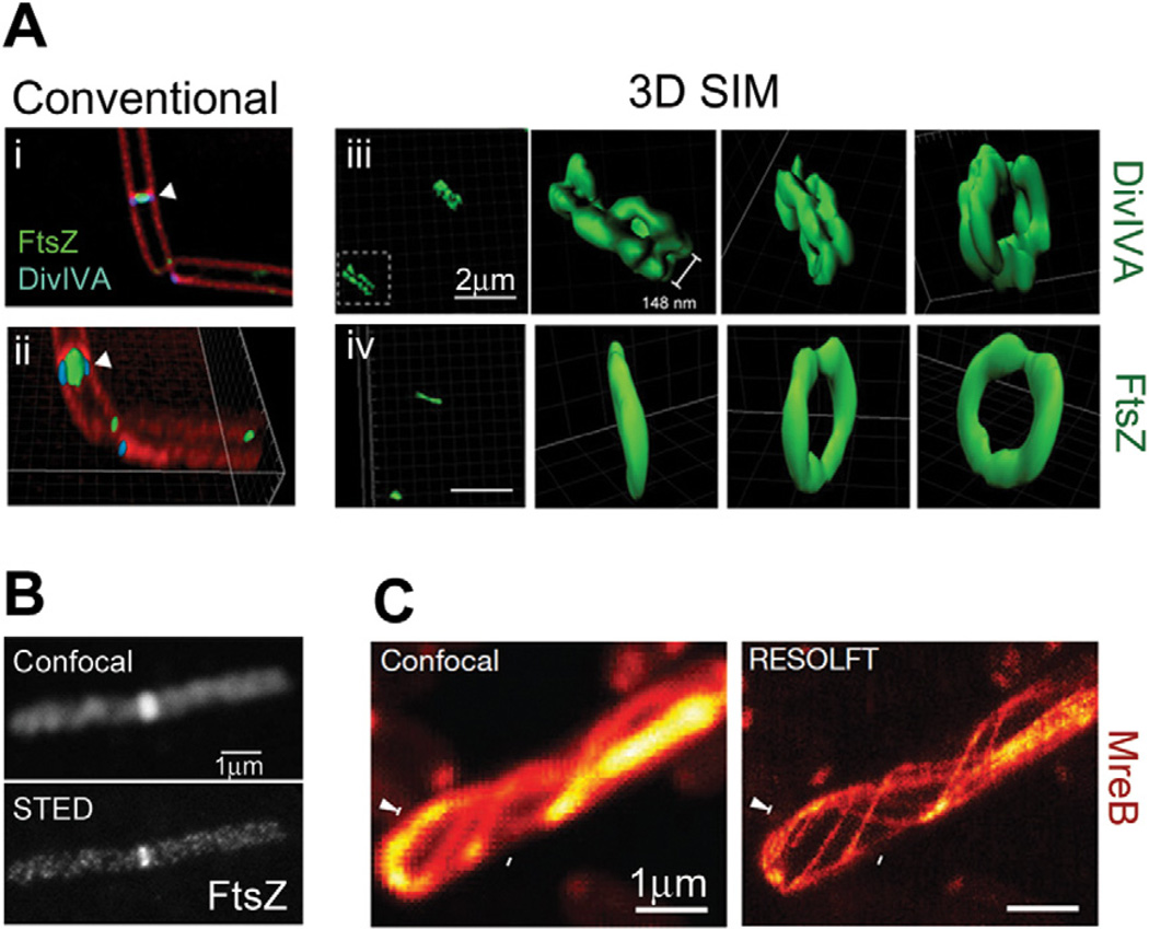
Fig. 3
SIM and STED superresolution images. A. Ring structures formed by DivIVA and FtsZ in B. subtilis cells. Diffraction-limited images generated by conventional deconvolution (i and ii) show the proximity...
Figure images are served from the NIH/NLM PubMed Central Open Access Subset or Europe PMC; copyright remains with the publishers and authors.
💬 Discussion
0 commentsNo comments yet. Be the first to start a discussion!
Leave a Comment