Abstract
The apical complex is the definitive cell structure of phylum Apicomplexa, and is the focus of the events of host cell penetration and the establishment of intracellular parasitism. Despite the importance of this structure, its molecular composition is relatively poorly known and few studies have experimentally tested its functions. We have characterized a novel Toxoplasma gondii protein, RNG2, that is located at the apical polar ring--the common structural element of apical complexes. During cell division, RNG2 is first recruited to centrosomes immediately after their duplication, confirming that assembly of the new apical complex commences as one of the earliest events of cell replication. RNG2 subsequently forms a ring, with the carboxy- and amino-termini anchored to the apical polar ring and mobile conoid, respectively, linking these two structures. Super-resolution microscopy resolves these two termini, and reveals that RNG2 orientation flips during invasion when the conoid is extruded. Inducible knockdown of RNG2 strongly inhibits host cell invasion. Consistent with this, secretion of micronemes is prevented in the absence of RNG2. This block, however, can be fully or partially overcome by exogenous stimulation of calcium or cGMP signaling pathways, respectively, implicating the apical complex directly in these signaling events. RNG2 demonstrates for the first time a role for the apical complex in controlling secretion of invasion factors in this important group of parasites.
🔬 Techniques
✨ Fluorophores
🧪 Sample Preparation
🏭 Microscope Brands
🧪 Reagent Suppliers
📷 Detectors
🏛️ Research Organizations (ROR)
Affiliated research institutions:
📋 Methods
Growth and generation of protein-tagged and knockdown parasites T. gondii tachyzoites were grown by serial passage in human foreskin fibroblast (HFF) cells as previously described [85] . RNG2-HA parasites were previously generated by 3′ endogenous tagging with 3XHA coding sequence of the rng2 gene (toxodb.org gene ID:TgME49_244470) [29] . To generate the conditional RNG2 knockdown parasite strain (iΔHA-RNG2), we began by amplifying 2015 bp upstream of the RNG2 start codon (5′ flank) using the primers 5′-CTGACATATGGAGACTGCCACAAAGGAAGGTACAC and 5′-GATCATCCATCGAAACGCTCCGTGACGGAAGTA . We digested the product with Nsi I and Nde I and ligated this into the equivalent sites of the vector pPR2-HA3 (Chris Tonkin and GvD, unpublished), a modified version of the vector pPR (a kind gift from Lilach Sheiner, U. Georgia; [42] ). We next amplified a 2042 bp fragment beginning at the start codon of RNG2 (3′ flank) with the primers 5′-GATCCCCGGGATGCACCCCCACCTTTCTTCCGCAG and 5′-CGATGCGGCCGCGACGGTGGTGTTATTGATTGGTTGC . We digested this with Xma I and Not I and ligated this into equivalent sites of the pPR2-HA3 vector that already contained the RNG2 5′ flank. The resulting vector positions the first RNG2 codon downstream of the ATc-regulatable t7s4 promoter and a 3xHA tag. We linearized the resulting vector with Not I and transfected this into TATiΔku80 parasites (a kind gift from Lilach Sheiner and Boris Striepen, U. Georgia; [42] ). Parasites were selected with pyrimethamine and cloned by limiting dilution. To identify parasite clones where the t7s4 promoter had successfully replaced the native RNG2 promoter, we utilized the primers P1 ( 5′- CAGATTCCGAATTCTTTGG ), P2 ( 5′-TGTAGAGCTGGTGCGTGAG ) and P3 ( 5′-AAGGGGACGCAGTTCTCGGA ) in the combinations described in Figure 5A . For RNG2 cMyc tagging, we PCR amplified a 3′ fragment of rng2 gene using the primers 5′- GATCAGATCTGCAGCTGACACACTCCTGACG and 5′- GCATTCTAGAGTTTGTTGATGCGTCCGAGACAAC , digested this with Bgl II and Xba I and ligated into the Bgl II and Avr II sites of the vector pgCM3, a vector that fuses the 3′ region of a gene-of-interest with a 3× cmyc tag (NK and GvD, unpublished). This vector was linearized with Avr II, transfected into the iΔHA-RNG2 strain, selected on chloramphenicol and cloned by limiting dilution. RNG2 knockdown was induced by culturing with 0.5 μg ml −1 of anhydrotetracycline (ATc). All PCRs were performed with Phusion polymerase (Thermo Scientific). To tag the C-terminus of RNG2 with GFP, we digested the pCTG vector [86] with Avr II and Bam HI and ligated this into the equivalent sites of the pHA 3 -LIC-DHFR(RNG2) vector [29] . This exchanged the 3xHA tag with a GFP tag in a RNG2 3′ replacement vector that we termed pGFP-LIC-DHFR(RNG2). We linearized the resultant vector with Nsi I, transfected this into the TATi/ΔKu80 parasites [42] and selected on pyrimethamine. We cloned the resulting drug resistant parasites and confirmed expression of RNG2-GFP by microscopy. We next tagged the N-terminus of RNG2-GFP with a mCherry-3× c-myc tag through a promoter replacement strategy. First, we digested the pPR2-HA 3 (RNG2) vector described above with Nhe I and Xma I to excise the 3x-HA tag. We then digested the mCherry-3× c-myc tag from the vector pCTChM3 (a kind gift from Chris Tonkin, Walter and Eliza Hall Institute) with Nhe I and Xma I and ligated the resulting fragment into the pPR2 (RNG2) vector to generate the vector we termed pPR2-GFP(RNG2). We next replaced the pyrimethamine-resistance cassette in this vector with a chloramphenicol-resistance cassette. We PCR amplified the chloramphenicol-resistance cassette from the vector pgCM 3 using the primers 5′-GATCATGCATAAAACCCTCGAAGGCTGCTAGTAC and 5′-GATCACTAGTGGATCCCCCTCGGG . The resulting PCR product was digested with Spe I and Nsi I and ligated into the equivalent sites of the pPR2-GFP(RNG2) vector to generate a vector we termed pPR2-CAT-GFP(RNG2). We linearized this vector with Not I and transfected this into the RNG2-GFP cell line, selecting on chloramphenicol. We cloned drug-resistant parasites, and confirmed integration through Western blotting. For C-terminal 3XcMyc-tagged RNG1 and CAM1 (via 3′ endogenous gene replacement) the coding sequence of each gene was amplified and cloned into pBTM3 (GvD, unpublished). The primers used to amplify the coding sequence of RNG1 were 5′- GATCAGATCTAAAATGGCGCTAATTCCCTCGC and 5′- GATCCCTAGGCGCCAGGTAGTAGACAGGTGGA , and CAM1 were 5′-GATCCCTAGGTTTATTCGCGGAAGGCAGAGAC and 5′-TGGACTGTGGTCGACGCAGAAG . For transient expression of RNG1-GFP, the 3XcMyc tag was removed from the RNG1-cMyc vector by digestion with Avr II and Not 1 and GFP coding sequence ligated in its place. The CAM1-GFP vector was a kind gift by Martin Blume, Bio21, Australia. To label MORN1 we transiently transfected parasites with a cMyc-tagged MORN1 plasmid (a kind gift from MJ Gubbels, Boston College).
Show full methods section
Growth and generation of protein-tagged and knockdown parasites T. gondii tachyzoites were grown by serial passage in human foreskin fibroblast (HFF) cells as previously described [85] . RNG2-HA parasites were previously generated by 3′ endogenous tagging with 3XHA coding sequence of the rng2 gene (toxodb.org gene ID:TgME49_244470) [29] . To generate the conditional RNG2 knockdown parasite strain (iΔHA-RNG2), we began by amplifying 2015 bp upstream of the RNG2 start codon (5′ flank) using the primers 5′-CTGACATATGGAGACTGCCACAAAGGAAGGTACAC and 5′-GATCATCCATCGAAACGCTCCGTGACGGAAGTA . We digested the product with Nsi I and Nde I and ligated this into the equivalent sites of the vector pPR2-HA3 (Chris Tonkin and GvD, unpublished), a modified version of the vector pPR (a kind gift from Lilach Sheiner, U. Georgia; [42] ). We next amplified a 2042 bp fragment beginning at the start codon of RNG2 (3′ flank) with the primers 5′-GATCCCCGGGATGCACCCCCACCTTTCTTCCGCAG and 5′-CGATGCGGCCGCGACGGTGGTGTTATTGATTGGTTGC . We digested this with Xma I and Not I and ligated this into equivalent sites of the pPR2-HA3 vector that already contained the RNG2 5′ flank. The resulting vector positions the first RNG2 codon downstream of the ATc-regulatable t7s4 promoter and a 3xHA tag. We linearized the resulting vector with Not I and transfected this into TATiΔku80 parasites (a kind gift from Lilach Sheiner and Boris Striepen, U. Georgia; [42] ). Parasites were selected with pyrimethamine and cloned by limiting dilution. To identify parasite clones where the t7s4 promoter had successfully replaced the native RNG2 promoter, we utilized the primers P1 ( 5′- CAGATTCCGAATTCTTTGG ), P2 ( 5′-TGTAGAGCTGGTGCGTGAG ) and P3 ( 5′-AAGGGGACGCAGTTCTCGGA ) in the combinations described in Figure 5A . For RNG2 cMyc tagging, we PCR amplified a 3′ fragment of rng2 gene using the primers 5′- GATCAGATCTGCAGCTGACACACTCCTGACG and 5′- GCATTCTAGAGTTTGTTGATGCGTCCGAGACAAC , digested this with Bgl II and Xba I and ligated into the Bgl II and Avr II sites of the vector pgCM3, a vector that fuses the 3′ region of a gene-of-interest with a 3× cmyc tag (NK and GvD, unpublished). This vector was linearized with Avr II, transfected into the iΔHA-RNG2 strain, selected on chloramphenicol and cloned by limiting dilution. RNG2 knockdown was induced by culturing with 0.5 μg ml −1 of anhydrotetracycline (ATc). All PCRs were performed with Phusion polymerase (Thermo Scientific). To tag the C-terminus of RNG2 with GFP, we digested the pCTG vector [86] with Avr II and Bam HI and ligated this into the equivalent sites of the pHA 3 -LIC-DHFR(RNG2) vector [29] . This exchanged the 3xHA tag with a GFP tag in a RNG2 3′ replacement vector that we termed pGFP-LIC-DHFR(RNG2). We linearized the resultant vector with Nsi I, transfected this into the TATi/ΔKu80 parasites [42] and selected on pyrimethamine. We cloned the resulting drug resistant parasites and confirmed expression of RNG2-GFP by microscopy. We next tagged the N-terminus of RNG2-GFP with a mCherry-3× c-myc tag through a promoter replacement strategy. First, we digested the pPR2-HA 3 (RNG2) vector described above with Nhe I and Xma I to excise the 3x-HA tag. We then digested the mCherry-3× c-myc tag from the vector pCTChM3 (a kind gift from Chris Tonkin, Walter and Eliza Hall Institute) with Nhe I and Xma I and ligated the resulting fragment into the pPR2 (RNG2) vector to generate the vector we termed pPR2-GFP(RNG2). We next replaced the pyrimethamine-resistance cassette in this vector with a chloramphenicol-resistance cassette. We PCR amplified the chloramphenicol-resistance cassette from the vector pgCM 3 using the primers 5′-GATCATGCATAAAACCCTCGAAGGCTGCTAGTAC and 5′-GATCACTAGTGGATCCCCCTCGGG . The resulting PCR product was digested with Spe I and Nsi I and ligated into the equivalent sites of the pPR2-GFP(RNG2) vector to generate a vector we termed pPR2-CAT-GFP(RNG2). We linearized this vector with Not I and transfected this into the RNG2-GFP cell line, selecting on chloramphenicol. We cloned drug-resistant parasites, and confirmed integration through Western blotting. For C-terminal 3XcMyc-tagged RNG1 and CAM1 (via 3′ endogenous gene replacement) the coding sequence of each gene was amplified and cloned into pBTM3 (GvD, unpublished). The primers used to amplify the coding sequence of RNG1 were 5′- GATCAGATCTAAAATGGCGCTAATTCCCTCGC and 5′- GATCCCTAGGCGCCAGGTAGTAGACAGGTGGA , and CAM1 were 5′-GATCCCTAGGTTTATTCGCGGAAGGCAGAGAC and 5′-TGGACTGTGGTCGACGCAGAAG . For transient expression of RNG1-GFP, the 3XcMyc tag was removed from the RNG1-cMyc vector by digestion with Avr II and Not 1 and GFP coding sequence ligated in its place. The CAM1-GFP vector was a kind gift by Martin Blume, Bio21, Australia. To label MORN1 we transiently transfected parasites with a cMyc-tagged MORN1 plasmid (a kind gift from MJ Gubbels, Boston College).
Microscopy Immunofluorescence assays
(IFAs) were performed as previously described [87] using antibodies and their concentrations listed in Supplemental Table S1 . 3D-SIM was implemented on a DeltaVision OMX V4 Blaze (Applied Precision) with samples prepared as described [88] , excited using 488 and 568 nm lasers and imaged using band pass filters at 528 and 608 with a 60× oil immersion lens (1.42 NA). Parasite pellicles were extracted in deoxycholate as previously described [66] . Briefly, we filtered parasites through a 3 μm filter and resuspended them in phosphate-buffered saline. We attached parasites to coverslips with 0.1% polyethyleneimine (PEI) and extracted in 10 mM deoxycholate for 10 min at room temperature. We fixed parasites in 4% paraformaldehyde for 10 min and then proceeded as for IFAs. Cells or cell extracts were analyzed on a Leica TCS SP2 confocal laser-scanning microscope. Only the brightness/contrast ratio of the images was modified, using Adobe Photoshop CS4. For live cell imaging, parasites were incubated in glass-bottomed dishes (MatTek) in phenol-red free Dulbecco's modified Eagle's medium supplemented with 1% foetal calf serum and antibiotics. During imaging, parasites were incubated in a 5% CO 2 /air atmosphere in a humidified 37°C chamber. Imaging was performed using a DeltaVision set-up with an inverted Olympus IX71 microscope, an Olympus objective lens (UPlanSApo, 100×/1.40 oil), and a Photometrics CoolSNAP HQ 2 camera. Images were acquired using 2×2 binning, and deconvolved prior to linear adjustment of contrast and brightness. For transmission electron microscopy, parasites were cultured for three days on 0.5 μg ml −1 ATc, fixed in PBS with 2.5% paraformaldehyde and 1% glutaraldehyde, post fixed in 1% OsO 4 , and pellet-embedded in 1% low-melting agarose. The agarose block was ethanol dehydrated, embedded in LR White resin and polymerized. Ultrathin sections were cut on a Leica Ultracut R microtome, lead and uranium stained and visualized with a Philips CM120 BioTWIN transmission electron microscope at 120 kV.
Western blot analysis
Antibodies and their concentrations used for Western blots are listed in Table S1 . Parasites were filtered through a 3 μm filter, counted by haemocytometer and solubilized in sample buffer (Invitrogen) at equivalent cell densities. Standard Western blot detection was performed, with Horse Radish Peroxidase conjugated secondary antibodies detected using SuperSignal West Pico Chemiluminescent Substrate (Pierce). Signal strength was quantified using a BioRad Chemidoc imager. Growth, replication and conoid extrusion assays For growth assays extracellular parasites were filtered, counted by haemocytometer, and 500 parasites added to 25 cm 2 tissue culture flasks containing a confluent monolayer of HFF cells. ATc (0.5 μg ml −1 ) was added from the outset of the experiment. To visualize plaque sizes, flasks were aspirated, fixed with 5 ml 100% ethanol (5 minutes), stained with 5 ml crystal violet solution (15 minutes) then washed once with 1× phosphate-buffered saline (PBS) and dried before imaging. For replication assays, parasites grown for three days with or without ATc (0.5 μg ml −1 ), harvested and filtered. Equal numbers were allowed to invade HFF cells on coverslips for two hours. Coverslips were washed three times with Dulbecco's modified Eagle's medium (DMEM) (supplemented with 1% FCS, 0.2 mM L-glutamine) to remove uninvaded parasites, and cultured for 24 hours with ongoing +/− ATc regimens. Cells were then fixed and processed for SAG1 IFA and parasite number per parasitophorous vacuole scored. To assess conoid extrusion ability, parasites were grown for three days with or without ATc, harvested, filtered and resuspened in DMEM to 2.5×10 7 parasites ml −1 . A23187 was added to samples at a final concentration of 5 μM (or equivalent volume of DMSO as a control), and parasites incubated for 30 seconds at 37 °C, then fixed with 1.25% glutaraldehyde and settled on PEI coated coverslips. Conoid extrusion was scored by phase microscopy with >200 cells counted per replicate (n = 3). Invasion, motility and evacuole assays Red/green invasion assays were performed as described previously [44] , [45] . Briefly, parasites were grown for two days with or without ATc, harvested within HFF cells by trypsinisation, then mechanically released by passage through a 26 gauge needle in Endo Buffer (44.7 mM K 2 SO 4 , 10 mM MgSO 4 , 106 mM sucrose, 5 mM glucose, 20 mM Tris-H 2 SO 4 , 3.5 mg/ml BSA, pH 8.2). Cells were counted and resuspended to 2.5×10 7 parasites ml −1 , and 200 μl allowed to settle onto HFF cells on coverslips in Endo buffer for 20 minutes. Endo Buffer was then aspirated, and replaced with 200 μl of Invasion Buffer (DMEM supplemented with 3% FCS, 10 mM HEPES, pH 7.4). After 10 minutes at 37 °C, cells were fixed with 2.5% Paraformaldehyde and 0.02% glutaraldehyde in PBS, blocked, then probed with anti-SAG1 (Abcam) to label uninvaded cells. Samples were then permeabilized (0.25%TX100 in 1xPBS) for 10 minutes and probed with anti-GAP45 to label all cells. IFAs were completed with secondary antibodies as normal, and then imaged using a Leica SP2 confocal microscope. Fields of view were selected observing the green (anti-GAP45) channel only to eliminate biased selection of parasites. Images were processed using the Leica SP2 software, and labeled cells scored according to invaded (red) or uninvaded (red and green). A minimum of 200 parasites were scored for each of three biological replicates, and invasion percentage calculated as invaded over total parasites. For motility assays, PEI coated coverslips were incubated with fetal calf serum for two hours. Parasite cultures grown for two days with or without ATc were needle passed, filtered and resuspended to 10 7 cells ml −1 . 1 ml of parasites was placed on coverslip with either no drug (plus DMSO to equivalent of drug samples), 1 μm cytochalasin D, or 5 μM A23187, and incubated at 37 °C for 90 minutes. Samples were then fixed and SAG1 IFAs performed. Evacuole assays were performed as previously described [47] .
Secretion assay
Parasite cultures grown for two days with or without ATc were harvested, pelleted, washed with Invasion Buffer and resuspended to 2.5×10 8 cells ml −1 . Cells were maintained at 20°C in all steps post harvesting. 50 μl of parasite suspensions were mixed separately with an equal volume of either Invasion Buffer alone (plus DMSO to equivalent of drug samples), 1.0 mM μM Zaprinast, 10 μM A23187 or 10 mM 8-Br-cGMP (final concentrations 0.5 mM, 5 μm, and 5 mM, respectively). Cells were incubated at 37°C for 20 minutes to allow secretion, then arrested on ice for 2 minutes before parasites were separated from secreted soluble proteins by centrifugation at 8000rpm at 4°C for 2 minutes. 85 μl of supernatant was removed, centrifuged at 8000rpm at 4°C for 2 minutes to remove any remaining cells, and 75 μl removed and boiled with Sample Buffer as the secreted protein fraction. The pelleted cells were washed with PBS and then boiled with Sample Buffer. Secreted protein samples were analyzed for MIC2 and AMA1 by Western blot, and cell pellets analyzed for Tom40 to verify equal cell numbers used for the different assay conditions.
Supporting Information Figure S1 Live cell imaging of the N- and C-termini of RNG2. (A-B) A Western blot depicting mCherry-cMyc-RNG2-GFP parasites probed with (A) anti-cMyc and (B) anti-GFP. In both blots, the masses of the tagged RNG2 protein are equivalent in size (>260 kDa), indicative of successful targeting of both termini of the gene. (C) Live cell imaging of mCherry-cMyc-RNG2-GFP intracellular parasites. Arrowheads depict the apical ring. In all parasites, N-terminal mCherry labeling is posterior to the C-terminal GFP labeling. (i) In the newly formed apical rings of daughter buds, the N-terminus of RNG2 is also posterior to the C-terminus (bottom panel, arrows). Note: fluorescence is also retained in the residual bodies. (D) Live cell imaging of mCherry-c-myc-RNG2-GFP parasites in extracellular parasites. (i) N-terminal mCherry labeling is posterior to the C-terminal GFP-labeling when the conoid is retracted. (ii) Treatment with Ca 2+ ionophore A23187 causes conoid extrusion, and relocation of the N-terminus of RNG2 to the anterior side of the C-terminus. Scale bars are 2 μm. (PDF) Click here for additional data file. Figure S2 Western blot and immunofluorescence assays for microneme maturity with RNG2 depletion. (A) In parasite total protein samples, microneme proteins MIC2 and AMA1, and mitochondrial protein Tom40, show equivalent amounts of protein in iΔHA-RNG2 cells treated with or without ATc for three days. Only HA-RNG2, detected by HA antibodies, shows depletion with ATc treatment. Equal cell numbers were used in all gel lanes. (B, C) IFA detection of (B) AMA1 and HA-RNG2, or (C) MIC2 in intracellular iΔHA-RNG2 cells treated with or without ATc for two days. Scale bar = 5 μm. (PDF) Click here for additional data file. Figure S3 8-Br-cGMP-stimulation of microneme secretion is muted in RNG2 minus cells. MIC2 secretion without RNG2 (iΔHA-RNG2 cells +ATc) or with RNG2 (iΔHA-RNG2 cells -ATc and parental cells). Constitutive MIC2 secretion, and secretion with exogenous cGMP (by analogue 8-Br-cGMP) (A), or calcium stimulation (by ionophore A23187) (B) is assayed by Western blot. Stimulated microneme secretion by exogenous calcium is strong in all cells, but by exogenous cGMP is reduced in RNG2 knockdown cells. (PDF) Click here for additional data file. Table S1 Antibodies used for microscopy and protein assays. (PDF) Click here for additional data file.
📊 Figures
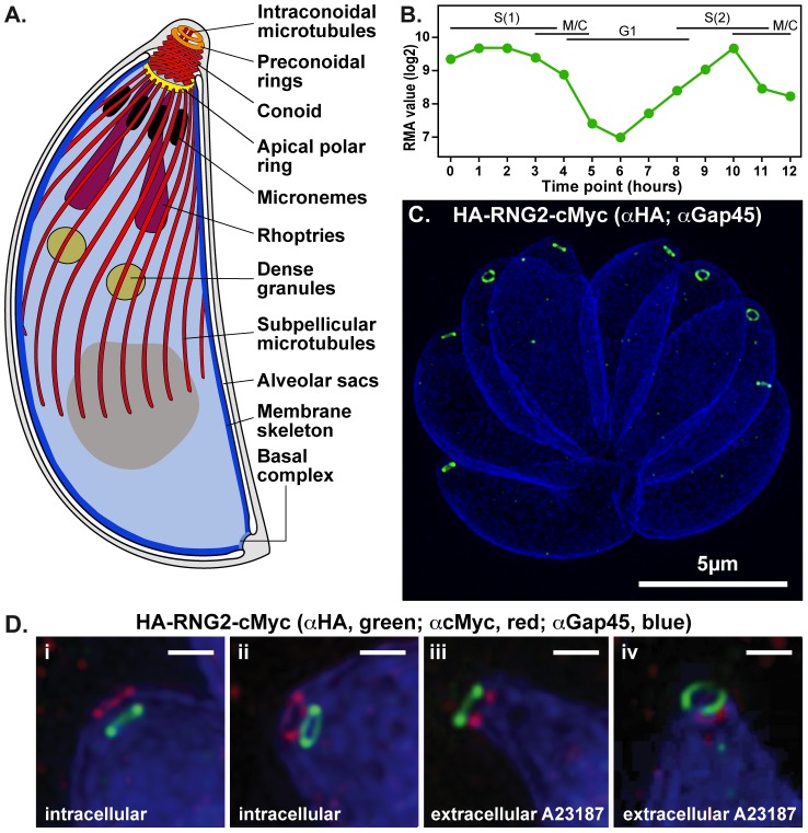
Figure 1
RNG2 apical rings.
(A) Schematic of Toxoplasma gondii cell representing the structural elements of the apical complex, the cell pellicle, and the secretory organelles. The conoid is shown extruded. (B) RNG2 expression t...
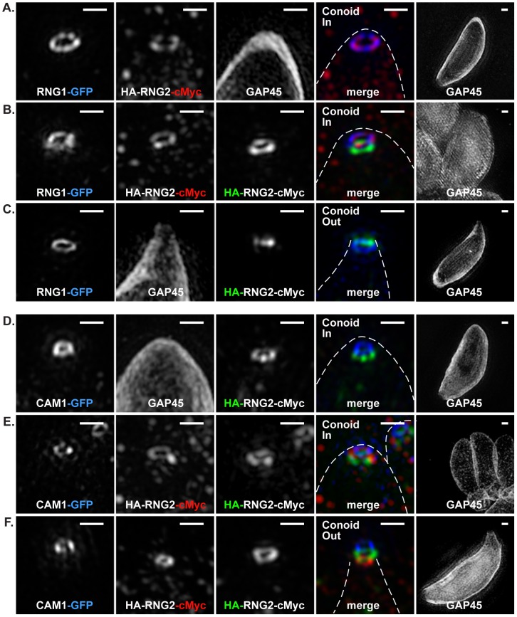
Figure 2
3D-SIM of RNG2 location relative apical polar ring marker RNG1 and conoid marker CAM1.
(A-C) RNG1-GFP (pseudo-colored blue), and (D-F) CAM1-GFP (pseudo-colored blue) colocalized with HA (green) and cMyc (red) of the HA-RNG2-cMyc fusion. Conoid position is indicated. Individual immuno-si...
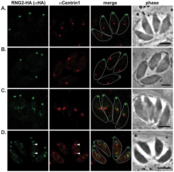
Figure 3
RNG2 appears in daughter cells after centrosome duplication.
RNG2-HA (green) cells immuno-labeled for centrin1 (red) show single centrosome duplication at the beginning of daughter cell formation (A, B), after which RNG2 appears in association with each centros...
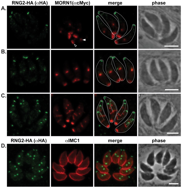
Figure 4
RNG2 appears before centrocone duplication or IMC1 association with daughter pellicles.
(A, B, C) RNG2-HA (green) co-expressed with MORN1-cMyc (red). MORN1 can be seen at the basal complex of both mother (e.g. A, open arrowhead) and daughter cell pellicles, and at the centrocone (e.g. A,...
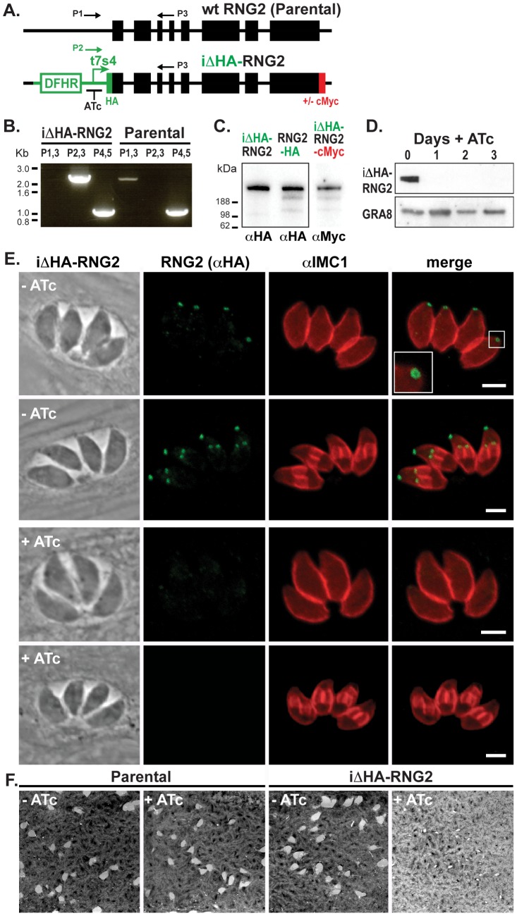
Figure 5
RNG2 is required for parasite growth.
(A) Schematic of the chromosomal locus of wild type RNG2 and the insertional mutant iu0394HA-RNG2 showing the tetracycline regulatable promoter (t7s4), N-terminal HA tag, C-terminal cMyc tag (integrat...
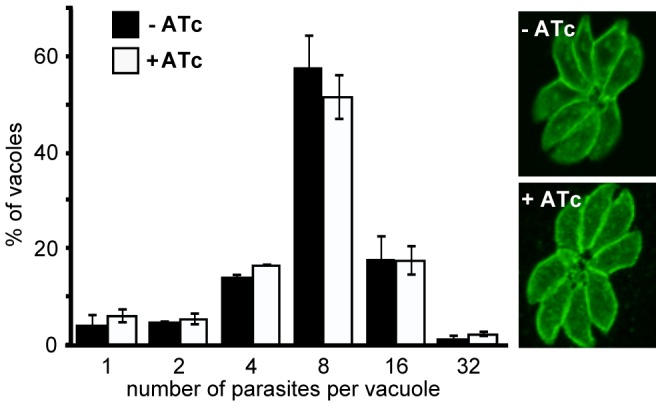
Figure 6
RNG2 is not required for intracellular parasite replication.
Replication rate of iu0394HA-RNG2 parasites grown with (+) or without (u2212) ATc treatment measured by parasite number per parasitophorous vacuole after 24 hours of growth post infection. >200 vacuol...
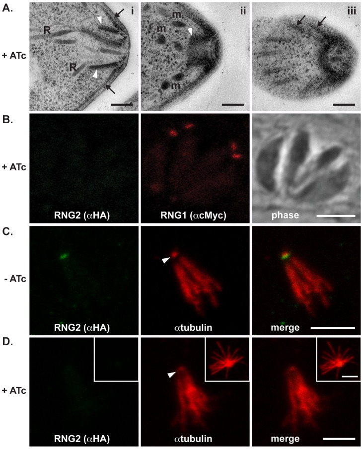
Figure 7
RNG2 knockdown results in no obvious structural change.
(A) Transmission electron microscopy of ATc-treated parasites show typical apical structures. (i) Transverse section of apical complex showing rhoptries (R) extending within the conoid (white arrowhea...
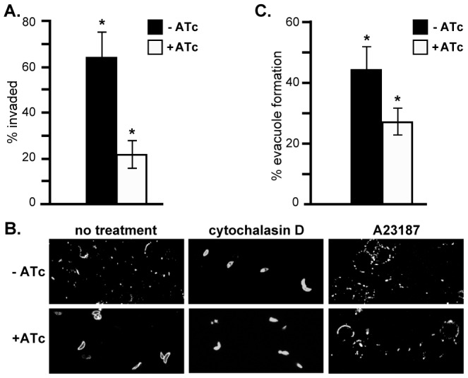
Figure 8
RNG2 is required for invasion, motility and rhoptry evacuole formation.
(A) Percentage parasite invasion success of iu0394HA-RNG2 cells grown either with or without ATc. >200 parasites were scored for each of three biological replicates. (B) Gliding motility detected by S...
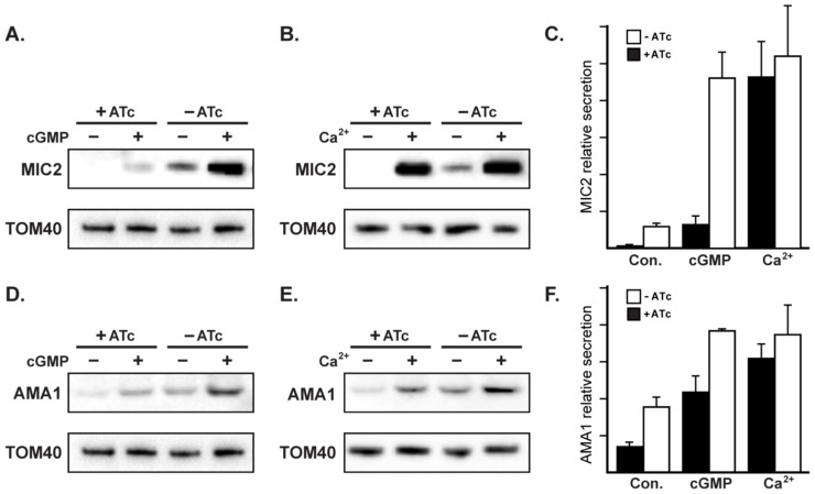
Figure 9
RNG2 has a role in regulated microneme secretion.
Constitutive secretion of microneme proteins MIC2 (A, B) and AMA1 (D, E) from extracellular iu0394HA-RNG2 cells grown with or without ATc. Secretion was also assessed with cGMP stimulation by Zaprinas...
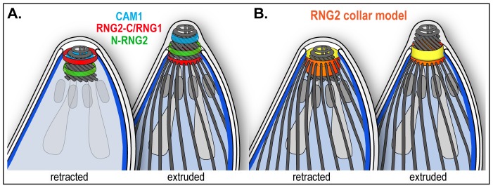
Figure 10
Schematic of RNG2 location within the apical complex.
(A) Inferred positions of the N and C termini of RNG2 (labeled N-RNG2 and RNG2-C, respectively), conoid marker CAM1, and apical polar ring marker RNG1 within the structures of the apical complex, and ...
Figure images are served from the NIH/NLM PubMed Central Open Access Subset or Europe PMC; copyright remains with the publishers and authors.
💬 Discussion
0 commentsNo comments yet. Be the first to start a discussion!
Leave a Comment