Abstract
Abstract Introduction Nomenclature Uterine contractions and the ‘functional syncytium theory’ Interstitial cells of Cajal in the gastrointestinal and urinary tracts Are there m‐ICLC in myometrium? The effect of c‐kit inhibition on myometrial contractility Electrophysiology of m‐ICLC Imaging of tissue level signals in myometrium Emerging concepts – the possible role of extracellular ATP in myometrial contractility Where do m‐ICLC fit in with a model for the generation of myometrial contractions? Future research directions Conclusion A strict regulation of contractility in the uterus and fallopian tube is essential for various reproductive functions. The uterus contributes, through either increased contractility or periods of relative quiescence, to: (i) expulsion of menstrual debris, (ii) sperm transport, (iii) adequate embryo placement during implantation, (iv) enlarging its capacity during pregnancy and (v) parturition. The dominant cell population of the uterine wall consists of smooth muscle cells that contain the contractile apparatus responsible for the generation of contractile force. Recent interest has focused on a new population of cells located throughout the myometrium on the borders of smooth muscle bundles. These cells are similar to interstitial cells of Cajal (ICC) in the gut that are responsible for the generation of electrical slow waves that control peristalsis. A precise role for myometrial Cajal‐like interstitial cells (m‐ICLC) has not been identified. m‐ICLC express the c‐kit receptor, involved in creating and maintaining the ICC phenotype in the gastrointestinal tract. However, both acute and prolonged inhibition of this receptor with the c‐kit antagonist imatinib mesylate does not appear to affect the spontaneous contractility of myometrium. Calcium imaging of live tissue slices suggests that contractile signalling starts on the borders of smooth muscle bundles where m‐ICLC are located and recently the possible role of extracellular ATP signalling from m‐ICLC has been studied. This manuscript reviews the evidence regarding tissue‐level signalling in the myometrium with a particular emphasis on the anatomical and possible functional aspects of m‐ICLC as new elements of the contractile mechanisms in the uterus.
🔬 Techniques
✨ Fluorophores
🧪 Sample Preparation
🔬 Cell Lines
🏭 Microscope Brands
🏛️ Research Organizations (ROR)
Affiliated research institutions:
📊 Figures
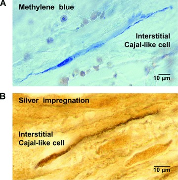
Fig 1
Human myometrium. (A) Methylene blue vital staining, before cryofixation (cryosectioning). Note the selective affinity of an m-ICLC for the blue dye. (B) Silver impregnation after fixation and paraffi...
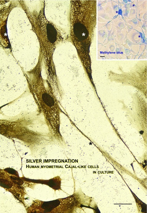
Fig 2
Photographic reconstruction of human m-ICLC in culture establishing contacts with smooth muscle cells. In compliance with our experience, silver impregnation is one of the choice methods for revealing...
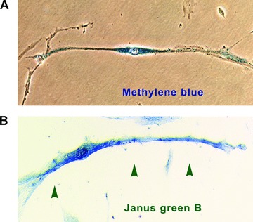
Fig 3
Vital staining methods (efficient in pointing out the very long processes) performed on m-ICLC in culture. Primary confluent cultures (day 8) from pregnant human myometrium. (A) Representative m-ICLC ...
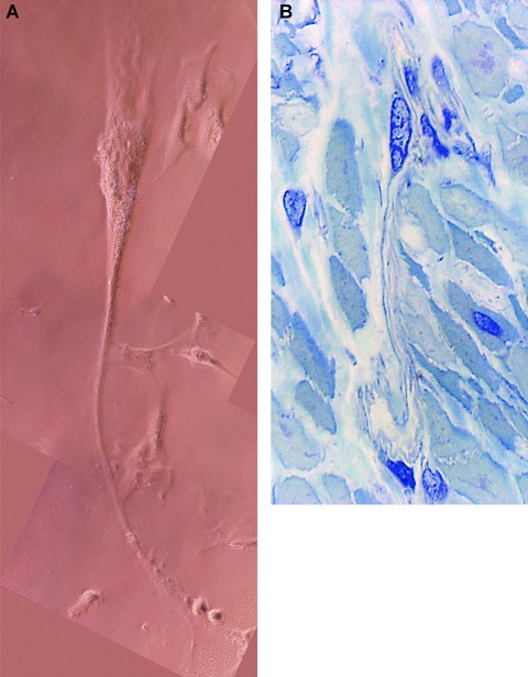
Fig 4
(A) Human myometrium. Primary semi-confluent cell culture (day 4), phase contrast microscopy. Photographic reconstruction of an m-ICLC with a very long, moniliform cytoplasmic processes (arrows) emerg...
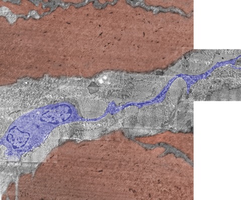
Fig 5
Electron micrograph of human non-pregnant myometrium. Note the presence of an m-ICLC (blue) with a long thin cytoplasmic process that suddenly comes out from the cellular body and runs between smooth ...
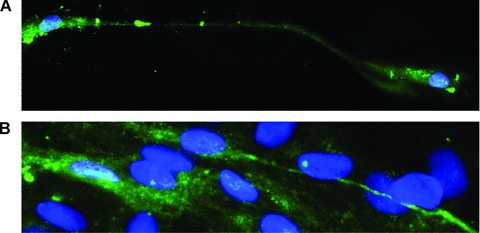
Fig 6
Human myometrium cells in culture (the second passage); immunofluorescence labelling for c-kit; FITC-conjugated secondary antibodies (green) were used to visualize the reaction (green), m-ICLC that di...
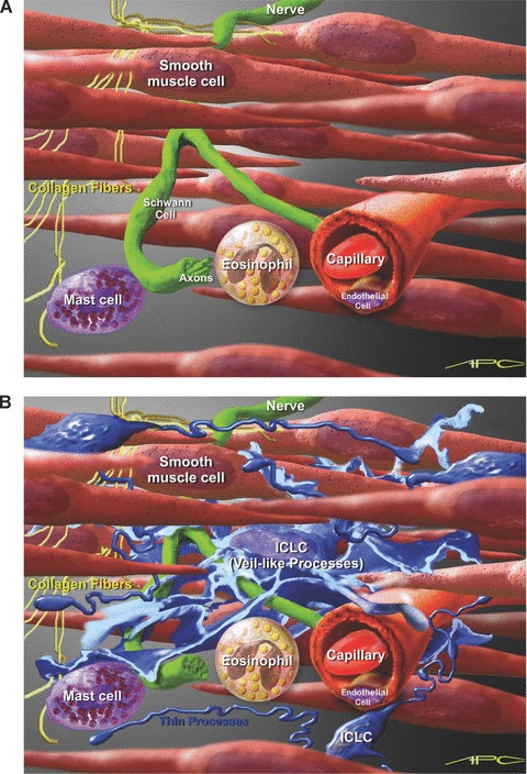
Fig 7
Artistic view of possible interconnectivity of m-ICLC in the uterine wall. (A) Myometrial background with smooth muscle cells, nerve fibers and some connective tissue cells. (B) Close proximity of m-I...
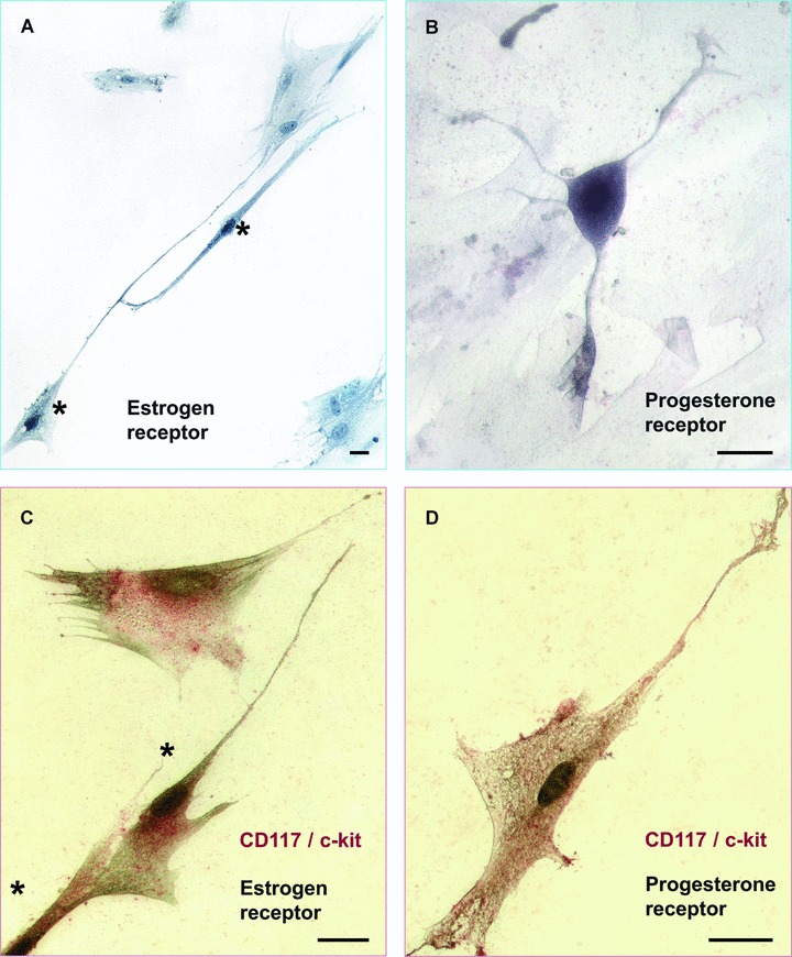
Fig 8
Human myometrial cell culture, fourth passage. Immunocytochemical staining for the estrogen and progesterone receptors. (A) Immunocytochemical detection of estrogen receptor u2013 dark stained nuclei ...
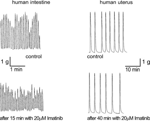
Fig 9
Representative traces of myometrial human smooth muscle inhibition by imatinib 20 u03bcM. Reprinted from Ref. [ 78 ], with permission from Elsevier.
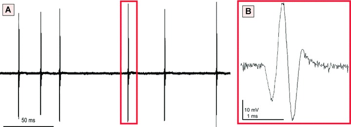
Fig 10
Extracellular single-unit recording performed on m-ICLC cultures (human pregnant myometrium), at the third passage. (A) Notice the spontaneous electrical activity in MIC. (B) Spontaneous field potenti...
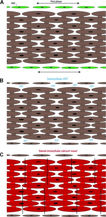
Fig 11
Schematic representation of a smooth muscle bundle. (A) Myometrial ICs (green) produce a synchronous signal along the border of the smooth muscle bundles. (B) ATP and possibly other messenger molecule...
Figure images are served from the NIH/NLM PubMed Central Open Access Subset or Europe PMC; copyright remains with the publishers and authors.
💬 Discussion
0 commentsNo comments yet. Be the first to start a discussion!
Leave a Comment