Abstract
Background: Platelets have distinct roles in the vascular system in that they are the major mediator of thrombosis, critical for restoration of tissue integrity, and players in vascular inflammatory conditions. In close spatiotemporal proximity, the complement system acts as the first line of defense against invading microorganisms and is a key mediator of inflammation. Whereas the fluid phase cross-talk between the complement and coagulation systems is well appreciated, the understanding of the pathophysiological implications of such interactions is still scant. Methods: We analyzed coexpression of the anaphylatoxin receptor C3aR with activated glycoprotein IIb/IIIa on platelets of 501 patients with coronary artery disease using flow cytometry; detected C3aR expression in human or murine specimen by polymerase chain reaction, immunofluorescence, Western blotting, or flow cytometry; and examined the importance of platelet C3aR by various in vitro platelet function tests, in vivo bleeding time, and intravital microscopy. The pathophysiological relevance of C3aR was scrutinized with the use of disease models of myocardial infarction and stroke. To approach underlying molecular mechanisms, we identified the platelet small GTPase Rap1b using nanoscale liquid chromatography coupled to tandem mass spectrometry. Results: We found a strong positive correlation of platelet complement C3aR expression with activated glycoprotein IIb/IIIa in patients with coronary artery disease and coexpression of C3aR with glycoprotein IIb/IIIa in thrombi obtained from patients with myocardial infarction. Our results demonstrate that the C3a/C3aR axis on platelets regulates distinct steps of thrombus formation such as platelet adhesion, spreading, and Ca 2+ influx. Using C3aR −/− mice or C3 −/− mice with reinjection of C3a, we uncovered that the complement activation fragment C3a regulates bleeding time after tail injury and thrombosis. Notably, C3aR −/− mice were less prone to experimental stroke and myocardial infarction. Furthermore, reconstitution of C3aR −/− mice with C3aR +/+ platelets and platelet depletion experiments demonstrated that the observed effects on thrombosis, myocardial infarction, and stroke were specifically caused by platelet C3aR. Mechanistically, C3aR-mediated signaling regulates the activation of Rap1b and thereby bleeding arrest after injury and in vivo thrombus formation. Conclusions: Overall, our findings uncover a novel function of the anaphylatoxin C3a for platelet function and thrombus formation, highlighting a detrimental role of imbalanced complement activation in cardiovascular diseases.
🔬 Techniques
🔭 Microscopes
✨ Fluorophores
🧪 Sample Preparation
🔬 Cell Lines
🏭 Microscope Brands
🧪 Reagent Suppliers
💾 Data Repositories
🏛️ Research Organizations (ROR)
Affiliated research institutions:
📋 Methods
We analyzed co-expression of the anaphylatoxin receptor C3aR with activated GPIIb/IIIa on platelets of 501 coronary artery disease patients using flow cytometry, detected C3aR expression in human or murine specimen by PCR, immunofluorescence, western blotting or flow cytometry and examined the importance of platelet C3aR by various in vitro platelet function tests, by in vivo bleeding time and intravital microscopy. The pathophysiological relevance of C3aR was scrutinized using disease models of myocardial infarction and stroke. To approach underlying molecular mechanisms, we identified the platelet small GTPase Rap1b using nanoscale liquid chromatography coupled to tandem mass spectrometry.
Methods Please see the online-only Data
Supplement for expanded methods. Requests by researchers to access the data, analytic methods, and study materials for the purposes of reproducing the results or replicating procedures can be made to the corresponding author who manages the information.
Study approval
For experiments with human material, written informed consent was received from participants prior to inclusion in the study (approval number 270/2011B01). All animal experiments were approved by governmental authorities and performed in accordance with the German law guidelines of animal care (approval number M10/12).
Immunofluorescence staining
Human thrombi harvested from aspirates of the coronary arteries of patients with myocardial infarction undergoing percutaneous coronary intervention (PCI) were analyzed by immunofluorescence microscopy. Samples were stained with primary antibodies: anti-human C3aR and anti-human or corresponding control IgG followed by a secondary antibody conjugated with ALEXA-fluorochromes.
Show full methods section
We analyzed co-expression of the anaphylatoxin receptor C3aR with activated GPIIb/IIIa on platelets of 501 coronary artery disease patients using flow cytometry, detected C3aR expression in human or murine specimen by PCR, immunofluorescence, western blotting or flow cytometry and examined the importance of platelet C3aR by various in vitro platelet function tests, by in vivo bleeding time and intravital microscopy. The pathophysiological relevance of C3aR was scrutinized using disease models of myocardial infarction and stroke. To approach underlying molecular mechanisms, we identified the platelet small GTPase Rap1b using nanoscale liquid chromatography coupled to tandem mass spectrometry.
Methods Please see the online-only Data
Supplement for expanded methods. Requests by researchers to access the data, analytic methods, and study materials for the purposes of reproducing the results or replicating procedures can be made to the corresponding author who manages the information.
Study approval
For experiments with human material, written informed consent was received from participants prior to inclusion in the study (approval number 270/2011B01). All animal experiments were approved by governmental authorities and performed in accordance with the German law guidelines of animal care (approval number M10/12).
Immunofluorescence staining
Human thrombi harvested from aspirates of the coronary arteries of patients with myocardial infarction undergoing percutaneous coronary intervention (PCI) were analyzed by immunofluorescence microscopy. Samples were stained with primary antibodies: anti-human C3aR and anti-human or corresponding control IgG followed by a secondary antibody conjugated with ALEXA-fluorochromes.
Flow cytometry analysis
Human platelets in freshly-collected blood were evaluated for the surface expression of C3aR and P-selectin (CD62P) after gating for the platelet specific marker CD42b. Isolated murine platelets for flow cytometric analysis were stained with the anti-mouse antibodies FITC-anti-P-selectin, FITC-anti-GPVI, FITC-anti-GPIbα, FITC-anti-integrin αIIbβIII, FITC-anti-integrin α5 and FITC-anti-integrin β3. Activated GPIIb/IIIa was detected using fibrinogen-Alexa488 or PE-anti-integrin GPIIb/IIIa. Samples were analyzed with a FACSCalibur flow cytometer (Becton Dickinson, Heidelberg, Germany).
Mice
C57BL/6 mice and C3 −/− mice were obtained from The Jackson Laboratory. C3aR −/− mice were a kind gift from Dr. Rick Wetsel (University of Houston, Texas, USA) and C5aR −/− mice were kindly provided by Dr. Craig Gerard (Harvard Medical School, Boston, USA). Spa.1 −/− mice were provided by Dr. Yasutoshi Agata (Kyoto University, Kyoto, Japan) and bred with the C3aR −/− mice to obtain a Spa.1 −/− x C3aR −/− double knockout strain. GPIbalpha −/− mice were kindly provided by Dr. Jerry Ware (University of Arkansas for Medical Sciences, USA). All strains were on a C57BL/6 background and genotypes were confirmed by PCR. Littermates were used in all experiments as control animals.
Isolation of human and murine platelets
Human platelets were isolated according to standard protocols. Murine platelet preparations were obtained using a modified protocol. For activation, isolated platelets were stimulated with 20 µM ADP or 0.01 U/mL thrombin and/or C3a (as indicated in figure legends) at room temperature.
Western blotting
Whole cell lysates were separated by SDS-polyacrylamide gel electrophoresis and subjected to western blot. Membranes were incubated with primary antibodies (anti-mouse C3aR, anti-human C3aR or anti-Spa-1, clone M-300 or anti-RAP1 or anti-GAPDH or anti-mouse phospho-PI3K or anti-mouse total PI3K or anti-Tubulin). Membranes were scanned with the Odyssey Infrared Imaging System (LI-COR, Bad Homburg, Germany) and analyzed.
Flow chamber assay
Isolated human platelets were stimulated with recombinant human C3a or scrambled C3a and perfused through a transparent flow chamber over a coated surface (collagen (20 µg/ml), vWF (20 µg/ml) or fibrinogen (50 µg/ml)) with high (1.700 s −1 ) shear rates for 10 min. 29 Experiments were recorded with a video recorder linked to a microscope (optical objectives ×20 and ×40; Carl Zeiss) and adherent platelets were quantified. Platelet Spreading Isolated murine platelets were incubated on fibrinogen-coated and BSA-blocked coverslides for 30 minutes at room temperature. Platelets were fixed and permeabilized with PHEM buffer containing PFA and NP-40, stained with rhodamin-phalloidin and analyzed with an Axio Observer Z.1 microscope (Zeiss). Scanning Ion Conductance Microscopy (SICM) Isolated WT and C3aR −/− platelets were stimulated with thrombin and allowed to adhere for 30 min to fibrinogen-coated culture plates. Platelets were then fixed with 4% PFA and mounted into the SICM setup. Platelet morphology was analysed using a custom-written software. Briefly, the cell contour was determined automatically using a height threshold of 50 nm and processed to calculate the morphology parameters (area A , height, and circularity C = 4π A / P 2 with perimeter P ).
Immunoprecipitation
Resting and stimulated platelets were lysed by adding an equal amount of lysis buffer. Samples were incubated with anti-C3aR antibody. Protein G-Sepharose was added and samples were incubated over night at 4 °C with rotation. Immunoblotting was performed as indicated. SDS PAGE and in-gel digestion Complete protein eluates of the pull-down experiments were submitted to a short run (1cm in length) on 1D SDS-PAGE. The proteins were visualized by staining and the corresponding gel sectors were excised and subjected to tryptic in-gel digestion. The resulting peptide mixtures were desalted before Liquid chromatography/Mass spectrometry (LC/MS) measurement.
Nanoscale liquid chromatography coupled to tandem mass spectrometry
(NanoLC-MS/MS) analysis LC-MS analysis was carried out on a nanoLC (Easy-nLC , Thermo Fisher Scientific, formerly Proxeon Biosystems) coupled to a LTQ-Orbitrap-XL (Thermo Fisher Scientific). MS data were processed using the MaxQuant software suite, peptide sequences were retrieved by using the Andromeda search engine, data were searched against the Uniprot mouse database.
Rap1 activation and pulldown assay
For detection and pulldown of activated Rap1 (GTP-Rap1), a commercially available kit (affinity precipitation assay, Merck Millipore) was used. Measurement of cytosolic Ca 2+ concentration Washed murine platelets were loaded with 5 μM fura-2 acetoxymethylester in the presence of 0.2 μg/ml pluronic F-127. Loaded platelets were activated with thrombin (0.01U) ± C3a (200 nM), ± C3aR Inhibitor SB290157 (10 µM). Calcium responses were measured under stirring with a spectrofluorimeter (LS 55; PerkinElmer) at alternate excitation wavelengths of 340 and 380 nm (37°C). The 340/380 nm ratio values were converted into concentrations of [Ca 2+ ]. Platelet aggregometry Aggregation of isolated human or murine platelets was estimated from light transmission determined with a luminoaggregometer (model 700; ChronoLog). Platelets were activated with 20 µM ADP, C3a in different doses or control protein (scrambled C3a) at the indicated concentrations. Analysis was performed with the Aggrolink8 software (ChronoLog). Bleeding time Mice were anesthetized and a 3-mm segment of the tail tip was removed with a scalpel. Tail bleeding was monitored by gentle absorption of the blood with filter paper at 20-s intervals without making contact with the wound site.
Intravital microscopy
Intravital microscopy and induction of platelet thrombus formation in vivo were carried out as described before. 29 Stroke model All stroke experiments were performed in accordance with the recently published ARRIVE guidelines (nc3rs) and performed as described before. 30 Myocardial infarction model All myocardial infarction experiments were performed as described before. 31 Statistics Data are provided as means±SD; n represents the number of experiments. All data were tested for significance using unpaired Student t-test and one-way or two-way ANOVA with Bonferroni’s post hoc test. Results with P < 0.05 were considered statistically significant.
📊 Figures
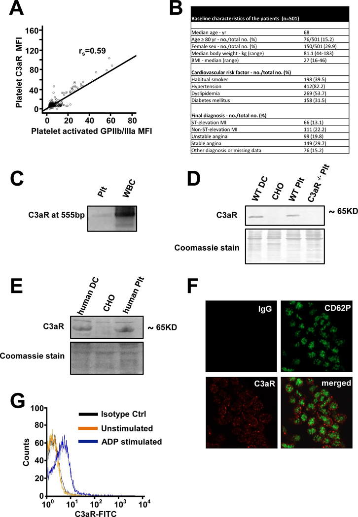
Figure 1
Platelets express the anaphylatoxin receptor C3aR.
(A) Analyzing patients with coronary artery disease, expression of C3aR and activated GPIIb/IIIa (activation specific Ab PAC-1) was measured by flow cytometry. The correlation of C3aR with activated G...
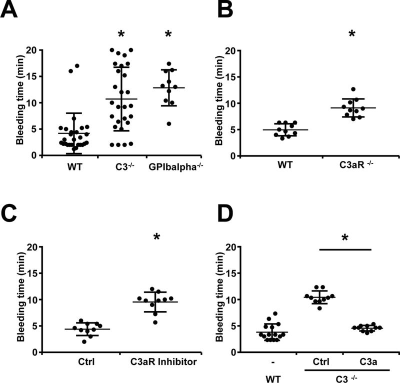
Figure 2
Prolonged bleeding time in C3 deficient mice is mediated by C3a.
To assess the role of the C3aR in hemostasis, tail bleeding time was assessed in anesthesized C3 u2212/u2212 mice, C3aR u2212/u2212 mice and WT mice after amputation of the tail tip. Data represent me...
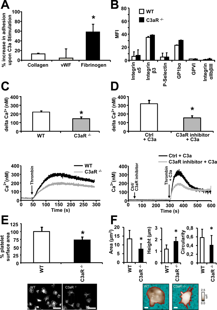
Figure 3
C3aR is important for platelet function.
(A) Human isolated platelets were perfused over coverslides coated with different extracellular matrix proteins using a parallel-plate flow chamber. Under flow conditions (shear rates 1700s u22121 ), ...
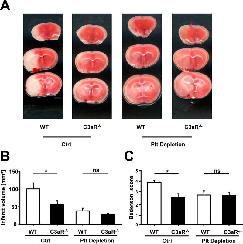
Figure 4
Platelet derived C3aR contributes to experimental stroke.
(A - C) Stroke was induced using the tMCAO model. (A) depicts sample images of TTC stainings for infarcted brain tissue 24h after stroke induction. (B) Brain infarct volumes in control and platelet-de...
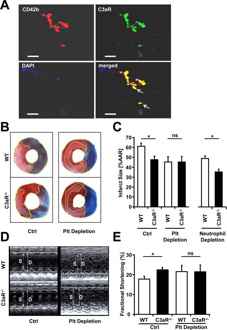
Figure 5
Platelet derived C3aR contributes to experimental myocardial infarction.
(A) Expression of C3aR on platelets in human thrombi from aspirates of the coronary arteries of patients with myocardial infarction undergoing PCI. Double immunofluorescence staining was carried out w...
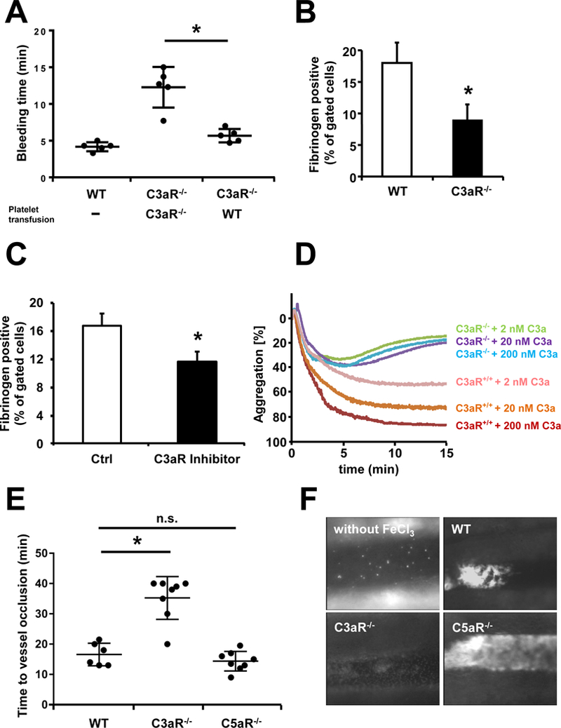
Figure 6
Platelet C3aR mediates thrombosis.
(A) Tail-bleeding time was measured in C3aR u2212/u2212 mice after i.v. injection of isolated WT or C3aR u2212/u2212 platelets (1 u00d7 10 8 / mouse). In C3aR u2212/u2212 mice transfused with WT plate...
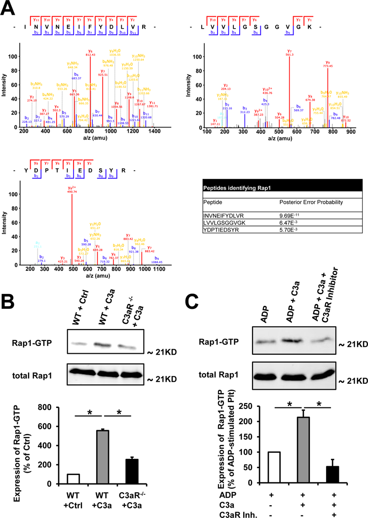
Figure 7
Treatment with C3a results in Rap1b activation.
(A) Rap1-detection after immuno-precipitation of platelet lysates using an anti-C3aR antibody. Protein eluates of pull-down experiments using isolated murine platelets and an anti-C3aR antibody were s...

Figure 8
Role of Rap1b in C3aR mediated thrombus formation.
(A) Generation of Spa.1 u2212/u2212 x C3aR u2212/u2212 double knockout (DKO) mice. Rap1 is activated (Rap1*GTP) by different ligands binding to G-Protein coupled receptors such as C3aR. Rap1 is inhibi...
Figure images are served from the NIH/NLM PubMed Central Open Access Subset or Europe PMC; copyright remains with the publishers and authors.
💬 Discussion
0 commentsNo comments yet. Be the first to start a discussion!
Leave a Comment