Abstract
The transient receptor potential canonical subfamily member 5 (TRPC5), one of seven mammalian TRPC members, is a nonselective calcium-permeant cation channel. TRPC5 is of considerable interest as a drug target in the treatment of progressive kidney disease, depression, and anxiety. Here, we present the 2.8-Å resolution cryo-electron microscopy (cryo-EM) structure of the mouse TRPC5 (mTRPC5) homotetramer. Comparison of the TRPC5 structure to previously determined structures of other TRPC and TRP channels reveals differences in the extracellular pore domain and in the length of the S3 helix. The disulfide bond at the extracellular side of the pore and a preceding small loop are essential elements for its proper function. This high-resolution structure of mTRPC5, combined with electrophysiology and mutagenesis, provides insight into the lipid modulation and gating mechanisms of the TRPC family of ion channels.
🔬 Techniques
✨ Fluorophores
🧪 Sample Preparation
🔬 Cell Lines
🏭 Microscope Brands
🧪 Reagent Suppliers
📷 Detectors
💻 Software Details
💾 Data Repositories
🏛️ Research Organizations (ROR)
Affiliated research institutions:
📋 Methods
Protein expression and purification
A synthetic gene fragment encoding residues 1 to 765 (excluding amino acids 766 to 975) of mouse TRPC5 was cloned into the pEGBacMam vector. The resulting protein contains a maltose-binding protein tag on its N terminus. P3 baculoviruses were produced in the Bac-to-Bac Baculovirus Expression System (Invitrogen). HEK293S GnTI − [from the American Type Culture Collection (ATCC)] cells were infected with 10% (v/v) P3 baculovirus at a density of 2.0 to 3.0 × 10 6 cells/ml for protein expression at 37°C. After 24 hours, 10 mM sodium butyrate was added, and the temperature was reduced to 30°C for 72 hours before harvesting. Cells were gently disrupted and resuspended in a solution containing 30 mM Hepes, 150 mM NaCl, and 1 mM DTT (pH 7.5) with EDTA-free protease inhibitor cocktail (Roche). Cells were solubilized for 2 to 3 hours in a solution containing 1.0% (w/v) N -dodecyl-β- d -maltopyranoside (Affymetrix), 0.1% (w/v) CHS (Sigma-Aldrich), 30 mM Hepes, 150 mM NaCl, and 1 mM DTT (pH 7.5) with EDTA-free protease inhibitor cocktail (Roche). After 30 min, the cell lysate was then centrifuged for 60 min at 100,000 g , and the supernatant was incubated in amylose resin (New England Biolabs) at 4°C overnight. The resin was washed with 20 column volumes of wash buffer [25 mM Hepes, 150 mM NaCl, 0.1% (w/v) digitonin, 0.01% (w/v) CHS, and 1 mM DTT (pH 7.5) with EDTA-free protease inhibitor cocktail (Roche)]. The protein was eluted with four column volumes of wash buffer with 40 mM maltose. The protein was then concentrated to 0.5 ml and mixed with PMAL-C8 (Anatrace) at 1:4 (w/w) with gentle agitation for 4 hours. Bio-Beads SM-2 (50 mg/ml, reconstitution mixture; Bio-Rad) were added to remove additional detergent; a disposable Poly-Prep column was used to remove Bio-Beads. After incubation at 4°C overnight, the protein was further purified on a Superose 6 size exclusion column in 25 mM Hepes, 150 mM NaCl, and 1 mM DTT (pH 7.5). All purification procedures were carried out either on ice or at 4°C. The peak fractions corresponding to tetrameric TRPC5 was concentrated to 7.0 mg/ml and used for preparation of cryo-EM sample grids.
Show full methods section
Protein expression and purification
A synthetic gene fragment encoding residues 1 to 765 (excluding amino acids 766 to 975) of mouse TRPC5 was cloned into the pEGBacMam vector. The resulting protein contains a maltose-binding protein tag on its N terminus. P3 baculoviruses were produced in the Bac-to-Bac Baculovirus Expression System (Invitrogen). HEK293S GnTI − [from the American Type Culture Collection (ATCC)] cells were infected with 10% (v/v) P3 baculovirus at a density of 2.0 to 3.0 × 10 6 cells/ml for protein expression at 37°C. After 24 hours, 10 mM sodium butyrate was added, and the temperature was reduced to 30°C for 72 hours before harvesting. Cells were gently disrupted and resuspended in a solution containing 30 mM Hepes, 150 mM NaCl, and 1 mM DTT (pH 7.5) with EDTA-free protease inhibitor cocktail (Roche). Cells were solubilized for 2 to 3 hours in a solution containing 1.0% (w/v) N -dodecyl-β- d -maltopyranoside (Affymetrix), 0.1% (w/v) CHS (Sigma-Aldrich), 30 mM Hepes, 150 mM NaCl, and 1 mM DTT (pH 7.5) with EDTA-free protease inhibitor cocktail (Roche). After 30 min, the cell lysate was then centrifuged for 60 min at 100,000 g , and the supernatant was incubated in amylose resin (New England Biolabs) at 4°C overnight. The resin was washed with 20 column volumes of wash buffer [25 mM Hepes, 150 mM NaCl, 0.1% (w/v) digitonin, 0.01% (w/v) CHS, and 1 mM DTT (pH 7.5) with EDTA-free protease inhibitor cocktail (Roche)]. The protein was eluted with four column volumes of wash buffer with 40 mM maltose. The protein was then concentrated to 0.5 ml and mixed with PMAL-C8 (Anatrace) at 1:4 (w/w) with gentle agitation for 4 hours. Bio-Beads SM-2 (50 mg/ml, reconstitution mixture; Bio-Rad) were added to remove additional detergent; a disposable Poly-Prep column was used to remove Bio-Beads. After incubation at 4°C overnight, the protein was further purified on a Superose 6 size exclusion column in 25 mM Hepes, 150 mM NaCl, and 1 mM DTT (pH 7.5). All purification procedures were carried out either on ice or at 4°C. The peak fractions corresponding to tetrameric TRPC5 was concentrated to 7.0 mg/ml and used for preparation of cryo-EM sample grids.
Electron microscopy data collection Purified
TRPC5 protein sample (3.5 μl) in PMAL-C8 at 7.0 mg/ml was applied to glow-discharged Quantifoil R1.2/1.3 holey carbon 400-mesh copper grids (Quantifoil). Grids were blotted for 7 s at 100% humidity and flash frozen by liquid nitrogen–cooled liquid ethane using a FEI Vitrobot Mark I (FEI). The grid was then loaded onto FEI TF30 Polara electron microscope operated at 300-kV accelerating voltage. Image stacks were recorded on a Gatan K2 Summit direct detector (Gatan) set in super-resolution counting mode using SerialEM ( 35 ), with a defocus range between 1.5 and 3.0 μm. The electron dose was set to 8 e −/physical pixel per s, and the subframe time was set to 0.2 s. A total exposure time of 8 s resulted in 40 subframes per image stack. The total electron dose was 42.3 e −/Å 2 (~1.1 e −/Å 2 per subframe).
Image processing and three-dimensional reconstruction
Image stacks were gain normalized and binned twice to a pixel size of 1.23 Å before drift and local movement correction using MotionCor2 ( 36 ), resulted in the sums of all frames of each image stack with and without dose weighting. The dose-weighting sum was binned by 8× and subjected to visual inspection; images of poor quality were removed before particle picking. Particle picking and subsequent bad particle elimination through two-dimensional (2D) classification were performed using Python scripts or programs ( 37 ) with minor modifications in the 8× binned images. The selected 2D class averages were used to build an initial model using the common lines approach implemented in SPIDER ( 38 ) through M. Liao’s Python scripts ( 37 ), which was applied to later 3D classification using RELION ( 39 ). Contrast transfer function (CTF) parameters were estimated using CTFFIND4 ( 40 ) using the sum of all frames without dose weighting. Quality particle images were then boxed out from the dose-weighted sum of all 40 frames and subjected to RELION 3D classification. RELION 3D refinements were then performed on selected classes for the final map. The resolution of this map was further improved by using the sum of subframes 1 to 14. The number of particles in each dataset and other details related to data processing are summarized in table S1. Model building, refinement, and validation At 2.8-Å resolution, the cryo-EM map was of sufficient quality for de novo atomic model building in Coot ( 41 ). Amino acid assignment was achieved mainly on the basis of the defined densities for bulky residues (Phe, Trp, Tyr, and Arg) and the absence of side-chain densities for glycine residues. The atomic model was further visualized in Coot; a few residues with side chains moving out of the density during the refinement were fixed manually, followed by further refinement. The TRPC5 model was then subjected to global refinement and minimization in real space using the module “phenix.real_space_refine” in PHENIX ( 42 ). The geometries of the model were assessed using MolProbity ( 43 ) in the “comprehensive model validation” section of PHENIX. The final model exhibited good geometry as indicated by the Ramachandran plot (preferred region, 98.25%; allowed region, 1.75%; outliers, 0%). The pore radius was calculated using HOLE ( 44 ). Local resolution estimation was from ResMap ( 45 ).
Electrophysiology
The full-length, truncated TRPC5 constructs or empty vector was transfected into HEK293T cells (from the ATCC), together with an mCherry plasmid. Cells with red fluorescence were selected for whole-cell patch recordings (HEKA EPC 10 USB amplifier, PATCHMASTER 2.90 software). A 1-s ramp protocol from −100 to +100 mV was applied at a frequency of 0.2 Hz. Signals were sampled at 10 kHz and filtered at 3 kHz. The pipette solution contained 130 mM CsCl, 1 mM MgCl 2 , 5.7 mM CaCl 2 , 10 mM EGTA, and 10 mM Hepes (calculated free Ca 2+ , 200 nM), and the pH was titrated to 7.2 using CsOH. The standard bath solution contained 140 mM NaCl, 5 mM KCl, 1 mM MgCl 2 , 2 mM CaCl 2 , 10 mM Hepes, and 10 mM d -glucose, and the pH was adjusted to 7.4 with NaOH. The recording chamber (150 μl) was perfused at a rate of ~2 ml/min. All recordings were performed at room temperature. Quantification of proteins at the plasma membrane TRPC5-tranfected 293T cells were incubated with Sulfo-NHS-Biotin [0.5 mg/ml in phosphate-buffered saline (PBS), Aladdin] for 30 min and washed thrice with 50 mM glycine in PBS. Cells were lysed and aliquoted for extraction of plasma membrane proteins and total proteins separately. Biotinylated plasma membrane proteins were captured with streptavidin magnetic beads (Invitrogen). Western blotting was then performed to detect TRPC5 in the plasma membrane and total proteins. The ratio between biotinylated and total TRPC5 proteins was used to represent the level of TRPC5 proteins expressed at the cell surface. TRPC4 constructs were analyzed in the same way. Primary antibodies against TRPC5 and TRPC4 were purchased from Sangon Biotech (Shanghai, China) and Proteintech (Wuhan, China), respectively.
Supplementary Material http://advances.sciencemag.org/cgi/content/full/5/7/eaaw7935/DC1 Download PDF
📊 Figures
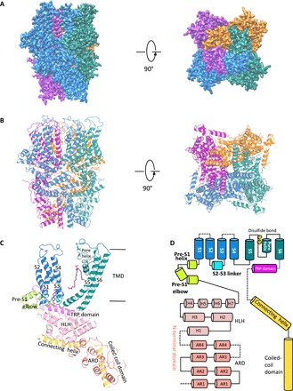
Fig. 1
Overall structure of mouse TRPC5.
( A ) Cryo-EM density map of mTRPC5 at 2.8 u00c5 overall resolution with each monomer represented in different colors (left, side view; right, top view). ( B ) Ribbon diagrams of the mouse TRPC5 model...
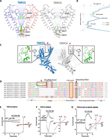
Fig. 2
TRPC5 ion conduction pathway compared with TRPC4 and other TRPCs.
( A ) Side view of TRPC5u2019s pore region with chains A and C compared with TRPC4 (gray). The ion conduction pathway is shown as dots and mapped using HOLE with key amino acid residues labeled. ( B )...
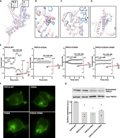
Fig. 3
Comparison of the TRPC5 structure with other TRP channel structures.
( A ) Superimposed side views of mouse TRPC5 subunit (blue) compared with other TRPC family members, including mouse TRPC4 [Protein Data Bank (PDB) ID code 5Z96, gray] ( 19 ), human TRPC3 (PDB ID 5ZBG...
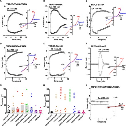
Fig. 4
Mutations in the loop preceding the disulfide bond of TRPC5 alter channel inactivation kinetics.
( A to F and I ) The time course of whole-cell currents measured at +80 and u221280 mV and representative I-V relationships in different conditions as labeled. TRPC5-C4motif, TRPC5 mutant carrying the...
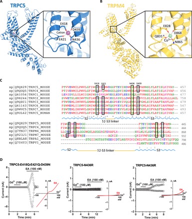
Fig. 5
The cation binding site in TRPC5.
( A ) A cation (purple sphere) on the cytosolic face is in the hydrophilic pocket of the S1-S4 domain, interacting with Glu 418 , Glu 421 , Asn 436 , and Asp 439 . Enlarged view of the cation binding ...
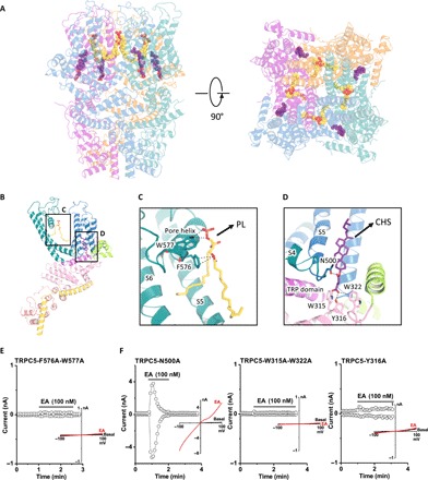
Fig. 6
Lipid coordination in TRPC5.
Lipid-channel interactions. ( A ) Side and top views of ribbon diagrams of the TRPC5 tetramer: four CHS molecules and four PLs (potentially ceramide-1-phosphate or phosphatidic acid) are shown as sphe...
Figure images are served from the NIH/NLM PubMed Central Open Access Subset or Europe PMC; copyright remains with the publishers and authors.
💬 Discussion
0 commentsNo comments yet. Be the first to start a discussion!
Leave a Comment