Abstract
It is widely recognized that animals respond to odors by generating or modulating specific motor behaviors. These reactions are important for daily activities, reproduction, and survival. In the sea lamprey, mating occurs after ovulated females are attracted to spawning sites by male sex pheromones. The ubiquity and reliability of olfactory-motor behavioral responses in vertebrates suggest tight coupling between the olfactory system and brain areas controlling movements. However, the circuitry and the underlying cellular neural mechanisms remain largely unknown. Using lamprey brain preparations, and electrophysiology, calcium imaging, and tract tracing experiments, we describe the neural substrate responsible for transforming an olfactory input into a locomotor output. We found that olfactory stimulation with naturally occurring odors and pheromones induced large excitatory responses in reticulospinal cells, the command neurons for locomotion. We have also identified the anatomy and physiology of this circuit. The olfactory input was relayed in the medial part of the olfactory bulb, in the posterior tuberculum, in the mesencephalic locomotor region, to finally reach reticulospinal cells in the hindbrain. Activation of this olfactory-motor pathway generated rhythmic ventral root discharges and swimming movements. Our study bridges the gap between behavior and cellular neural mechanisms in vertebrates, identifying a specific subsystem within the CNS, dedicated to producing motor responses to olfactory inputs.
🔬 Techniques
💻 Software
✨ Fluorophores
🧪 Sample Preparation
🏭 Microscope Brands
🧪 Reagent Suppliers
📷 Detectors
💻 Software Details
💾 Data Repositories
🏛️ Research Organizations (ROR)
Affiliated research institutions:
📋 Methods
Ethics Statement
All surgical and experimental procedures conformed to the guidelines of the Canadian Council on Animal Care (CCAC) and were approved by the Université de Montréal and Université du Québec à Montréal animal care committees, and the University of Windsor animal care committee, where some of the anatomical experiments were performed.
Animal Preparation
Experiments were performed on 98 reproductive adult sea lampreys ( Petromyzon marinus ) of both sexes provided by the Great Lakes Fishery Commission and the Department of Fisheries and Oceans Canada. In addition, some of the electrophysiology studies were carried out on larval ( n = 62), newly transformed ( n = 102), and parasitic ( n = 7) lampreys. All animals were kept in aerated fresh water maintained at 7°C until used.
Electrophysiology
For the CNS attached to the intact olfactory epithelium preparation, the animals were anesthetized with tricaine methanesulphonate (MS-222, 100 mg/l, Sigma-Aldrich, Oakville, ON, Canada) and decapitated at the level of the 7 th branchiopore. The surgery and all experiments were performed in cold oxygenated Ringer's (8–10°C) of the following composition (in mM): 130 NaCl, 2.1 KCl, 2.6 CaCl 2 , 1.8 MgCl 2 , 4.0 HEPES, 4.0 dextrose, and 1.0 NaHCO 3 , at pH 7.4. The branchial apparatus and the myotomal musculature were removed, with all the soft tissue attached to the ventral side of the cranium. A dorsal incision was made to expose the rostral spinal cord and the brain. The nasal cavity was left attached to the brain through the intact ONs. Care was taken to keep a maximum of the olfactory epithelium intact by simply performing a small window opening on its dorsal aspect, to simply permit faster perfusion and washout of odorants. The preparation was placed into a recording chamber continuously perfused with cold oxygenated Ringer's at a rate of ∼4 ml/min. A minimum of 1 h was allowed for recovery after surgery prior to recording. Using sharp glass microelectrodes filled with 4 M potassium acetate (80–130 MΩ), intracellular recordings were made from RS neurons in the MRRN under visual guidance through a binocular microscope ( Figure 1A ). The signals were amplified with an Axoclamp 2A (Axon Instruments, Foster City, CA). Only RS neurons displaying a stable membrane potential lower than −70 mV for at least 15 min were considered in this study. For electric stimulations, we used homemade glass-coated tungsten electrodes (4–5 MΩ with a 10 µm tip exposure) and a Grass S88 stimulator (Astro-Med, Longueuil, QC, Canada). Stimulation was applied every 10 s as single, double, or triple pulses (2–50 µA intensity, 1–2 ms duration, and 20 ms pulse interval). Synaptic responses are presented as a mean of eight consecutive responses to the same stimulation. To observe “fictive locomotion” ( Figure 3 ), we recorded from ventral roots in newly transformed lampreys, using suction electrodes filled with Ringer's solution. The signals were amplified using AM systems 1800 dual channel amplifiers (A-M systems Inc., Sequim, WA). Semi-intact preparations ( Figure 6B–D ) were dissected as follows: the brain and rostral spinal cord were exposed like previously described [17] , [19] , whereas the caudal two-third of the body was kept intact to freely swim behind. In this case, because of the presence of cutaneous sensory inputs, the brain was transected at the level of the diencephalon for decerebration purposes before the experiment. The preparation was then transferred into a double compartment recording chamber. Teflon-coated stainless steel microwires (50 µm diameter) were inserted into the segmental muscles for EMG recording.
Show full methods section
Ethics Statement
All surgical and experimental procedures conformed to the guidelines of the Canadian Council on Animal Care (CCAC) and were approved by the Université de Montréal and Université du Québec à Montréal animal care committees, and the University of Windsor animal care committee, where some of the anatomical experiments were performed.
Animal Preparation
Experiments were performed on 98 reproductive adult sea lampreys ( Petromyzon marinus ) of both sexes provided by the Great Lakes Fishery Commission and the Department of Fisheries and Oceans Canada. In addition, some of the electrophysiology studies were carried out on larval ( n = 62), newly transformed ( n = 102), and parasitic ( n = 7) lampreys. All animals were kept in aerated fresh water maintained at 7°C until used.
Electrophysiology
For the CNS attached to the intact olfactory epithelium preparation, the animals were anesthetized with tricaine methanesulphonate (MS-222, 100 mg/l, Sigma-Aldrich, Oakville, ON, Canada) and decapitated at the level of the 7 th branchiopore. The surgery and all experiments were performed in cold oxygenated Ringer's (8–10°C) of the following composition (in mM): 130 NaCl, 2.1 KCl, 2.6 CaCl 2 , 1.8 MgCl 2 , 4.0 HEPES, 4.0 dextrose, and 1.0 NaHCO 3 , at pH 7.4. The branchial apparatus and the myotomal musculature were removed, with all the soft tissue attached to the ventral side of the cranium. A dorsal incision was made to expose the rostral spinal cord and the brain. The nasal cavity was left attached to the brain through the intact ONs. Care was taken to keep a maximum of the olfactory epithelium intact by simply performing a small window opening on its dorsal aspect, to simply permit faster perfusion and washout of odorants. The preparation was placed into a recording chamber continuously perfused with cold oxygenated Ringer's at a rate of ∼4 ml/min. A minimum of 1 h was allowed for recovery after surgery prior to recording. Using sharp glass microelectrodes filled with 4 M potassium acetate (80–130 MΩ), intracellular recordings were made from RS neurons in the MRRN under visual guidance through a binocular microscope ( Figure 1A ). The signals were amplified with an Axoclamp 2A (Axon Instruments, Foster City, CA). Only RS neurons displaying a stable membrane potential lower than −70 mV for at least 15 min were considered in this study. For electric stimulations, we used homemade glass-coated tungsten electrodes (4–5 MΩ with a 10 µm tip exposure) and a Grass S88 stimulator (Astro-Med, Longueuil, QC, Canada). Stimulation was applied every 10 s as single, double, or triple pulses (2–50 µA intensity, 1–2 ms duration, and 20 ms pulse interval). Synaptic responses are presented as a mean of eight consecutive responses to the same stimulation. To observe “fictive locomotion” ( Figure 3 ), we recorded from ventral roots in newly transformed lampreys, using suction electrodes filled with Ringer's solution. The signals were amplified using AM systems 1800 dual channel amplifiers (A-M systems Inc., Sequim, WA). Semi-intact preparations ( Figure 6B–D ) were dissected as follows: the brain and rostral spinal cord were exposed like previously described [17] , [19] , whereas the caudal two-third of the body was kept intact to freely swim behind. In this case, because of the presence of cutaneous sensory inputs, the brain was transected at the level of the diencephalon for decerebration purposes before the experiment. The preparation was then transferred into a double compartment recording chamber. Teflon-coated stainless steel microwires (50 µm diameter) were inserted into the segmental muscles for EMG recording.
Calcium Imaging Experiments
RS cells were retrogradely labeled in Ringer's solution for 24 to 36 h by placing Calcium-Green dextran crystals (3000 MW, Invitrogen, Eugene, OR) on the rostral stump of the spinal cord, transected at the first segment. Labeled cells were observed on a Nikon epifluorescent microscope equipped with a 20× (0.75 NA) objective. A fluorescein isothiocyanate (FITC) excitation/emission filter set was used to visualize the neurons. The emitted light was captured with an intensified CCD video camera (Photometrics CoolSNAP HQ, Roper Scientific, Tucson, AZ) and recorded at a rate of two images per second, using Metafluor imaging software (Molecular Devices, Sunnyvale, CA). Calcium responses are expressed as relative changes in fluorescence (ΔF/F%). Anatomical Experiments Anterograde and retrograde labeling was obtained after unilateral injections of Texas Red-conjugated dextran amines (3000 MW, Molecular Probe). A period of 24 to 36 h was allowed for the transport of the tracer. The preparations were then immersed in a solution of 4% paraformaldehyde/0.4% picric acid in phosphate-buffered saline for 5 to 6 h. They were then transferred overnight into a solution of 20% sucrose in phosphate buffer. Transverse sections of 25 µm thickness were made with a cryostat and mounted on microscope slides with Vectashield (Vector laboratories, Burlington, ON, Canada) for observation under epifluorescence microscopy (Nikon E600 microscope equipped with a DXM1200 digital camera, Nikon, Montreal, QC, Canada; or with a Nikon E800 microscope in Windsor, ON, Canada).
Chemical Stimulations and Drugs
All drugs were purchased from Sigma-Aldrich (Oakville, ON, Canada), except sex pheromones (graciously provided by Dr. W. Li, Michigan State University, MI). They were kept as frozen concentrated stock solutions (at −80°C for pheromones) and dissolved to their final concentration in Ringer's solution prior to their use. For all local ejections, the inactive dye Fast Green was added to the drug solution to monitor the extent of application. Drug application was performed by pressure ejections of concentrated substances through a glass micropipette in the nasal cavity, or the brain tissue, using a Picospritzer (General Valve, Fairfield, NJ). Ejection of Ringer's with Fast Green at the same location was used as control in each experiment. Chemical stimulations of the olfactory epithelium were performed on reproductive adult lampreys during the first days after their capture in the wild.
Statistics
Data were analyzed using paired t test or a Mann-Whitney test (Sigmastat, SPSS, Chicago, IL, USA). Significance was set at p
📊 Figures
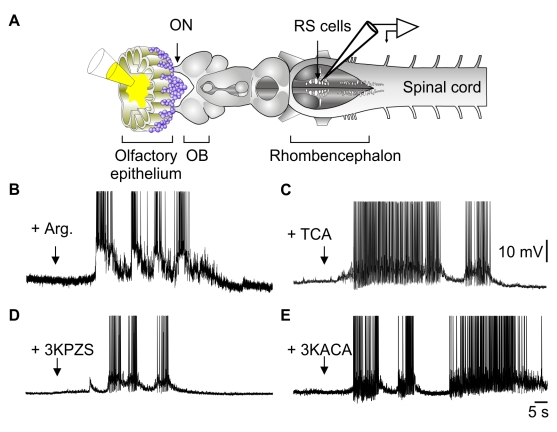
Figure 1
Olfactory epithelium stimulation activates RS cells.
(A) Illustration of the experimental procedure in an isolated olfactory epithelium-brain-spinal cord preparation. (B) Responses of RS cell to the application of L-arginine over the olfactory epitheliu...
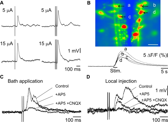
Figure 2
Olfactory nerve stimulation activates RS cells.
(A) Responses of RS cells following electrical stimulation of the ON with 5 or 15 u00b5A (top versus bottom traces); single shocks or trains of stimulation (left versus right traces). Each trace is a ...
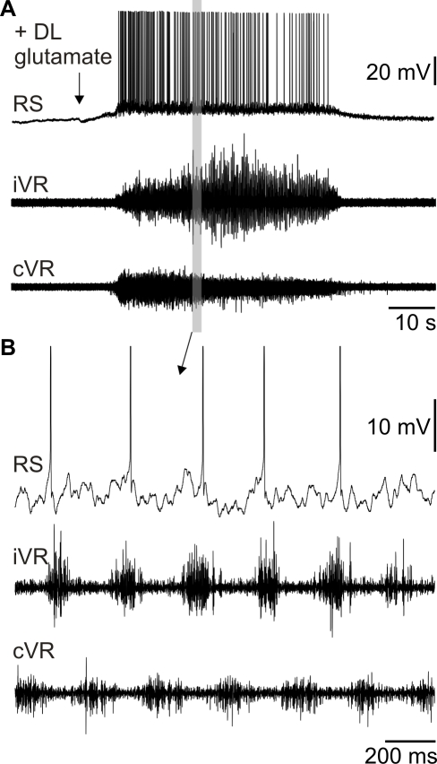
Figure 3
Glutamate injection into the OB induces fictive locomotion.
(A) Top trace: Intracellular recording of a RS cell. Note the large excitation induced by the injection of 3 mM glutamate in the ipsilateral OB. Bottom traces: Ventral root (VR) discharges on both sid...
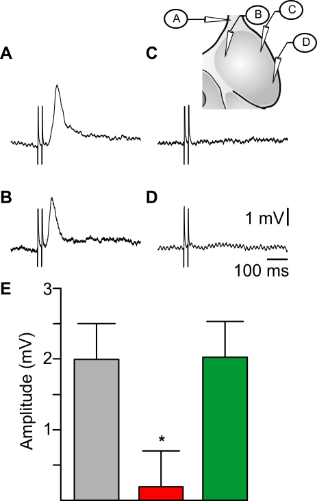
Figure 4
Olfactory-locomotor information transits through the medial region of the OB.
(Au2013D) Responses in a single ipsilateral RS neuron to 30 u00b5A stimulation of the ON and OB. The schematic (inset) indicates the location of stimulating electrodes. Note that a synaptic response w...
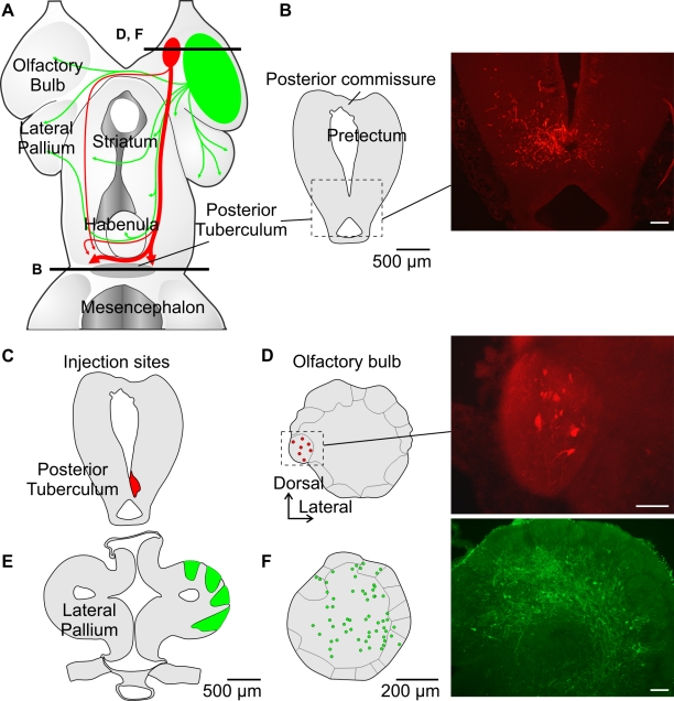
Figure 5
The medial region of the OB projects to the PT.
(A) Schematic dorsal view of the forebrain summarizing the efferent OB projections in the lamprey. Projections from OB regions other than the medial region are shown in green. (B) Anterograde labeling...
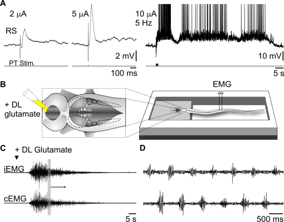
Figure 6
Stimulation of the PT activates RS neurons and locomotion.
(A) PT stimulation induces RS responses. Raising the stimulation intensity from 2 to 5 u00b5A increases the amplitude of the evoked synaptic responses (left two traces), while a short stimulation trai...
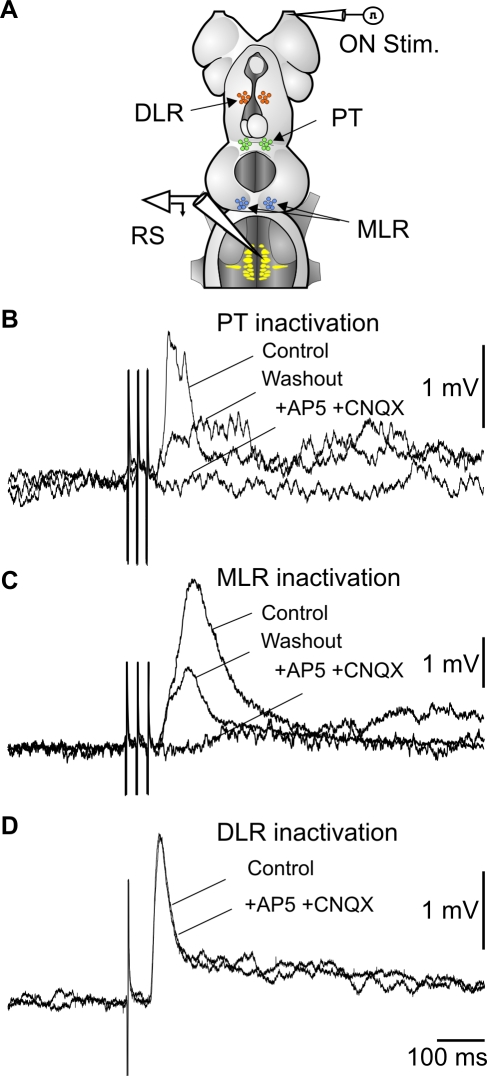
Figure 7
Olfactory inputs are relayed via the PT and MLR.
(A) Schematic illustration showing the experimental procedure where glutamate receptor antagonists were injected in different sites indicated by the arrows. (B) RS cell responses to ON stimulation are...
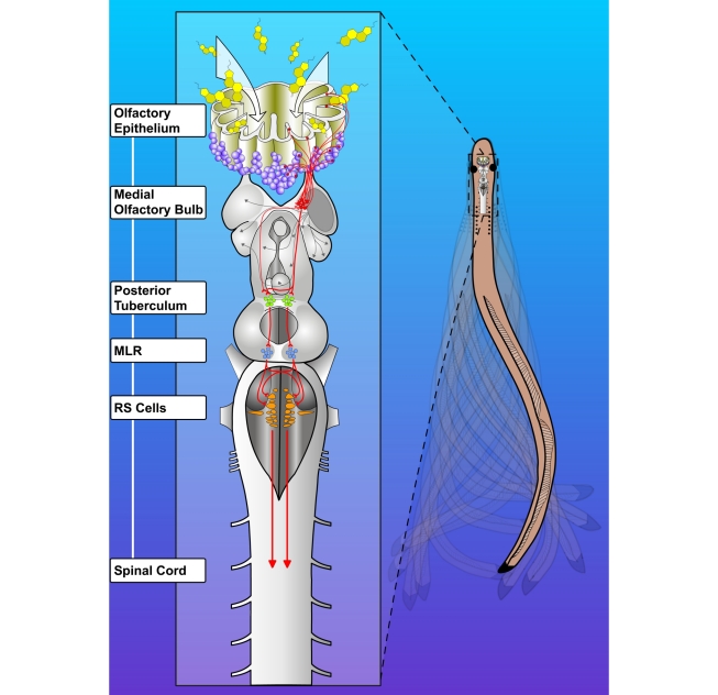
Figure 8
Schematic representation of the olfactory-locomotor circuitry in lampreys.
Stimulation of the olfactory sensory neurons in the periphery activates neurons in the OB. There are two distinct projections from the OB, one from the lateral and another from the medial part. The la...
Figure images are served from the NIH/NLM PubMed Central Open Access Subset or Europe PMC; copyright remains with the publishers and authors.
💬 Discussion
0 commentsNo comments yet. Be the first to start a discussion!
Leave a Comment