Abstract
Abstract Background The human T-cell leukemia virus type I (HTLV-I) basic leucine-zipper factor (HBZ) has previously been shown to modulate transcriptional activity of Jun family members. The presence of a novel isoform of HBZ, termed HBZ-SP1, has recently been characterized in adult T-cell leukemia (ATL) cells and has been found to be associated with intense nuclear spots. In this study, we investigated the role of these nuclear bodies in the regulation of the transcriptional activity of JunB. Results Using fluorescence microscopy, we found that the HBZ-SP1 protein localizes to intense dots corresponding to HBZ-NBs and to nucleoli. We analyzed the relative mobility of the EGFP-HBZ-SP1 fusion protein using fluorescence recovery after photobleaching (FRAP) analysis and found that the deletion of the ZIP domain perturbs the association of the HBZ-SP1 protein to the HBZ-NBs. These data suggested that HBZ needs cellular partners, including bZIP factors, to form HBZ-NBs. Indeed, by cotransfection experiments in COS cells, we have found that the bZIP factor JunB is able to target delocalized form of HBZ (deleted in its nuclear localization subdomains) into the HBZ-NBs. We also show that the viral protein is able to entail a redistribution of JunB into the HBZ-NBs. Moreover, by transfecting HeLa cells (known to express high level of JunB) with a vector expressing HBZ-SP1, the sequestration of JunB to the HBZ-NBs inhibited its transcriptional activity. Lastly, we analyzed the nuclear distribution of HBZ-SP1 in the presence of JunD, a Jun family member known to be activated by HBZ. In this case, no NBs were detected and the HBZ-SP1 protein was diffusely distributed throughout the nucleoplasm. Conclusion Our results suggest that HBZ-mediated sequestration of JunB to the HBZ-NBs may be causing the repression of JunB activity in vivo.
🔬 Techniques
✨ Fluorophores
🧪 Sample Preparation
🔬 Cell Lines
🏭 Microscope Brands
🧪 Reagent Suppliers
🏛️ Research Organizations (ROR)
Affiliated research institutions:
📋 Methods
Plasmids
The vectors pcDNA-HBZ-SP1-Myc, pEGFP-HBZ-bZIP (Fig. 1A ), pcDNA-JunB, pCMV-JunD-Flag have already been described [ 23 , 25 , 30 ]. To generate the EGFP fusion proteins, HBZ-SP1, HBZ-SP1ΔZIP, HBZΔAD, and HBZΔADΔbZIP DNA (Fig. 1A ) were PCR amplified from the pcDNA-HBZ-SP1-Myc vector, digested with Eco RI and Bam HI, and subcloned in frame into similarly digested pEGFP-C2 (Clontech). pEGFP-HBZΔAD and pEGFP-HBZΔADΔbZIP encode for the HBZ-SP1 protein from residues 77 to 206 and 77 to 132, respectively. pLCR-Luc and pNLS-EGFP (corresponding to EGFP under the control of the nuclear localization signal of SV40) is a gift from G. Steger and M. Piechaczyk, respectively.
Fluorescence microscopy analysis
COS cells were cultured in DMEM supplemented with 10% FCS. 24 h before transfection, cells were seeded onto glass slides. They were transfected using the jetPEI™ transfection reagent (Qbiogene) according to the manufacturer's instructions and, after 48 h, they were washed with PBS, fixed, and permeabilized with 4% paraformaldehyde and 0.1% Triton ×-100 for 30 min at room temperature. If necessary, cells were incubated with primary antibody (mouse anti-Myc antibody 9E10 or anti-Flag, Sigma-Aldrich; mouse anti-nucleolin, anti-JunB, or anti-RNA polymerase II antibody, Santa Cruz Biotechnology Inc.; the mouse anti-SC35 antibody is a gift from J. Tazi) for 1 h at room temperature. Samples were washed with PBS and then incubated with secondary FITC- or Texas Red-labelled antibodies (Pierce) for 1 h at room temperature. Coverslips were mounted with Vectashield containing DAPI (Abcys) for direct observation. Fluorescence images were acquired by fluorescence microscopy (model DM R; Leica) at room temperature with a 63x, NA 1.32, oil immersion objective at pinhole size 1 Airy (observation with immersion oil type DF, Cargille Laboratories Inc.). DAPI, GFP, FITC, and Texas Red were excited by 365-, 488-, 492-, and 596-nm laser light and emission was detected at 420, 507, 520, and 620 nm, respectively. The photographs were taken with a Photo Leika DC 250 camera and the images were analyzed with QFluoro software (Leica).
Show full methods section
Plasmids
The vectors pcDNA-HBZ-SP1-Myc, pEGFP-HBZ-bZIP (Fig. 1A ), pcDNA-JunB, pCMV-JunD-Flag have already been described [ 23 , 25 , 30 ]. To generate the EGFP fusion proteins, HBZ-SP1, HBZ-SP1ΔZIP, HBZΔAD, and HBZΔADΔbZIP DNA (Fig. 1A ) were PCR amplified from the pcDNA-HBZ-SP1-Myc vector, digested with Eco RI and Bam HI, and subcloned in frame into similarly digested pEGFP-C2 (Clontech). pEGFP-HBZΔAD and pEGFP-HBZΔADΔbZIP encode for the HBZ-SP1 protein from residues 77 to 206 and 77 to 132, respectively. pLCR-Luc and pNLS-EGFP (corresponding to EGFP under the control of the nuclear localization signal of SV40) is a gift from G. Steger and M. Piechaczyk, respectively.
Fluorescence microscopy analysis
COS cells were cultured in DMEM supplemented with 10% FCS. 24 h before transfection, cells were seeded onto glass slides. They were transfected using the jetPEI™ transfection reagent (Qbiogene) according to the manufacturer's instructions and, after 48 h, they were washed with PBS, fixed, and permeabilized with 4% paraformaldehyde and 0.1% Triton ×-100 for 30 min at room temperature. If necessary, cells were incubated with primary antibody (mouse anti-Myc antibody 9E10 or anti-Flag, Sigma-Aldrich; mouse anti-nucleolin, anti-JunB, or anti-RNA polymerase II antibody, Santa Cruz Biotechnology Inc.; the mouse anti-SC35 antibody is a gift from J. Tazi) for 1 h at room temperature. Samples were washed with PBS and then incubated with secondary FITC- or Texas Red-labelled antibodies (Pierce) for 1 h at room temperature. Coverslips were mounted with Vectashield containing DAPI (Abcys) for direct observation. Fluorescence images were acquired by fluorescence microscopy (model DM R; Leica) at room temperature with a 63x, NA 1.32, oil immersion objective at pinhole size 1 Airy (observation with immersion oil type DF, Cargille Laboratories Inc.). DAPI, GFP, FITC, and Texas Red were excited by 365-, 488-, 492-, and 596-nm laser light and emission was detected at 420, 507, 520, and 620 nm, respectively. The photographs were taken with a Photo Leika DC 250 camera and the images were analyzed with QFluoro software (Leica).
FRAP
FRAP was performed using the Zeiss LSM Meta 510 confocal microscope. FRAP recoveries were acquired at 37°C on EGFP-HBZ-SP1- or EGFP-HBZ-SP1ΔZIP-expressing cells plated on glass coverslips. The 488-nm line of the Ar+ laser was used for the excitation of EGFP. Cells were observed using a 63x, NA 1.2, oil immersion objective at pinhole size 1 Airy (observation with immersion oil Immersol™, Zeiss). After 5 prebleach scans, a region of interest was bleached and fluorescence recovery was analyzed for 5 min. Experimental recoveries were normalized and corrected for z-position fluctuation of cells using another region as an internal standard.
HeLa cell transfection and luciferase assay
HeLa cells were cultured in DMEM supplemented with 10% FCS and were transfected using the jetPEI™ transfection reagent (Qbiogene). 1 μg of pcDNA3.1(-)/Myc-His/ lacZ (β-galactosidase-containing reference plasmid) was included in each transfection to control for transfection efficiency. The total amount of DNA in each transfection was the same (3.6 μg) through the adequate added amount of pcDNA3.1(-)/Myc-His. Cell extracts normalized for protein content were used for luciferase and β-galactosidase assays as already described [ 30 ].
Cell cycle analysis by flow cytometry
Cell cycle analysis was based on the measurement of nuclear DNA content using flow cytometry and propidium iodide. Briefly, HeLa cells were trypsinized, washed with PBS, and fixed in cold 70% ethanol. After being washed in PBS, cells were resuspended in PBS containing 0.1% Triton ×-100, 40 μg/ml propidium iodide, and 50 μg/ml RNase A. Stained cells were then processed with an EPICS XL4C cytofluorometer (Coulter) and analyzed with the CellCycle software. For flow cytometric analysis of transiently transfected cells, 10 6 HeLa cells were transfected with 5 μg of pNLS-EGFP or pEGFP-HBZ-SP1 using the BD Calphos™ mammalian transfection kit (BD Biosciences) according to the manufacturer's instructions. At 24 h posttransfection, cells were harvested and GFP-positive cells were then analyzed for DNA content with a UV laser by the EPICS XL4C cytofluorometer (Coulter). For analyses of S-phase entry, the cells were arrested by using a double thymidine block. The HeLa cells were blocked for 12h with 2,5 mM thymidine, washed with DMEM without serum, transfected with the indicated vector and, at 12 h posttransfection, cells were again blocked with 2,5 mM thymidine for another 12 h to arrest all cells at the beginning of S phase. The cells were released from the thymidine block by three washes in fresh medium and allowed to progress through G 1 and into S phase by stimulation with 10% FCS. The cell cycle positioning of serum-stimulated cells was determined at different time points by propidium iodide staining and flow cytometry analysis as described above.
📊 Figures

Figure 1
Subcellular localization of HBZ-SP1 . (A) Subnuclear localization of the HBZ-SP1 protein in transfected COS cells. The original HBZ (a) and HBZ-SP1 (b and c) isoforms fused to the Myc epitope were tra...
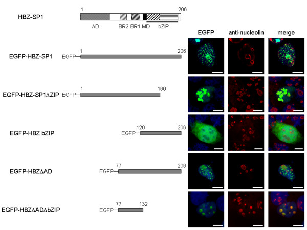
Figure 2
Molecular structures and nuclear localizations of HBZ-SP1 and its deletion mutants fused to EGFP . HBZ is composed of an N-terminal activation domain (AD), two basic regions (BR1 and BR2) involved in ...
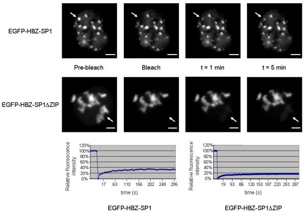
Figure 3
The HBZ-SP1 protein dynamically associates with subnuclear foci in living cells in a C-terminus-dependent mechanism . COS cells were transiently transfected with EGFP-HBZ-SP1 or EGFP-HBZ-SP1u0394ZIP. ...

Figure 4
Stimulation of HBZ-NBs formation by JunB . COS cells were transiently transfected with EGFP (a), HBZ bZIP fused to EGFP (b), or JunB (c), cultivated on glass slides, fixed, and then were analyzed by f...

Figure 5
JunB and HBZ bZIP are involved in the formation of HBZ-NBs . COS cells were cotransfected with pcDNA-JunB and pEGFP-HBZ bZIP in the absence (b) or in the presence of pcDNA-HBZ-SP1-Myc (c-e). Analysis ...
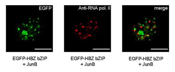
Figure 6
The HBZ-NBs do not colocalize with endogenous RNA polymerase II . COS cells cotransfected with pcDNA-JunB and pEGFP-HBZ bZIP were labelled with a mouse anti-RNA polymerase II antibody and detected usi...

Figure 7
Effects of the HBZ-SP1 protein on HPV-18 transcription in HeLa cells . (A) The HBZ-SP1 protein inhibits HPV-18 transcription. HeLa cells (6 u00d7 10 5 ) were cotransfected with 0.1 u03bcg of LCR-Luc, ...

Figure 8
The bZIP domain of the HBZ-SP1 protein is involved in the cell cycle arrest induced by transfection of HeLa cells with pEGFP-HBZ-SP1 . (A) Cell cycle of HeLa cells transfected with different mutants o...
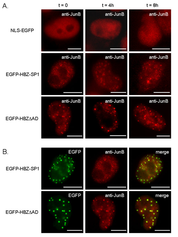
Figure 9
Targeting of endogenous JunB to HBZ-NBs in HeLa cells transfected with pEGFP-HBZ-SP1 . (A) Nuclear localization of endogenous JunB in HeLa cells transfected with different mutants of the HBZ-SP1 prote...

Figure 10
Immunofluorescence microscopy analysis of the colocalization of JunD and the HBZ-SP1 protein in vivo . COS cells cotransfected with pCMV-JunD-Flag and pEGFP-HBZ-SP1 were labelled with a mouse anti-Fla...
Figure images are served from the NIH/NLM PubMed Central Open Access Subset or Europe PMC; copyright remains with the publishers and authors.
💬 Discussion
0 commentsNo comments yet. Be the first to start a discussion!
Leave a Comment