Abstract
Munc18-1 forms a template to organize assembly of the neuronal SNARE complex that triggers neurotransmitter release, binding first to a closed conformation of syntaxin-1 where its amino-terminal region interacts with the SNARE motif, and later binding to synaptobrevin. However, the mechanism of SNARE complex assembly remains unclear. Here, we report two cryo-EM structures of Munc18-1 bound to cross-linked syntaxin-1 and synaptobrevin. The structures allow visualization of how syntaxin-1 opens and reveal how part of the syntaxin-1 amino-terminal region can help nucleate interactions between the amino termini of the syntaxin-1 and synaptobrevin SNARE motifs, while their carboxyl termini bind to distal sites of Munc18-1. These observations, together with mutagenesis, SNARE complex assembly experiments, and fusion assays with reconstituted proteoliposomes, support a model whereby these interactions are critical to initiate SNARE complex assembly and multiple energy barriers enable diverse mechanisms for exquisite regulation of neurotransmitter release.
🔬 Techniques
🧬 Organisms
✨ Fluorophores
🔬 Cell Lines
🏭 Microscope Brands
🧪 Reagent Suppliers
📷 Detectors
💻 Software Details
💾 Data Repositories
🏛️ Research Organizations (ROR)
Affiliated research institutions:
📋 Methods
Protein expression and purification
Escherichia coli expression and purification of full-length rat syntaxin-1A, the cytoplasmic fragment of rat syntaxin-1A (residues 2–253), a cysteine-free variant of full-length rat SNAP-25a, a full-length rat synaptobrevin-2, rat synaptobrevin-2 (residues 29–83), rat synaptobrevin (residues 1–96), full-length rat Munc18-1, full-length Cricetulus griseus NSF, full-length Bos taurus αSNAP, and a rat Munc13-1 fragment spanning the C 1 , C 2 B, MUN, and C 2 C domains (residues 529–1725, Δ1408–1452) (referred to as Munc13C) were described previously ( 9 , 10 , 13 , 50 – 52 ). The following mutants were also described previously ( 10 , 31 , 47 ) and were purified through the same protocols used for the WT proteins: full-length syntaxin-1 S186C, C145A, C271A, C272A; SNAP-25a R136C C84S, C85S, C90S, C92S; SNAP-25a M71D, L78D C84S, C85S, C90S, C92S (SNAP-25m); full-length synaptobrevin-2 L26C; synaptobrevin-1 1–96 L26C; Munc18-1 P335A; Munc18-1 D326K; and Munc18-1 L348R. Briefly, full-length syntaxin-1 was expressed overnight at 25°C upon induction with 0.4 mM isopropyl-β- d -thiogalactoside (IPTG). Cell pellets were resuspended with 20 mM Hepes (pH 7.4), 500 mM NaCl, 8 mM imidazole, and 1 mM tris(2-carboxyethyl)phosphine (TCEP). The protein was initially purified via affinity chromatography using HisPur Ni–nitrilotriacetic acid (NTA) resin (Thermo Fisher Scientific) in 20 mM tris (pH 7.4), 500 mM NaCl, 8 mM imidazole, 2% (v/v) Triton X-100, and 6 M urea. Upon extensive washes, the protein was eluted in 20 mM tris (pH 7.4), 500 mM NaCl, 400 mM imidazole, and 0.1% dodecylphosphocholine (DPC). The polyhistidine tag was removed using thrombin protease, followed by size exclusion chromatography on a Superdex 200 column (GE 10/300) equilibrated in 20 mM tris (pH 7.4), 125 mM NaCl, 1 mM TCEP, and 0.2% DPC. Expression of syntaxin-1 2–253 was induced with 0.4 mM IPTG and expressed overnight at 25°C. Upon cell lysis, purification was done using Glutathione Sepharose 4B resin (GE) in phosphate-buffered saline (PBS; pH 7.4), PBS with 1% (v/v) Triton X-100, and PBS with 1 M NaCl. The glutathione S -transferase (GST)–tag was cleaved by thrombin protease, and the eluted protein was further purified by anion exchange chromatography on a HiTrap Q column (GE) in 25 mM tris (pH 7.4) and 1 mM TCEP using a linear gradient from 0 to 1 M NaCl. Please note that the purification of syntaxin-1 2–253 C145A, L165A, E166A, L205C (SyxLE) was carried out with the same procedure except that the final purification was done by size exclusion chromatography using a Superdex 75 (GE 10/300) column equilibrated with 100 mM phosphate buffer at pH 7.0 with 100 mM NaCl, 6 M urea, 0.2 mM EDTA, and 0.5 mM TCEP. Cysteine-free SNAP-25 was expressed overnight at 25°C upon induction with 0.4 mM IPTG. Upon cell lysis, protein purification was performed using HisPur Ni-NTA resin (Thermo Fisher Scientific) in 50 mM tris (pH 8.0), 500 mM NaCl, 20 mM imidazole, and 1% (v/v) Triton X-100. The His6-tag was cleaved by thrombin protease, and the protein was purified by size exclusion chromatography using a Superdex 75 column (GE 16/60) in 50 mM tris (pH 8.0) and 150 mM NaCl. Full-length synaptobrevin-2 was expressed overnight at 25°C upon induction with 0.4 mM IPTG. Cells were resuspended in PBS buffer containing 1% (v/v) Triton X-100. Purification was done using Glutathione Sepharose 4B resin (GE) at 4°C. The bound proteins were treated with PBS with 1% (v/v) Triton X-100, followed by the addition of thrombin to cleave the GST-tag. The protein was further purified by cation exchange chromatography on a HiTrap S column (GE) in 25 mM NaAc (pH 5.5), 1 mM TCEP, and 1% (w/v) using a linear gradient from 0 to 1 M NaCl.
Show full methods section
Protein expression and purification
Escherichia coli expression and purification of full-length rat syntaxin-1A, the cytoplasmic fragment of rat syntaxin-1A (residues 2–253), a cysteine-free variant of full-length rat SNAP-25a, a full-length rat synaptobrevin-2, rat synaptobrevin-2 (residues 29–83), rat synaptobrevin (residues 1–96), full-length rat Munc18-1, full-length Cricetulus griseus NSF, full-length Bos taurus αSNAP, and a rat Munc13-1 fragment spanning the C 1 , C 2 B, MUN, and C 2 C domains (residues 529–1725, Δ1408–1452) (referred to as Munc13C) were described previously ( 9 , 10 , 13 , 50 – 52 ). The following mutants were also described previously ( 10 , 31 , 47 ) and were purified through the same protocols used for the WT proteins: full-length syntaxin-1 S186C, C145A, C271A, C272A; SNAP-25a R136C C84S, C85S, C90S, C92S; SNAP-25a M71D, L78D C84S, C85S, C90S, C92S (SNAP-25m); full-length synaptobrevin-2 L26C; synaptobrevin-1 1–96 L26C; Munc18-1 P335A; Munc18-1 D326K; and Munc18-1 L348R. Briefly, full-length syntaxin-1 was expressed overnight at 25°C upon induction with 0.4 mM isopropyl-β- d -thiogalactoside (IPTG). Cell pellets were resuspended with 20 mM Hepes (pH 7.4), 500 mM NaCl, 8 mM imidazole, and 1 mM tris(2-carboxyethyl)phosphine (TCEP). The protein was initially purified via affinity chromatography using HisPur Ni–nitrilotriacetic acid (NTA) resin (Thermo Fisher Scientific) in 20 mM tris (pH 7.4), 500 mM NaCl, 8 mM imidazole, 2% (v/v) Triton X-100, and 6 M urea. Upon extensive washes, the protein was eluted in 20 mM tris (pH 7.4), 500 mM NaCl, 400 mM imidazole, and 0.1% dodecylphosphocholine (DPC). The polyhistidine tag was removed using thrombin protease, followed by size exclusion chromatography on a Superdex 200 column (GE 10/300) equilibrated in 20 mM tris (pH 7.4), 125 mM NaCl, 1 mM TCEP, and 0.2% DPC. Expression of syntaxin-1 2–253 was induced with 0.4 mM IPTG and expressed overnight at 25°C. Upon cell lysis, purification was done using Glutathione Sepharose 4B resin (GE) in phosphate-buffered saline (PBS; pH 7.4), PBS with 1% (v/v) Triton X-100, and PBS with 1 M NaCl. The glutathione S -transferase (GST)–tag was cleaved by thrombin protease, and the eluted protein was further purified by anion exchange chromatography on a HiTrap Q column (GE) in 25 mM tris (pH 7.4) and 1 mM TCEP using a linear gradient from 0 to 1 M NaCl. Please note that the purification of syntaxin-1 2–253 C145A, L165A, E166A, L205C (SyxLE) was carried out with the same procedure except that the final purification was done by size exclusion chromatography using a Superdex 75 (GE 10/300) column equilibrated with 100 mM phosphate buffer at pH 7.0 with 100 mM NaCl, 6 M urea, 0.2 mM EDTA, and 0.5 mM TCEP. Cysteine-free SNAP-25 was expressed overnight at 25°C upon induction with 0.4 mM IPTG. Upon cell lysis, protein purification was performed using HisPur Ni-NTA resin (Thermo Fisher Scientific) in 50 mM tris (pH 8.0), 500 mM NaCl, 20 mM imidazole, and 1% (v/v) Triton X-100. The His6-tag was cleaved by thrombin protease, and the protein was purified by size exclusion chromatography using a Superdex 75 column (GE 16/60) in 50 mM tris (pH 8.0) and 150 mM NaCl. Full-length synaptobrevin-2 was expressed overnight at 25°C upon induction with 0.4 mM IPTG. Cells were resuspended in PBS buffer containing 1% (v/v) Triton X-100. Purification was done using Glutathione Sepharose 4B resin (GE) at 4°C. The bound proteins were treated with PBS with 1% (v/v) Triton X-100, followed by the addition of thrombin to cleave the GST-tag. The protein was further purified by cation exchange chromatography on a HiTrap S column (GE) in 25 mM NaAc (pH 5.5), 1 mM TCEP, and 1% (w/v) using a linear gradient from 0 to 1 M NaCl.
Expression of synaptobrevin-2 1–96 or synaptobrevin-2 29–83 Q36C
(Syb) was induced with 0.4 mM IPTG and expressed overnight at 23°C. Purification was done using Glutathione Sepharose 4B resin (GE), followed by cleavage of the GST-tag. The final purification of synaptobrevin-2 1–96 was performed using size exclusion chromatography on a Superdex 75 column (GE 16/60) equilibrated in 20 mM tris (pH 7.4) and 125 mM NaCl. The final purification of synaptobrevin-2 29–83 Q36C was carried out using size exclusion chromatography on a Superdex 75 column (GE 10/300) equilibrated with 100 mM phosphate buffer at pH 7.0 with 100 mM NaCl, 6 M urea, 0.2 mM EDTA, and 0.5 mM TCEP. Expression of αSNAP was induced by the addition of 0.4 mM ITPG and continued overnight at 25°C. Protein purification was performed using Glutathione Sepharose 4B resin (GE) by washing bound proteins with PBS, PBS with 1% (v/v) Triton X-100, and PBS with 1 M NaCl. Upon GST-tag cleavage in the presence of thrombin, the protein was purified by size exclusion chromatography using a Superdex 75 column (GE 16/60) in 20 mM tris (pH 7.4), 150 mM KCl, and 1 mM TCEP. Expression of NSF was induced with 0.4 mM IPTG and continued overnight at 20°C. Purification was performed using HisPur Ni-NTA resin (Thermo Fisher Scientific), followed by size exclusion chromatography of hexameric NSF on a Superdex S200 column (GE 16/60) in 50 mM tris (pH 8.0), 100 mM NaCl, 1 mM adenosine triphosphate (ATP), 1 mM EDTA, 1 mM dithiothreitol (DTT), and 10% (v/v) glycerol. Removal of the His6-tag and monomerization of NSF were performed using Tobacco Etch Virus (TEV) protease and apyrase, respectively, while dialyzing with nucleotide-free buffer for 36 hours. To separate the hexameric form of NSF from the monomeric, three rounds of size exclusion chromatography on a Superdex S200 column (GE 16/60) in 50 mM NaPi (pH 8.0), 100 mM NaCl, and 0.5 mM TCEP were performed by reinjecting fractions with hexameric NSF. Final reassembly of monomers and gel filtration chromatography of reassembled hexameric NSF were done using a Superdex S200 column (GE 16/60) in 50 mM tris (pH 8.0), 100 mM NaCl, 1 mM ATP, 1 mM EDTA, 1 mM TCEP, and 10% (v/v) glycerol.
Expression of full-length
Munc18-1 was induced with 0.4 mM IPTG and continued overnight at 20°C. Upon cell lysis and centrifugation, the supernatant was loaded on Glutathione Sepharose 4B resin (GE) at 4°C, and the bound proteins were washed with PBS, PBS with 1% (v/v) Triton X-100, and PBS with 1 M NaCl. The GST-tag was cleaved by a thrombin treatment, followed by immediate size exclusion chromatography using a Superdex 200 column (GE 16/60) in a buffer containing 20 mM tris (pH 7.4), 200 mM KCl, and 1 mM TCEP.
Expression of the rat
Munc13C (residues 529–1725, Δ1408–1452) was induced with 0.5 mM IPTG and performed overnight at 16°C. The protein was purified by affinity chromatography using HisPur Ni-NTA resin (Thermo Fisher Scientific) with extensive washes with 50 mM tris (pH 8), 10 mM imidazole, 750 mM NaCl, 1 mM TCEP, and 10% (v/v) glycerol. The protein was eluted with 50 mM tris (pH 8), 250 mM NaCl, 1 mM TCEP, 10% (v/v) glycerol, and 500 mM imidazole and dialyzed overnight at 4°C in 50 mM tris (pH 8), 250 mM NaCl, 1 mM TCEP, 2.5 mM CaCl 2 , and 10% (v/v) glycerol in the presence of thrombin. The protein was further purified by anion exchange chromatography on a HiTrap Q column (GE) in 20 mM tris (pH 8.0), 1 mM TCEP, and 10% (v/v) glycerol using a linear gradient from 0 to 1 M NaCl. The following mutants were generated using the QuikChange site-directed mutagenesis and custom-designed primers and purified as the unmodified constructs: full-length syntaxin-1A (1–288) M138A; full-length syntaxin-1A (1–288) D184P; syntaxin-1A (2–253) M183A; syntaxin-1A (2–253) D184P; syntaxin-1A (2–253) C145A, L165A, E166A, L205C; syntaxin-1A (2–253) C145A, L165A, E166A, M183A, L205C; full-length syntaxin-1A (1–288) C145A, L165A, E166A, M183A, L205C; full-length syntaxin-1A (1–288) C145A, L165A, E166A, D184P, L205C; synaptobrevin-2 29–83 Q36C; Munc18-1 S42Q; Munc18-1 L307R; Munc18-1 Q301D; and Munc18-1 E352K. Template complex formation Before complex formation, TCEP was removed from each protein preparation using Superdex 75 10/300 GL column. TCEP-free Syb (100 to 200 μM) was mixed with 400 to 500 μM 2,2-dithiodipyridine (Sigma-Aldrich). The reaction was monitored at 343 nm using an Agilent 8453 ultraviolet-visible spectrometer. Upon completion, the excess of 2,2-dithiodipyridine was removed using a PD MiniTrap G-25 Sephadex column with 100 mM phosphate at pH 7.0, 100 mM NaCl, 6 M urea, and 0.2 mM EDTA as the elution buffer. An excess of activated Syb was then mixed with TCEP-free SyxLE and left incubated at room temperature overnight. The efficiency of disulfide bond formation between Syb and SyxLE was estimated on the basis of the absorbance at 343 nm. The SyxLE/Syb conjugate was purified by size exclusion chromatography using Superdex 75 10/300 GL and 20 mM Hepes (pH 7.4), 150 mM KCl as the running buffer. TCEP-free Munc18-1 D326K was then mixed with purified SyxLE/Syb and left rotated overnight at room temperature. The template complex was finally purified by size exclusion chromatography using a Superdex 75 10/300 GL column in 20 mM Hepes (pH 7.4) with 150 mM KCl.
EM data acquisition
Fluorinated fos-choline-8 was added into purified template complex formed by SyxLE and Munc18-1 D326K (7.5 mg/ml) to a final concentration of 5 mM, and 3 μl of the sample was applied to glow-discharged (30 mA, 80 s) Quantifoil R1.2/1.3 300-mesh gold holey carbon grids (Quantifoil, Micro Tools GmbH, Germany). Grids were blotted for 4.0 s under 100% humidity at 4°C before being plunged into the liquid ethane using Mark IV Vitrobot (FEI). Micrographs were acquired on a Titan Krios microscope (FEI) operated at 300 kV with a K3 direct electron detector (Gatan), using a slit width of 20 eV on a GIF-Quantum energy filter. SerialEM was used for data collection. A calibrated magnification of 46,296 was used for imaging of the samples, yielding a pixel size of 1.08 Å on the images. The defocus range was set from −1.6 to −2.6 μm. Each micrograph was dose-fractionated to 30 frames with a total dose of about 60 e − /Å 2 .
Image processing
The cryo-EM refinement statistics are summarized in table S1. A total of 7401 movie frames of the SyxLE/Syb–Munc18-1 D326K complex were motion-corrected, binned twofold, resulting in a pixel size of 1.08 Å, and dose-weighted using MotionCor2. The contrast transfer function (CTF) parameters were estimated using Gctf. RELION3 ( 53 ) was used for the following processing. Particles were first roughly picked by using the Laplacian-of-Gaussian blob method and then subjected to 2D classification. Class averages representing projections of the SyxLE/Syb-Munc18-1 D326 complex in different orientations were used as templates for reference-based particle picking. Extracted particles were binned three times and subjected to 2D classification. Particles from the classes with fine structural features were selected for 3D classification using an initial model generated from a subset of the particles in RELION3. Particles from one of the resulting 3D classes showing good secondary structural features were selected and reextracted into the original pixel size of 1.08 Å. Subsequently, we performed finer 3D classification imposed by using local search in combination with small angular sampling (3.75°), resulting in new classes showing two distinct conformations of the SyxLE/Syb–Munc18-1 D326 complex. The particles corresponding to two different conformations were selected and refined separately, leading to two different cryo-EM maps at 3.5-Å (class2) and 3.7-Å (class1) resolution, respectively.
Model building and refinement
Model building of both template complexes was started by docking the individual chains from the previously solved crystal structure of the syntaxin-1–Munc18-1 complex [Protein Data Bank (PDB) ID: 3C98] using Chimera 1.15 ( 54 ). The models were improved by iterative manual building in Coot 0.9 ( 55 ) and real-space refinement in the software package Phenix 1.19.1 ( 56 ). The density for Hd in syntaxin in both tc1 and tc2 is rather weak, preventing the assignment of individual residues. This part was therefore built as a polyalanine helix, with the register of the residues undetermined. The quality of the model stereochemistry and fit to density was evaluated with the comprehensive validation method MolProbity ( 57 ) as implemented in the Phenix package. The statistics are shown in table S1. The PyMOL Molecular Graphics System, Version 4.6.0 (Schrödinger LLC) was used for visualization of the structures, preparation of molecular diagrams for the figures, and calculations of buried surface areas. Gel filtration–binding assay Samples (6 to 20 μM) containing Munc18-1 D326K, Munc18-1 D326K incubated with SyxLE and Syb, or Munc18-1 D326K incubated with SyxLE/Syb were injected into a size exclusion chromatography column (Superdex 30 Increase 10/300 GL) using 20 mM Hepes (pH 7.4), 150 mM KCl as the running buffer. To form potential complexes, selected proteins were incubated overnight at 4°C before injection. Selected eluted fractions were loaded into SDS-PAGE gel and stained with InstantBlue Coomassie protein stain to confirm protein coelution.
SDS-PAGE SNARE complex assembly assay
To detect the SDS-resistant SNARE complex, the template complex was formed as described above, and the SyxLE/Munc18-1 D326K complex was preformed (overnight incubation at 4°C). SNAP-25 (5 μM) was added to 5 μM template complex; 5 μM Syb and 5 μM SNAP-25 were added to the SyxLE/Munc18-1 D326K complex; or 5 μM Syb, 5 μM SNAP-25, and 5 μM SyxLE were mixed. The reaction was done in 25 mM Hepes (pH 7.4), 150 mM KCl, and 10% (v/v) glycerol at room temperature. After 3 min, the reaction was stopped by addition of the SDS-PAGE gel-loading buffer. The samples were then loaded into SDS-PAGE gels and stained with InstantBlue Coomassie protein stain. The gels were imaged using a Bio-Rad ChemiDoc imaging system. Mass photometry Samples containing 1000 nM SyxLE/Syb, or SyxLE M183A/Syb, or SyxLE D184P/Syb, or syntaxin-1 (2–253), or syntaxin-1 (2–253) M183A, syntaxin-1 (2–253) D184P were incubated overnight at room temperate with TCEP-free 1000 nM or 2000 nM WT or mutant Munc18-1. Before the measurements, the samples were diluted 10-fold using PBS at pH 7.4. High-precision microscope cover glasses were rinsed with Milli-Q water, isopropanol, Milli-Q water, isopropanol, and Milli-Q water and dried using a stream of nitrogen gas. Clean coverslips with attached silicon gaskets were then mounted on immersion oil (refractive index of 1.518)–covered lenses of a Refeyn’s second-generation mass photometer. All the measurements were performed using PBS at pH 7.4. Data were collected using AcquireMP software. A single gasket was filled with 13 to 17.1 μl of PBS to enable focusing of the coverslip. Once the focus signal was stable, 0.9 to 7 μl of the 100 nM sample were added and quickly mixed to achieve the desired final protein concentrations. A movie was recorded for 60 s (2819 frames) and processed using DiscoverMP. Contrast-to-mass calibration was achieved using a bovine serum albumin (BSA) standard (0.002 mg/ml). The contrasts observed for BSA monomer (66 kDa), dimer (132 kDa), and trimer (198 kDa) were used to generate a standard calibration curve. Each measurement is displayed as normalized histograms with Gaussian fitting of the binding event counts with a loaded standard calibration curve. K D was calculated using a standard one-ligand binding model.
Solution SNARE complex assembly assay
To monitor SNARE complex assembly in solution, synaptobrevin (1–96) L26C and SNAP-25 R136C (50 to 150 μM) were respectively labeled with Alexa Fluor 488 maleimide and tetramethylrhodamine (TMR) maleimide at room temperature with 10- to 20-fold excess of the dyes for 2 hours. The excess of the reagents was removed using a Superdex 75 (10/300) column equilibrated with 20 mM tris (pH 7.4), 150 mM NaCl, and 1 mM TCEP. SNARE complex assembly was monitored by detecting Alexa Fluor 488 donor fluorescence intensity (excitation at 468 nm and emission at 518 nm) as a function of time at 37°C using a PTI Quantamaster 400 spectrofluorometer (T-format) equipped with a rapid Peltier temperature–controlled four-position sample holder with a GG495 long-pass filter mounted. To start the reactions, 0.1 μM synaptobrevin (1–96) L26C–Alexa Fluor 488 was mixed with 1 μM SNAP-25 R136C-TMR and 1 μM syntaxin-1 (2–253)/Munc18-1 complexes (preformed for 1 hour at room temperature). The reaction buffer was 25 mM Hepes (pH 7.4), 150 mM KCl, 1 mM TCEP, and 10% (v/v) glycerol.
Trans-SNARE complex formation assay
Single-cysteine variants of syntaxin-1A S186C and synaptobrevin-2 L26C were labeled with TMR and Alexa Fluor 488, respectively, as described above. V-liposomes were prepared similarly to those used for content mixing fusion assays (see below) except that they contained synaptobrevin-2 L26C–Alexa Fluor 488 with a 1:10,000 protein-to-lipid ratio. S-liposomes were prepared as the T-liposomes used for the content mixing assays but using syntaxin S186C-TMR without SNAP-25. Trans-SNARE complex formation was measured by the development of FRET between Alexa Fluor 488–synaptobrevin on V-liposomes (0.0625 mM total lipid) and TMR–syntaxin-1A on S-liposomes (0.25 mM total lipid) at 37°C using a PTI Quantamaster 400 spectrofluorometer (T-format) equipped with a rapid Peltier temperature–controlled four-position sample holder. Before the measurements, the S-liposomes were incubated with 0.37 μM WT or mutant Munc18-1 for 1 hour at room temperature. The Alexa Fluor 488 donor fluorescence of V-liposomes at 518 nm (excitation at 468 nm) was recorded to monitor the development of FRET over time upon mixing with S-liposomes containing the preformed syntaxin-1/Munc18-1 complexes, 2 μM SNAP-25a M71D, L78D, 0.2 μM Munc13C, and 0.1 mM EGTA. At 1100 s, the reaction was paused and 0.6 mM CaCl 2 was quickly added to each reaction. The reaction buffer contained 25 mM Hepes (pH 7.4), 150 mM KCl, 1 mM TCEP, and 10% (v/v) glycerol. A GG495 long-pass filter (Edmund Optics) was used to filter scattered light.
Content mixing assays
The assay was performed as previously described ( 17 ). Briefly, V-liposomes containing full-length synaptobrevin-2 (protein-to-lipid ratio of 1:500) were prepared with 39% 1-palmitoyl, 2-oleoyl phosphatidylcholine (POPC), 19% 1,2-dioleoyl-sn-glycero-3-phospho- l -serine (DOPS), 19% 1-palmitoyl-2-oleoyl-sn-phosphatidylethanolamine (POPE), 20% cholesterol, 1.5% N -(7-nitrobenz-2-oxa-1,3-diazol-4-yl)-1,2-dihexadecanoyl-sn-glycero-3-phosphoethanolamine, triethylammonium salt (NBD-PE), and 1.5% 1,2-dihexadecanoyl-sn-glycero-3-phosphoethanolamine (Marina Blue DHPE). T-liposomes containing syntaxin-1 (WT or M183, or D184P; syntaxin-1–to–lipid ratio of 1:800) and 25 μM SNAP-25 were prepared with 38% POPC, 18% DOPS, 20% POPE, 20% cholesterol, 2% phosphatidylinositol 4,5-biphosphate (PIP 2 ), and 2% diacylglycerol (DAG). Dried lipid films were resuspended in 25 mM Hepes (pH 7.4), 150 mM KCl, 1 mM TCEP, and 2% octyl β-glucopyranoside (βOG). Lipid solutions were then mixed with the respective proteins and with 4 μM phycoerythrin-biotin for T-liposomes or with 8 μM Cy5-streptavidin for V-liposomes in 25 mM Hepes (pH 7.4), 150 mM KCl, 1 mM TCEP, and 10% (v/v) glycerol. Proteoliposomes were prepared by detergent removal using dialysis with Amberlite XAD-2 beads (2 g/liter; Sigma-Aldrich) three times at 4°C and subsequent co-floatation on a three-layer histodenz gradient (35, 25, and 0%) and harvested from the topmost layer. Content mixing signals were measured from the development of FRET between Cy5-streptavidin trapped in V-liposomes and phycoerythrin-biotin trapped in T-liposomes (excitation at 565 nm and emission at 670 nm). Each reaction was prepared in a total volume of 200 μl with V-liposomes (0.125 mM total lipid), T-liposomes (0.25 mM total lipids), 2.5 mM MgCl 2 , 2 mM ATP, 0.1 mM EGTA, 5 μM streptavidin, and the following proteins: 1 μM SNAP-25, 0.4 μM NSF purified in ATP-containing buffer, 2 μM αSNAP, 1 μM Munc18-1 (WT or other tested mutants), and 0.2 μM Munc13C. At 300 s, the reaction was paused and 0.6 mM CaCl 2 was added to each reaction mixture and quickly mixed. All experiments were performed at 30°C using a PTI Quantamaster 400 spectrofluorometer (T-format) equipped with a rapid Peltier temperature–controlled four-position sample holder. Content mixing measurements were normalized to control reactions collected without streptavidin in the presence of 1% βOG to measure the maximal Cy5 fluorescence attainable.
Statistics
To analyze the statistical significance of the results obtained in various assays ( Figs. 3 to 5 ), all experiments were performed at least in triplicates and one-way analysis of variance (ANOVA) was performed through an all-pairwise multiple comparison procedure using the Holm-Sidak test (*** P < 0.001 and ** P < 0.01). For the analysis of binding of WT and Munc18-1 mutants to SyxLE/Syb by mass photometry ( Fig. 3I ), the difference between the K D ’s obtained for WT and S42Q mutant Munc18-1 was obviously statistically significant ( P < 0.001), but the very large K D calculated for the S42Q mutant and the correspondingly large SD hinder the statistical analysis of significance for other pairwise comparisons within the group. Hence, all other comparisons within the group were performed after removing the S42Q data.
Supplementary Materials This PDF file includes: Figs. S1 to S12 Tables S1 to S3 Click here for additional data file.
📊 Figures
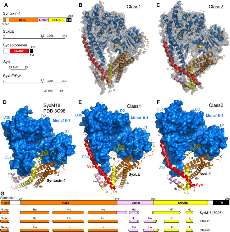
Fig. 1.
Two cryo-EM structures of the template complex.
( A ) Domain diagrams of syntaxin-1 and synaptobrevin, and summary of the fragments used to prepare SyxLE/Syb. N-pep, N-peptide; SNARE, SNARE motif. ( B and C ) 3D reconstructions of two structures of...
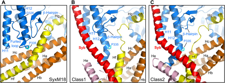
Fig. 2.
Structural changes in Munc18-1 that lead to synaptobrevin binding and template complex formation.
( A to C ) Close-up views of the area where the Munc18-1 loop unfurls to allow synaptobrevin binding in the syntaxin-1u2013Munc18-1 complex (SyxM18) before unfurling (A) and in class1 (B) and class2 (...
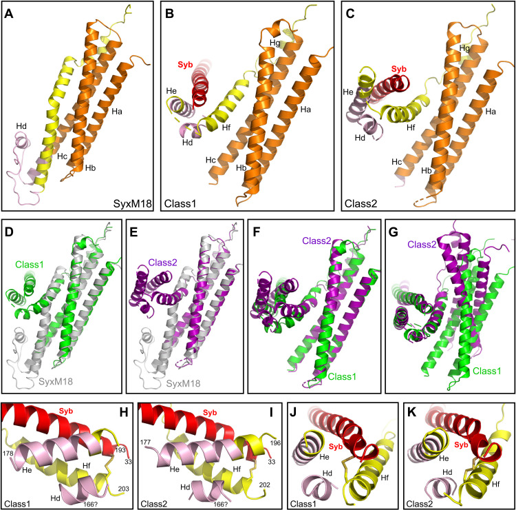
Fig. 3.
Conformational changes in syntaxin-1 that lead to template complex formation.
( A to C ) Close-up views of the region where the syntaxin-1 SNARE motif interacts with the H abc domain and the linker in SyxM18 (A) as well as with synaptobrevin in class1 (B) and class2 (C). ( D to...
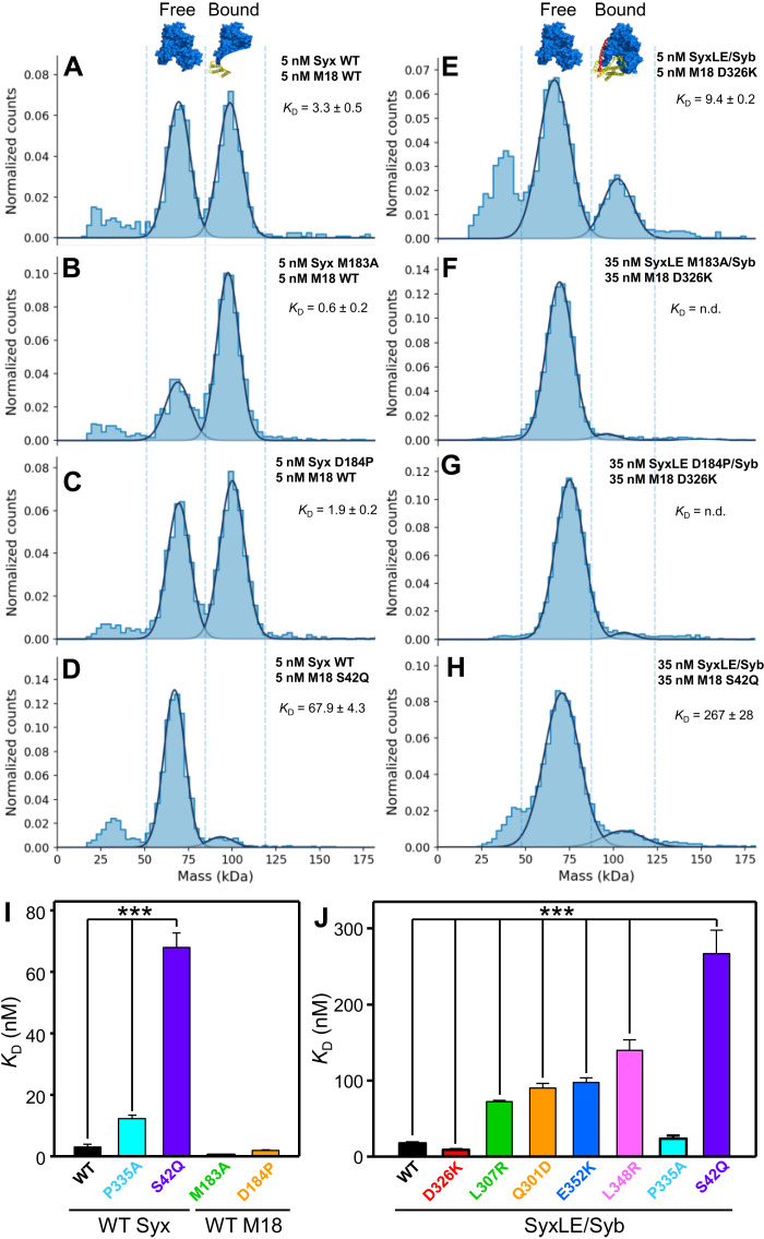
Fig. 4.
Analysis of Munc18-1u2013SNARE interactions by mass photometry.
( A to H ) Normalized histograms of mass distributions observed for samples containing the indicated concentrations of WT or mutant Munc18-1 (M18) plus WT or mutant syntaxin-1 (2u2013253) (Syx) (A to ...
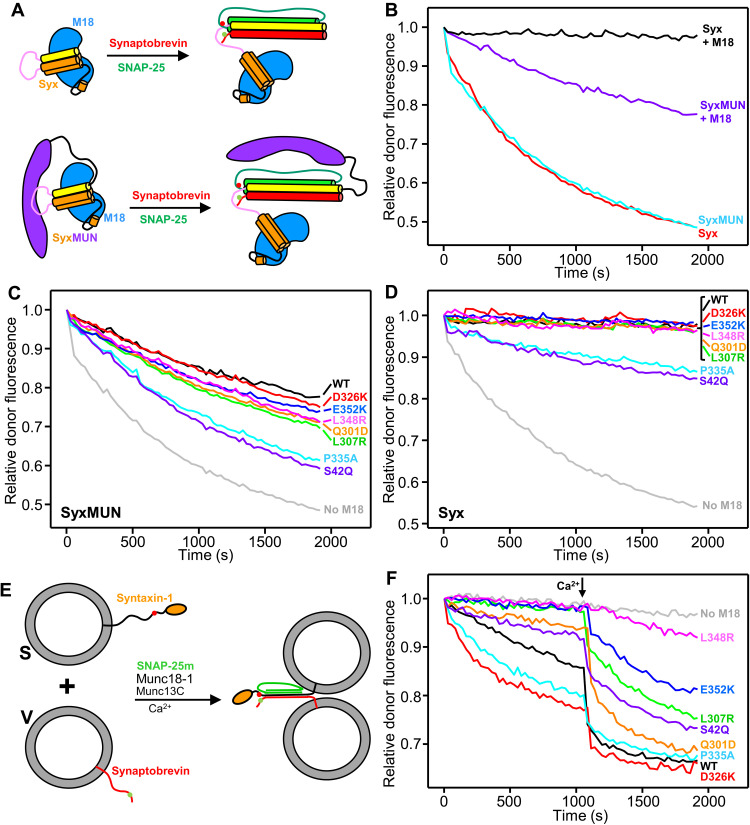
Fig. 5.
Effects of mutations in Munc18-1 on SNARE complex assembly rates.
( A ) Diagram summarizing the assays used to monitor SNARE complex assembly in solution starting with Munc18-1 (blue) bound to the syntaxin-1 cytoplasmic region (H abc domain, orange; linker, pink; SN...
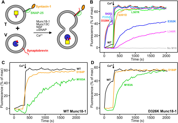
Fig. 6.
Effects of Munc18-1 and syntaxin-1 mutations on liposome fusion.
( A ) Diagram summarizing the content mixing between T-liposomes containing syntaxin-1 and SNAP-25, which were preincubated with Munc18-1, NSF, and u03b1SNAP, and V-liposomes containing synaptobrevin ...
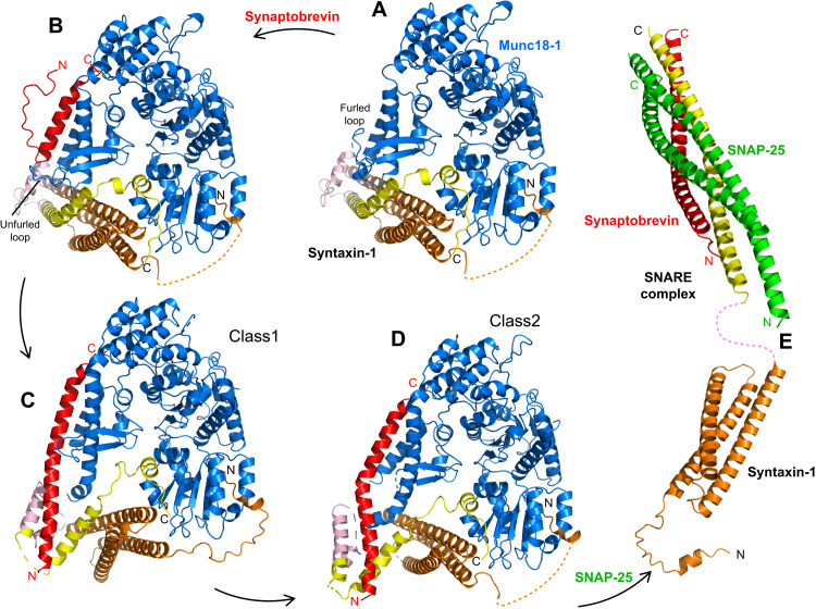
Fig. 7.
Model of SNARE complex assembly templated by Munc18-1 and the syntaxin-1 N-terminal region.
The model postulates that assembly starts with Munc18-1 bound to closed syntaxin-1 ( A ) (PDB code 3C98) and is initiated when the Munc18-1 loop unfurls to allow synaptobrevin binding ( B ) (model bui...
Figure images are served from the NIH/NLM PubMed Central Open Access Subset or Europe PMC; copyright remains with the publishers and authors.
💬 Discussion
0 commentsNo comments yet. Be the first to start a discussion!
Leave a Comment