Abstract
There is significant evidence that brain-infiltrating CD8+ T cells play a central role in the development of experimental cerebral malaria (ECM) during Plasmodium berghei ANKA infection of C57BL/6 mice. However, the mechanisms through which they mediate their pathogenic activity during malaria infection remain poorly understood. Utilizing intravital two-photon microscopy combined with detailed ex vivo flow cytometric analysis, we show that brain-infiltrating T cells accumulate within the perivascular spaces of brains of mice infected with both ECM-inducing (P. berghei ANKA) and non-inducing (P. berghei NK65) infections. However, perivascular T cells displayed an arrested behavior specifically during P. berghei ANKA infection, despite the brain-accumulating CD8+ T cells exhibiting comparable activation phenotypes during both infections. We observed T cells forming long-term cognate interactions with CX3CR1-bearing antigen presenting cells within the brains during P. berghei ANKA infection, but abrogation of this interaction by targeted depletion of the APC cells failed to prevent ECM development. Pathogenic CD8+ T cells were found to colocalize with rare apoptotic cells expressing CD31, a marker of endothelial cells, within the brain during ECM. However, cellular apoptosis was a rare event and did not result in loss of cerebral vasculature or correspond with the extensive disruption to its integrity observed during ECM. In summary, our data show that the arrest of T cells in the perivascular compartments of the brain is a unique signature of ECM-inducing malaria infection and implies an important role for this event in the development of the ECM-syndrome.
🔬 Techniques
🧬 Organisms
💻 Software
✨ Fluorophores
🧪 Sample Preparation
🔬 Cell Lines
🏭 Microscope Brands
🧪 Reagent Suppliers
📷 Detectors
💻 Software Details
💾 Data Repositories
🏛️ Research Organizations (ROR)
Affiliated research institutions:
📋 Methods
Ethics Animal work in New
York was carried out in strict accordance with the recommendation in the Guide for the Care for the Care and Use of Laboratory Animals of the Public Health Service (National Institutes of Health) and was approved by New York University School of Medicine Institutional Animal Care and Use Committee (IACUC). Animal work in the U.K. was approved following local ethical review by the Universities of Manchester and Glasgow Animal Procedures and Ethics Committees and was performed in strict accordance with the U. K Home Office Animals (Scientific Procedures) Act 1986 (approved H.O Project Licenses 70/6995 and 70/7293). All surgery was performed under anesthesia: ketamine (50 mg/kg), xylazine (10 mg/kg), acepromazine (1.7 mg/kg); or isoflurane (2% in O 2 at 0.2 L/min).
Mice
The following mice were used in the study: At Skirball Institute Animal facility, C57BL/6 (H-2b) from the National Cancer Institute or Taconic Labs, OT-I [ 71 ], P14 [ 52 ], CFP [ 72 ], DsRed [ 73 ], CX 3 CR1 CreER X Rosa26 iDTR mice [ 51 ]. Both CFP and DsRed were expressed under the control of a chicken β-actin promoter and CMV enhancer cassette. CX 3 CR1 CreER X Rosa26 iDTR (referred to as CX 3 CR1-iDTR) mice express Cre in CX 3 CR1 + cells upon treatment with tamoxifen, inducing expression of the diphtheria toxin receptor (DTR) and, thus, sensitivity to diphtheria toxin (DT) in those cells. At the Universities of Glasgow and Manchester, C57BL/6 mice from Harlan and Charles River, UK, hCD2-DsRed [ 74 ], CX 3 CR1 GFP/GFP [ 45 ], CX 3 CR1 GFP/GFP X C57BL/6 (F1) and CX 3 CR1 GFP/GFP X hCD2-DsRed +/+ mice (F1). In all cases, transgenic mice were fully backcrossed to a C57BL/6 background and were used between 6 and 12 weeks of age. Mice were maintained in specific pathogen-free conditions. Parasites GFP-expressing P . berghei ANKA parasites (GFP expressed under control of elogation factor 1a [eEF1a] promoter) were a kind gift from Chris Janse (Leiden University Medical Center) [ 75 ]. GFP/SIINFEKL-expressing P . berghei ANKA ( Pb -TG) parasites were a kind gift from William Heath (University of Melbourne) [ 22 ]. P . berghei NK65 parasites expressed GFP under control of the circumsporozoite promoter [ 76 ]. Parasites were maintained in liquid nitrogen and passaged through naive mice prior to being used to infect experimental animals. Experimental infections were initiated by i.v. inoculation with 10 4 or 10 6 pRBCs, depending upon the experiment, and infected mice were monitored for neurological symptoms (paralysis, ataxia, convulsions, and coma occurring between day 6 and 10 post-infection). Parasitemia was measured daily from day 3 p.i. by either examination of Giemsa-stained thin blood smears or by flow cytometric detection of DAPI stained GFP + parasites ( S11 Fig ) . Classification of experimental cerebral malaria The induction and severity of ECM was assessed using the following well-defined grading system [ 77 ] 1: no signs; 2: ruffled fur/and or abnormal posture; 3: lethargy; 4: reduced responsiveness to stimulation and/or ataxia and/or respiratory distress/hyperventilation; 5: prostration and/or paralysis and/or convulsions. Stages 2/3 were classified as prodromal signs of ECM and stages 4/5 were classified as ECM. Adoptive transfer Splenic DsRed +/+ OT-I CD8 + T cells were purified (>95% purity) from a naïve DsRed +/+ OT-I TCR Tg mouse using a Dynal Mouse CD8 + Negative Isolation Kit (Invitrogen). 10 4 and 10 6 DsRed +/+ OT-I CD8 + T cells were transferred i.v. into C57BL/6 and P14 TCR Tg recipients, respectively, one day prior to infection with 10 6 Pb -TG-pRBCs, as described above.
Show full methods section
Ethics Animal work in New
York was carried out in strict accordance with the recommendation in the Guide for the Care for the Care and Use of Laboratory Animals of the Public Health Service (National Institutes of Health) and was approved by New York University School of Medicine Institutional Animal Care and Use Committee (IACUC). Animal work in the U.K. was approved following local ethical review by the Universities of Manchester and Glasgow Animal Procedures and Ethics Committees and was performed in strict accordance with the U. K Home Office Animals (Scientific Procedures) Act 1986 (approved H.O Project Licenses 70/6995 and 70/7293). All surgery was performed under anesthesia: ketamine (50 mg/kg), xylazine (10 mg/kg), acepromazine (1.7 mg/kg); or isoflurane (2% in O 2 at 0.2 L/min).
Mice
The following mice were used in the study: At Skirball Institute Animal facility, C57BL/6 (H-2b) from the National Cancer Institute or Taconic Labs, OT-I [ 71 ], P14 [ 52 ], CFP [ 72 ], DsRed [ 73 ], CX 3 CR1 CreER X Rosa26 iDTR mice [ 51 ]. Both CFP and DsRed were expressed under the control of a chicken β-actin promoter and CMV enhancer cassette. CX 3 CR1 CreER X Rosa26 iDTR (referred to as CX 3 CR1-iDTR) mice express Cre in CX 3 CR1 + cells upon treatment with tamoxifen, inducing expression of the diphtheria toxin receptor (DTR) and, thus, sensitivity to diphtheria toxin (DT) in those cells. At the Universities of Glasgow and Manchester, C57BL/6 mice from Harlan and Charles River, UK, hCD2-DsRed [ 74 ], CX 3 CR1 GFP/GFP [ 45 ], CX 3 CR1 GFP/GFP X C57BL/6 (F1) and CX 3 CR1 GFP/GFP X hCD2-DsRed +/+ mice (F1). In all cases, transgenic mice were fully backcrossed to a C57BL/6 background and were used between 6 and 12 weeks of age. Mice were maintained in specific pathogen-free conditions. Parasites GFP-expressing P . berghei ANKA parasites (GFP expressed under control of elogation factor 1a [eEF1a] promoter) were a kind gift from Chris Janse (Leiden University Medical Center) [ 75 ]. GFP/SIINFEKL-expressing P . berghei ANKA ( Pb -TG) parasites were a kind gift from William Heath (University of Melbourne) [ 22 ]. P . berghei NK65 parasites expressed GFP under control of the circumsporozoite promoter [ 76 ]. Parasites were maintained in liquid nitrogen and passaged through naive mice prior to being used to infect experimental animals. Experimental infections were initiated by i.v. inoculation with 10 4 or 10 6 pRBCs, depending upon the experiment, and infected mice were monitored for neurological symptoms (paralysis, ataxia, convulsions, and coma occurring between day 6 and 10 post-infection). Parasitemia was measured daily from day 3 p.i. by either examination of Giemsa-stained thin blood smears or by flow cytometric detection of DAPI stained GFP + parasites ( S11 Fig ) . Classification of experimental cerebral malaria The induction and severity of ECM was assessed using the following well-defined grading system [ 77 ] 1: no signs; 2: ruffled fur/and or abnormal posture; 3: lethargy; 4: reduced responsiveness to stimulation and/or ataxia and/or respiratory distress/hyperventilation; 5: prostration and/or paralysis and/or convulsions. Stages 2/3 were classified as prodromal signs of ECM and stages 4/5 were classified as ECM. Adoptive transfer Splenic DsRed +/+ OT-I CD8 + T cells were purified (>95% purity) from a naïve DsRed +/+ OT-I TCR Tg mouse using a Dynal Mouse CD8 + Negative Isolation Kit (Invitrogen). 10 4 and 10 6 DsRed +/+ OT-I CD8 + T cells were transferred i.v. into C57BL/6 and P14 TCR Tg recipients, respectively, one day prior to infection with 10 6 Pb -TG-pRBCs, as described above.
Detection of vascular leakage
Vascular leakage was detected as described previously, with some modifications [ 78 ]. Briefly, 50 μL 3% Evans blue/PBS (w/v) was injected i.v. into anesthetized mice and allowed to circulate for 5–6 hours. 100–150 μL blood was collected just before perfusion for serum sample. Mice were exsanguinated under KXA anesthesia by intracardial perfusion with 20 mL ice cold PBS. Each brain was removed, weighed, deposited in 500 μL formamide and incubated in darkness at 37°C for 48 hours to extract Evans blue. Formamide was then aspirated and Evans blue absorbance at 610 nm was measured for serum and brain samples with EnVision 2104 Multilabel Reader plate reader (PerkinElmer). Leakage was calculated as Evans blue concentration(brain) multiplied by extraction volume (500 μL), divided by Evans blue concentration(serum), divided by brain mass, divided by hours of circulation, yielding μL serum/g brain/hr.
Intravital 2-photon microscopy
Non-recoverable intravital transcranial imaging was performed utilizing adapted published protocols [ 79 – 81 ]. Infected mice were imaged on days 5, 6 or 7 post infection. For the imaging of mice with ECM, mice were selected only when they scored 3 or above using the grading system described above. Mice imaged on day 5 post infection showed no signs of ECM. The following numbers of mice were imaged to assess the polyclonal T cell response within the subarachnoid and perivascular spaces:—2 mice infected with Pb ANKA on day 5 p.i., before ECM development, 4 mice with ECM (score > 3) on day 7 p.i., 4 mice infected with Pb NK65 on day 7 p.i. and 4 uninfected mice. To visualize blood vessels, mice were injected (i.v.) with either 20 ng Evans blue in PBS or 10 μL Qtracker 705 non-targeted quantum dots (Invitrogen) in PBS prior to imaging. After exposure of the skull by removal of the scalp and periosteum, mice were immobilized in a stereotaxic apparatus. An imaging window (∼3-mm diameter) was created on the right parietal bone 2–3 mm lateral and posterior to bregma by thinning the bone with a micro-drill, under a dissecting microscope. Two-photon transcranial microscopy was performed with either a LSM-7 MP or LSM-710 system (Zeiss), using a 20x W Plan-Apochromat water immersion objective (NA 1.0, Zeiss). Excitation wavelengths between 910 and 940 nm were generated by a tunable Ti-sapphire femtosecond pulsed laser. Fluorescent emission signals were detected using a combination of non-descanned fluorescence detectors. Emission signals were sequentially separated by dichroic mirrors and bandpass filters arranged in two configurations: 1) 740 and 625 dichroic mirrors (Qtracker 705), 490-nm dichroic mirror with a 485-nm shortpass (SHG), and 593-nm dichroic mirror in combination with 525/25 (GFP) and 585/22 (DsRed) bandpass filters (Semrock). 2) 442/45 (SHG) filter, 465-nm dichroic mirror with 483/35 (CFP/GFP) filter, 505-nm dichroic mirror with 538/36 (GFP/CellEvent) filter, 555-nm dichroic mirror with 610/70 (DsRed) filter and 660-nm dichroic mirror with 710/100 (Evans blue-albumin) filter (Chroma or Semrock). CFP and GFP signals were distinguished by assessing the ratio of 483/35 signal to 538/36 signal. Mice were maintained under anaesthesia throughout surgical and transcranial imaging procedures. Core body temperature was maintained at 37°C, thermostatically controlled by a rectal temperature probe. Perfusion of the cranial window with an isotonic solution was maintained throughout the imaging and provided a meniscus for the dipping lens objective. Preparation and imaging of an individual animal lasted up to 3 hours. Individual movies lasted between 15 and 35 minutes for uninfected and Plasmodium infected mice. For LCMV infected mice, movies lasted up to 1 hour. Imaging sessions started with the generation of a tile scan (1416x1416 μm) of the area in the center of the imaging window. This ‘map’ of the visible area included an average of 8.8±2.1 large vessels (30–100 μm) along with their associated smaller ancillary vessels. Further image acquisition was centered on these larger vessels, in non-overlapping regions of the tile scan. Vessels with T cells visible in the initial tile scan were selected for further image acquisition for motility analyses.
Two-photon image processing and analysis
Two-photon time-lapse sequences acquisition (283x283x30 μm, 2 μm Z step) (Zen software, Zeiss) was performed with low laser power and short pixel dwell time. As a result, significant dark current shot noise was present in some of our images, which had a detrimental impact on our chosen automated tracking software. In order to eliminate this high frequency speckled noise, a multiscale, -undecimated "A Trous" wavelet transform [ 82 ] based on a 3x3x3 linear kernel was applied on each volume of interest. Each channel and each time-point were decomposed separately into 7 additive layers plus final residual layer. Noise was mostly contained in the first layer, and cells were best detected in the 4th layer ( S12 Fig ) . By selecting the layers containing only cells, we produced time-lapse sequences which were devoid of noise and therefore suitable for cell tracking. Similarly, the blood vessel stacks were filtered by summing layers 3 to 7 of the wavelet decomposition. Values below 0 were ignored and a volumetric distance map was obtained by successive greyscale dilations and summations of this new image stack using a 3x3x3 structuring element. In the distance map, pixel values are proportional to the distance from the blood vessels and 0 inside the blood vessels. The volumetric distance map was used both to discard any cells inside the blood vessels, and for the remaining cells, to measure their distance from the blood vessels and perform motility analyses ( S13 Fig ) . Imaris (Bitplane) or Volocity (Improvision) software packages were used track cells using a combination of automated and manual processing. Motility analyses were subsequently exported and processed in Excel. Mean speed was calculated as path length/time (μm/min). Mean instantaneous speed was calculated by determining speed of a tracked cell at each consecutive time-point. Arrest coefficient of a cell was defined as the percentage of time points when its instantaneous speed was
📊 Figures
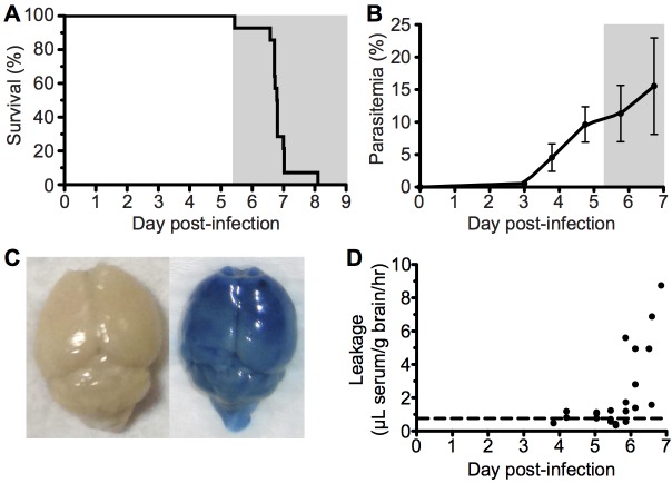
Fig 1
ECM with associated late stage vascular leakage.
C57BL/6 (n = 14) mice were intravenously infected with 10 6 Pb ANKA-pRBCs. (A) Survival and (B) peripheral parasitemia u00b1 SD were monitored daily during development of ECM (grey area). (n = 14, poo...
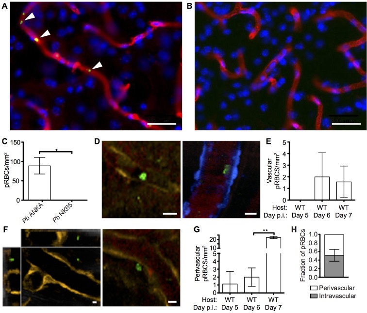
Fig 2
pRBCs make transient adhesive contact with endothelial cells and are deposited within the perivascular space of the meninges of mice with ECM.
C57BL/6 mice were infected with 10 4 Pb ANKA-GFP (n = 3) or Pb NK65-GFP (n = 3) pRBCs. Representative images demonstrating the (A) presence and (B) absence of cytoadherent GFP + pRBCs (green) within t...
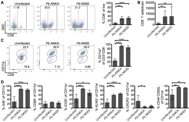
Fig 3
Parasite specific CD8 + T cells are comparably activated within the brains of mice infected with Pb ANKA and Pb NK65 parasites.
C57BL/6 mice were infected with 10 4 Pb ANKA or Pb NK65 pRBCs. (A) Representative flow cytometric plots and calculated percentages showing frequencies of CD8 + T cells (gated on live leukocytes) withi...
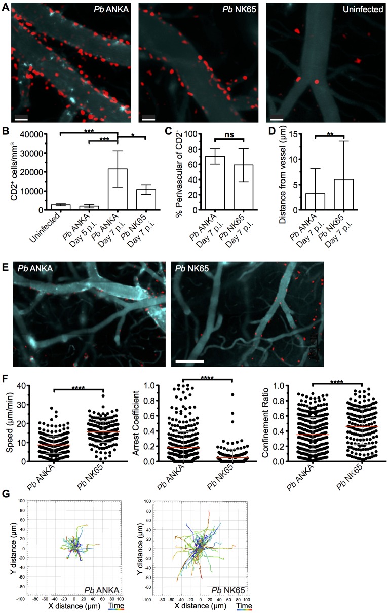
Fig 4
T cells exhibit equivalent perivascular compartmentalisation but distinct behaviours during Pb ANKA and Pb NK65 infections.
hCD2-DsRed C57BL/6 mice were infected with 10 4 Pb ANKA or Pb NK65 pRBCs or left uninfected. Transcranial two-photon microscopy of the meninges was performed on days 5 p.i. ( Pb ANKA), and 7 p.i. ( Pb...
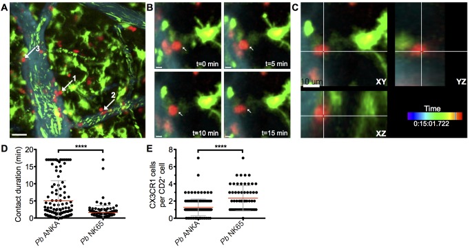
Fig 5
Perivascular T cells form long-lasting interactions with CX 3 CR1 +/GFP in the brains of mice infected with Pb ANKA.
hCD2-DsRed X CX 3 CR1 GFP/GFP dual reporter mice were infected with 10 4 Pb GFP ANKA (n = 5 from three experiments). (A) Maximum intensity projections from intravital two-photon microscopy movies (283...
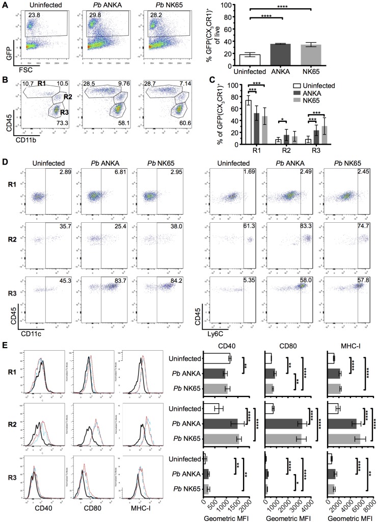
Fig 6
The intracerebral CX 3 CR1 +/GFP cellular response is comparable during Pb ANKA and Pb NK65 infections.
CX 3 CR1 +/GFP mice were infected with 10 4 Pb ANKA or Pb NK65 pRBCs. Brains from uninfected and infected (day 7 p.i.) mice were analyzed by flow cytometry. (A) Representative plots and calculated per...
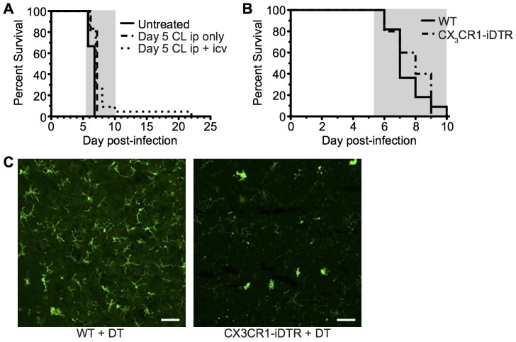
Fig 7
Depletion of systemically and perivascularly located phagocytic cells from day 5 p.i. does not prevent ECM.
(A) C57BL/6 mice were infected with 10 4 Pb ANKA pRBCs. On day 5 p.i. mice were injected i.p. with 300 u03bcL clodronate liposomes with (n = 22) or without (n = 6) 8 u03bcL clodronate liposomes i.c.v....
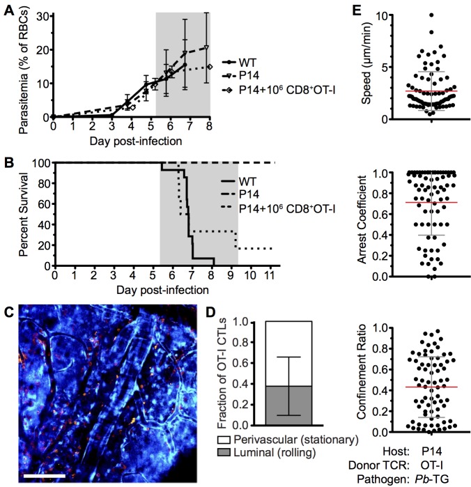
Fig 8
Parasite specific OT-I CD8 + T cells that directly cause ECM are perivascular and are highly arrested in the brain.
C57BL/6 (n = 9) and P14 (n = 10) mice were infected with 10 6 SIINFEKL-expressing Pb -TG pRBCs. Prior to infection, 10 6 nau00efve DsRed-expressing OT-I CD8 + T cells were adoptively transferred into ...
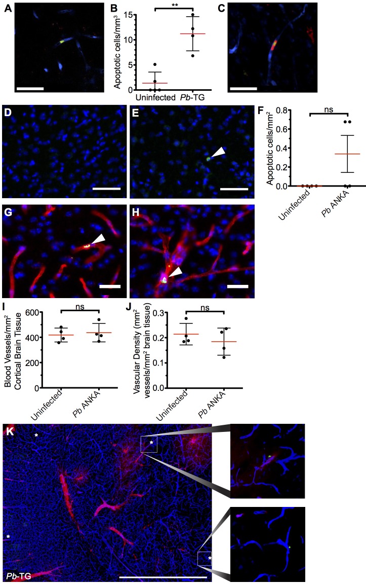
Fig 9
Widespread apoptosis or loss of vasculature does not occur in the brains of mice with ECM.
(A-C and K) 10 6 DsRed + CD8 + OT-I T cells were adoptively transferred into CFP + P14 host mice, which were subsequently infected with 10 6 SIINFEKL-expressing Pb -TG pRBCs (n = 4) or left uninfected...
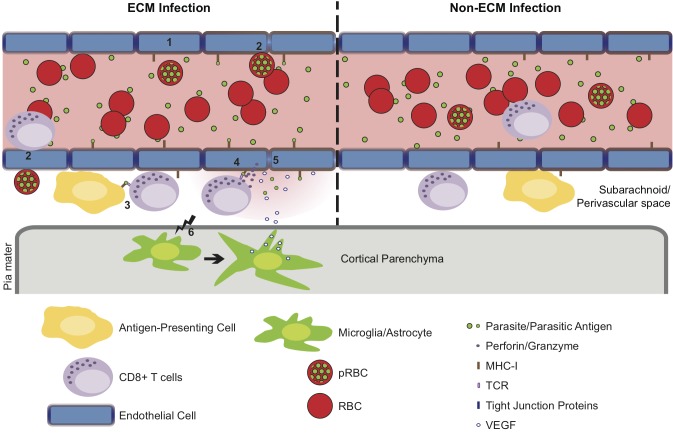
Fig 10
Hypothetical model for the CD8 + T cell-dependent development of ECM.
(1) Pb ANKA infection leads to the upregulation of adhesion molecules and cross-presentation of parasite antigen by MHC-I on brain microvascular endothelial cells [ 85 u2013 87 ]. (2) This promotes tr...
Figure images are served from the NIH/NLM PubMed Central Open Access Subset or Europe PMC; copyright remains with the publishers and authors.
💬 Discussion
0 commentsNo comments yet. Be the first to start a discussion!
Leave a Comment