Abstract
Giardia intestinalis is a ubiquitous parasitic protist that is the causative agent of giardiasis, one of the most common protozoan diarrheal diseases in the world. Giardia trophozoites attach to the intestinal epithelium using a specialized and elaborate microtubule structure, the ventral disc. Surrounding the ventral disc is a less characterized putatively contractile structure, the lateral crest, which forms a continuous perimeter seal with the substrate. A better understanding of ventral disc and lateral crest structure, conformational dynamics, and biogenesis is critical for understanding the mechanism of giardial attachment to the host. To determine the components comprising the ventral disc and lateral crest, we used shotgun proteomics to identify proteins in a preparation of isolated ventral discs. Candidate disc-associated proteins, or DAPs, were GFP-tagged using a ligation-independent high-throughput cloning method. Based on disc localization, we identified eighteen novel DAPs, which more than doubles the number of known disc-associated proteins. Ten of the novel DAPs are associated with the lateral crest or outer edge of the disc, and are the first confirmed components of this structure. Using Fluorescence Recovery After Photobleaching (FRAP) with representative novel DAP::GFP strains we found that the newly identified DAPs tested did not recover after photobleaching and are therefore structural components of the ventral disc or lateral crest. Functional analyses of the novel DAPs will be central toward understanding the mechanism of ventral disc-mediated attachment and the mechanism of disc biogenesis during cell division. Since attachment of Giardia to the intestine via the ventral disc is essential for pathogenesis, it is possible that some proteins comprising the disc could be potential drug targets if their loss or disruption interfered with disc biogenesis or function, preventing attachment.
🔬 Techniques
🔭 Microscopes
🧬 Organisms
💻 Software
✨ Fluorophores
🧪 Sample Preparation
🏭 Microscope Brands
🧪 Reagent Suppliers
📷 Detectors
🔎 Objectives
💻 Software Details
🏛️ Research Organizations (ROR)
Affiliated research institutions:
📋 Methods
Strains and culture conditions
Giardia intestinalis strain WBC6 (ATCC 50803) trophozoites were maintained at 37°C in modified TYI-S-33 medium with bovine bile [41] in 16 ml screw cap tubes (Fisher Scientific).
Detergent extraction of ventral discs for proteomic analysis
The primary goal in the isolation of intact ventral discs for proteomic analysis was the maintenance of microtubule-associated proteins, by removing radicals and metal ions that could damage disc structure, and by stabilizing microtubules using drugs like Taxol. We modified a cytoskeletal preparation from Holberton et al. [42] to isolate disc and flagellar cytoskeletons from Giardia . First, TYI-S-33 medium was decanted from one confluent 12 ml culture of trophozoites. Cells were demembranated and cytoskeletons were extracted by adding 1 ml of 1% Triton X-100 in 1X PHEM plus Taxol (60 mM PIPES, 25 mM HEPES, 10 mM EGTA, 1 mM MgCl 2 , pH 7.4, 1 mM DTT, 10 µM paclitaxel (Sigma)) and vortexing continuously at the highest setting for 3 minutes. To prevent proteolysis, protease inhibitors (Roche) were added to the preparation. Ventral disc cytoskeletons were then pelleted by centrifugation at 16,000×g, and the pellets were washed four times in 1X PHEM+Taxol lacking 1% Triton X-100. Sufficient extraction of cytoskeletons was confirmed by wet mount using phase contrast or DIC microscopy (see Figure 1 ). 10.1371/journal.pntd.0001442.g001 Figure 1 Structure of the cytoskeleton and ventral disc in Giardia. Panels A (DIC) and B (anti-alpha-tubulin immunostaining) illustrate the primary microtubule structures of interphase trophozoites including the ventral disc (vd), and the four pair of flagella: anterior (afl), ventral (vfl), posteriolateral (pfl) and caudal (cfl). Scale = 5 µm. The structures of the ventral disc and flagellar basal bodies are illustrated in the schematic in panel C, including the ventral disc microtubule (MT) array (vdMTs) and the lateral crest (lc), the overlap zone of the ventral disc MT spiral (oz), the supernumerary MT array (snMTs), and the four pair of flagella and eight basal bodies (bb). Other areas of the ventral side of the giardial cell, including the lateral shield (ls) and the marginal groove (mg) are also shown. The detergent-extracted disc preparation used for proteomic analysis is shown in panel D (phase contrast), and a similarly extracted cytoskeletal preparation is shown in panel E. The ventral disc (vd), ventrolateral flange (vlf), lateral crest (lc) and overlap zone (oz) are shown, as well as the bare area (ba) and underlying flagellar basal bodies (SEM courtesy of Joel Mancuso). Scale = 2 µ m .
Show full methods section
Strains and culture conditions
Giardia intestinalis strain WBC6 (ATCC 50803) trophozoites were maintained at 37°C in modified TYI-S-33 medium with bovine bile [41] in 16 ml screw cap tubes (Fisher Scientific).
Detergent extraction of ventral discs for proteomic analysis
The primary goal in the isolation of intact ventral discs for proteomic analysis was the maintenance of microtubule-associated proteins, by removing radicals and metal ions that could damage disc structure, and by stabilizing microtubules using drugs like Taxol. We modified a cytoskeletal preparation from Holberton et al. [42] to isolate disc and flagellar cytoskeletons from Giardia . First, TYI-S-33 medium was decanted from one confluent 12 ml culture of trophozoites. Cells were demembranated and cytoskeletons were extracted by adding 1 ml of 1% Triton X-100 in 1X PHEM plus Taxol (60 mM PIPES, 25 mM HEPES, 10 mM EGTA, 1 mM MgCl 2 , pH 7.4, 1 mM DTT, 10 µM paclitaxel (Sigma)) and vortexing continuously at the highest setting for 3 minutes. To prevent proteolysis, protease inhibitors (Roche) were added to the preparation. Ventral disc cytoskeletons were then pelleted by centrifugation at 16,000×g, and the pellets were washed four times in 1X PHEM+Taxol lacking 1% Triton X-100. Sufficient extraction of cytoskeletons was confirmed by wet mount using phase contrast or DIC microscopy (see Figure 1 ). 10.1371/journal.pntd.0001442.g001 Figure 1 Structure of the cytoskeleton and ventral disc in Giardia. Panels A (DIC) and B (anti-alpha-tubulin immunostaining) illustrate the primary microtubule structures of interphase trophozoites including the ventral disc (vd), and the four pair of flagella: anterior (afl), ventral (vfl), posteriolateral (pfl) and caudal (cfl). Scale = 5 µm. The structures of the ventral disc and flagellar basal bodies are illustrated in the schematic in panel C, including the ventral disc microtubule (MT) array (vdMTs) and the lateral crest (lc), the overlap zone of the ventral disc MT spiral (oz), the supernumerary MT array (snMTs), and the four pair of flagella and eight basal bodies (bb). Other areas of the ventral side of the giardial cell, including the lateral shield (ls) and the marginal groove (mg) are also shown. The detergent-extracted disc preparation used for proteomic analysis is shown in panel D (phase contrast), and a similarly extracted cytoskeletal preparation is shown in panel E. The ventral disc (vd), ventrolateral flange (vlf), lateral crest (lc) and overlap zone (oz) are shown, as well as the bare area (ba) and underlying flagellar basal bodies (SEM courtesy of Joel Mancuso). Scale = 2 µ m .
Proteomic analysis of a detergent extracted ventral disc preparation
We identified the proteins present in the ventral disc preparation using liquid chromatography tandem mass spectrometry (LC-MS/MS LTQ) [37] . All MS/MS samples were analyzed using X! Tandem ( www.thegpm.org ; version TORNADO (2008.02.01.2)). X! Tandem was set up to search protein sequences downloaded from Genbank ( Giardia intestinalis ) assuming the digestion enzyme trypsin. X! Tandem was searched with a fragment ion mass tolerance of 0.40 Da and a parent ion tolerance of 1.8 Da. Iodoacetamide derivative of cysteine was specified in X! Tandem as a fixed modification. Deamidation of asparagine, oxidation of methionine, sulphone of methionine, tryptophan oxidation to formylkynurenin of tryptophan and acetylation of the N-terminus were specified in X! Tandem as variable modifications. Scaffold (version Scaffold_2_03_01, Proteome Software Inc., Portland, OR) was used to validate MS/MS based peptide and protein identifications. Peptide identifications were accepted if they could be established at greater than 80.0% probability as specified by the Peptide Prophet algorithm [43] . Protein identifications were accepted if they could be established at greater than 95.0% probability and contained at least one identified peptide. Protein probabilities were assigned by the Protein Prophet algorithm [44] . Proteins that contained similar peptides and could not be differentiated based on MS/MS analysis alone were grouped to satisfy the principles of parsimony. C-terminal GFP-tagging of candidate disc-associated proteins (DAPs) using Gateway cloning Fifty-eight of the 102 candidate DAPs identified in the proteomic survey were chosen for localization; candidates that appeared to be metabolic, flagellar-associated or chromatin-associated proteins were excluded. All candidate DAP PCR forward primers (see Table S1 ) were designed to bind approximately 200–250 bp upstream of the gene to include the Giardia native promoter and contained the sequence CACC at the 5′ end to facilitate directional cloning. Blunt-ended PCR amplicons were generated by PCR using PfuTurbo Hotstart PCR Mastermix (Stratagene) with Giardia intestinalis strain WBC6 genomic DNA. The candidate DAP PCR amplicons were subsequently subcloned into the Invitrogen pENTR/D-TOPO backbone to generate Gateway entry clones. Inserts in entry clones were sequenced to confirm the identity and correct orientation of the gene. To construct DAP::GFP fusions, positive entry clones were then recombined, via LR reaction, with a 1-fragment GFP tagging E. coli / Giardia shuttle destination vector (pcGFP1F.pac, [39] ) using LR Clonase II Plus (Invitrogen). LR reactions were performed using 100 ng pcGFP1F.pac and 150 ng of DAP entry clone plasmid DNA. Positive clones were screened by digestion with Asc I, and bulk plasmid DNA was prepared using Qiagen's Endofree Plasmid Maxi Kit. To create C-terminal GFP-tagged candidate DAP strains, Giardia intestinalis strain WBC6 was electroporated with roughly 20 µg of plasmid DNA (above) using the GenePulserXL (BioRad) under previously described conditions [45] . Episomal DAP::GFP constructs were maintained in transformants using antibiotic selection (50 µg/ml puromycin) [46] .
Immunofluorescence microscopy and image data analysis Immunostaining of the GFP-tagged
DAP strains was performed as previously described [45] . To confirm disc localization, Metamorph image acquisition software (MDS Technologies) was used to collect 3D images using a Leica DMI 6000 wide-field inverted fluorescence microscope with a PlanApo 100X, NA 1.40 oil immersion objective. Serial sections of DAP::GFP strains were acquired at 0.2 µm intervals, and deconvolved using Huygens Professional deconvolution software (SVI). Two dimensional maximum intensity projections were created from the 3D data sets for presentation purposes. Assessment of DAP::GFP fusion protein turnover using FRAP We used laser fluorescence photobleaching of specific regions to measure the movement and steady state turnover of the new DAPs in Giardia , a technique that has been used extensively in other organisms [47] . Three DAP::GFP-expressing strains (whole disc, DAP5374; lateral crest, DAP13981 ; and disc plus axonemes, DAP17090 ) were selected as representative examples of different ventral disc localizations. The media in a confluent 12 ml culture was replaced with 1X HBS for 1 hour at 37°C. The culture was then iced for 15 minutes to detach cells, and 3 ml of the cell suspension were transferred to a coverslip placed in an 8-well plastic plate. Cells were incubated for 30 minutes at 37°C under nitrogen gas to allow them to attach to the coverslip. After 1 hour, 1 µl of CellMask orange (Invitrogen) was added to the cell suspension. Stained cells were incubated for 5 minutes at 37°C then rinsed twice with warmed 1X HBS. The edges of the coverslip were blotted and the coverslip was inverted onto a slide with double stick tape. Warmed 2% low-melt agarose (Sigma) in 1X HBS was added under the coverslip to embed the attached cells and the prep was sealed on all sides with VALAP. An Olympus FV1000 scanning laser confocal microscope equipped with a four channel PMT was used for imaging and simultaneous 405 nm bleaching. The pre-bleach image of the cell was acquired using a 60×, 1.42 NA objective and a 488 nm laser (at 5% with 4 µs/pixel scan speed). To photobleach a specific region of DAP localization, we used the 405 nm laser (90% power for 200 milliseconds).
Fluorescence recovery in the GFP-tagged
DAP strains was assessed by imaging once every minute, for up to 10 minutes, using the 488 nm low power laser excitation. Normalized GFP fluorescence recovery was calculated by subtracting the PMT background noise from the ROI intensity measurement; the background-subtracted intensity measurement was then divided by a fluorescent control ROI intensity measurement to normalize for photobleaching due to imaging. Negative staining and immunolabeling of isolated discs from DAP::GFP fusions Detergent extracted cytoskeletons containing GFP-tagged DAPs were isolated (see above). Immunolabeling and negative staining of the isolated discs was performed as previously described [48] in 1.5 ml Eppendorf tubes with gentle shaking at room temperature. Cytoskeletons were placed in a blocking buffer of 3% nonfat dry milk in PHEM buffer (60 mM PIPES, 25 mM HEPES, 10 mM EGTA, 2 mM MgCl 2 ) for 1 hour. Cytoskeletons were then labeled with an anti-GFP antibody in blocking buffer for 1.5 hours, and then rinsed 3 times, for 15 minutes each, in PHEM. Pelleting between steps was done at 2,000×g for 5 minutes with a short vortexing step for resuspension. Samples were incubated with 5 nm goat-anti-rabbit F(ab′)2 IgG antibody (BB International) in blocking buffer for 1 hour, then rinsed 3 times in PHEM for 15 minutes. For negative controls (not shown), we used secondary antibody only. For negative staining of DAP::GFP fusion strains, 300 mesh copper grids (EMS) were Formvar-coated, carbon-coated, then glow-discharged to make them more hydrophilic. A 5 µl droplet of cytoskeleton solution was placed on the grid, blotted, and then negative stained with a 5 µl droplet of 2% aqueous uranyl acetate (Ted Pella) and blotted. Grids were imaged with an AMT digital camera in a CM100 (FEI) transmission electron microscope operating at 80 kV.
Supporting Information Figure S1 Multiple sequence alignment of disc-associated Nek kinase homologs. Nek kinases identified in the ventral disc proteome were aligned to Nek kinases in other representative eukaryotes using MUSCLE [75] and presented using JalView [76] . (PDF) Click here for additional data file. Figure S2 Multiple sequence alignment of DIP13 homologs. The Giardia DIP13 homolog was aligned to DIP13 homologs in other representative eukaryotes using MUSCLE [75] . Alignment is presented using JalView [76] . (PDF) Click here for additional data file. Figure S3 Beta-, gamma-, and delta-giardin localize to the microribbons. Negative staining using anti-GFP immunogold labeling of beta-giardin::GFP, delta-giardin::GFP and gamma-giardin::GFP strains show the association of these proteins with the ventral disc microribbons. Scale bar = 200 nm. (TIF) Click here for additional data file. Table S1 Gateway and other oligonucleotide PCR primers for disc-associated proteins (DAPs). List of forward and reverse oligonucleotide PCR primers used in the construction of the DAP::GFP Giardia strains. (PDF) Click here for additional data file. Table S2 Proteins identified in the detergent-extracted disc preparation. Candidate disc-associated proteins identified in the proteomic analysis of the disc preparation are summarized with protein domain information and GFP localization, if tagged. (MB = median body, AFL = anterior flagella; CFL = caudal flagella; VFL = ventral flagella; and PFL = posteriolateral flagella). (XLSX) Click here for additional data file. Video S1 Z-stack of DAP17090 ::GFP immunostained with anti-beta-giardin. Fluorescence microscopy 3D stack of DAP17090 ::GFP (green) with the ventral disc microribbons localized using an anti-beta-giardin antibody (red). Nuclei are stained with DAPI. (MOV) Click here for additional data file. Video S2 Z-stack of DAP16263 ::GFP immunostained with anti-beta-giardin. Fluorescence microscopy 3D stack of DAP16263 ::GFP (green) with microribbons localized using an anti-beta-giardin antibody (red). Nuclei are stained with DAPI. (MOV) Click here for additional data file.
📊 Figures
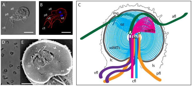
Figure 1
Structure of the cytoskeleton and ventral disc in Giardia.
Panels A (DIC) and B (anti-alpha-tubulin immunostaining) illustrate the primary microtubule structures of interphase trophozoites including the ventral disc (vd), and the four pair of flagella: anteri...
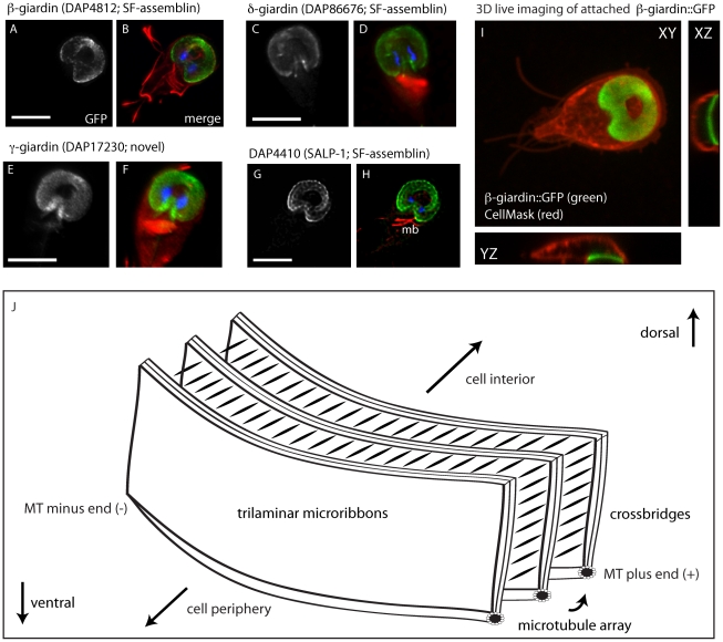
Figure 2
GFP tagging of known disc-associated proteins does not affect subcellular localization, disc conformation, or attachment.
Panels Au2013H show the ventral disc localization of C-terminal GFP-tagged, previously described disc-associated proteins beta-giardin (A,B), delta-giardin (C,D), gamma-giardin (E,F) and SALP-1 (G,H) ...
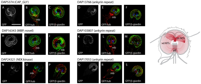
Figure 3
Novel disc-associated proteins associated with the entire disc.
Six new DAPs localize to the entire ventral disc spiral in a manner similar to the previously described microribbon-associated proteins (see Figure 2 and Figure S3 ) as visualized by C-terminal GFP ta...
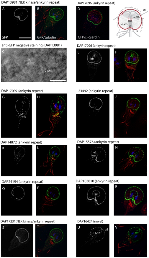
Figure 4
A diversity of disc-associated proteins localize to the lateral crest.
Ten new DAPs localize to the lateral crest (lc) structure that surrounds the ventral disc (see red, schematic) and comes in close contact with the intestinal epithelium during attachment [12] . In all...
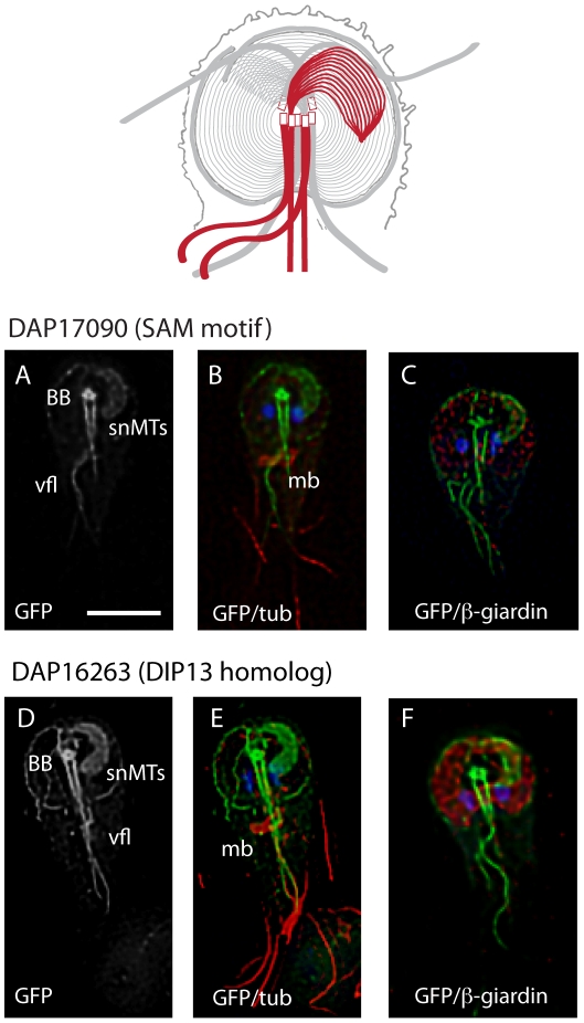
Figure 5
DAP17090 ::GFP and DAP16263 ::GFP localize to the supernumerary microtubules and axonemes .
Both DAP17090 (Au2013C), which contains a SAM protein-protein interaction motif, and DAP16263 (Du2013F), a DIP13 microtubule binding protein homolog, localize to the supernumerary MTs that are slightl...
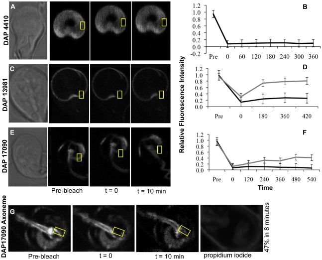
Figure 6
DAPs recover only at non-disc, axoneme or basal body regions.
Cells stably transformed with expression plasmids encoding full-length DAP::GFPs were subjected to quantitative FRAP analysis. Panel A shows representative images of DAP4410::GFP, from the group of pr...
Figure images are served from the NIH/NLM PubMed Central Open Access Subset or Europe PMC; copyright remains with the publishers and authors.
💬 Discussion
0 commentsNo comments yet. Be the first to start a discussion!
Leave a Comment