Abstract
Melt electrospinning is a promising approach to manufacture biocompatible scaffolds for tissue engineering. In this study, melt electrospinning of poly(ε-caprolactone) onto structured, metallic collectors resulted in scaffolds with an average pore size of 250-300 μm and an average fibre diameter of 15 μm. Scaffolds were seeded with ovine osteoblasts in vitro. Cell proliferation and deposition of mineralised extracellular matrix was assessed using PicoGreen® (Thermo Fisher Scientific, Scoresby, Australia) and WAKO® HR II (WAKO, Osaka, Japan) calcium assays. Biocompatibility, cell infiltration and the growth pattern of osteoblasts on scaffolds was investigated using confocal microscopy and scanning electron microscopy. Osteoblasts proliferated on the scaffolds over an entire 40-day culture period, with excellent survival rates and deposited mineralized extracellular matrix. In general, the 3D environment of the structured melt electrospun scaffold was favourable for osteoblast cultures.
🔬 Techniques
🔭 Microscopes
✨ Fluorophores
🧪 Sample Preparation
🔬 Cell Lines
🏭 Microscope Brands
🧪 Reagent Suppliers
💻 Software Details
🏛️ Research Organizations (ROR)
Affiliated research institutions:
📋 Methods
4.1.
Scaffold Fabrication and Preparation
Melt electrospun scaffolds were fabricated with PCL (MW) 80,000 g/mol (Sigma Aldrich, Castle Hill, Australia), onto structured electroconductive collectors as described previously [ 4 , 17 , 57 ]. Briefly, PCL pellets were placed into a 3 mL Luer lock syringe (B-Braun, Bella Vista, Australia) and heated to 80 °C with the syringe held upright ( Figure 5 A). A blunt 23G needle attached to the syringe was used as a spinneret and placed into a water-circulating system heated to 80 °C. The PCL melt was electrospun using a spinneret-collector distance of 50 mm, a flow rate of 10 μL/h and a voltage of 20 kV applied to the spinneret. The melt electrospun PCL-fibres were collected onto an array of 20 dome-shaped wire mesh collectors upon a grounded moving plate. Each collector possessed a similar 250 μm × 250 μm square void template architecture with rubber rings around the base of each filter to provide a separating air gap to minimise disturbing influences on the jet’s path from the adjacent collector ( Figure 5 B,C). The steel mesh of each collector was the highest point on the collector and was positioned directly under the spinneret for the 20 min duration of fibre collection to produce a 3D scaffold ( Figure 5 C,D). Figure 5 E,F reveals the heterogeneous structure on the concave of the scaffold, where the open square shaped 250 μm pores match the architecture of the collector mesh. After 20 min of fibre collection, an automated x-y-stage was used to move the molten PCL jet to the next collector. The produced scaffolds were weighed, punched and microscopically analysed to ensure a consistent scaffold structure and mass. To ameliorate surface properties, PCL scaffolds were incubated with 5 M NaOH for 4 h at 37 °C, washed thoroughly with ddH 2 O and stored in 70% ethanol. Prior to experiments, scaffolds were incubated with fresh 70% ethanol for 2 h, transferred to 24-well tissue culture plates (Corning, New York, NY, USA) under a sterile work bench and UV sterilised for 30 min. 4.2.
Show full methods section
4.1.
Scaffold Fabrication and Preparation
Melt electrospun scaffolds were fabricated with PCL (MW) 80,000 g/mol (Sigma Aldrich, Castle Hill, Australia), onto structured electroconductive collectors as described previously [ 4 , 17 , 57 ]. Briefly, PCL pellets were placed into a 3 mL Luer lock syringe (B-Braun, Bella Vista, Australia) and heated to 80 °C with the syringe held upright ( Figure 5 A). A blunt 23G needle attached to the syringe was used as a spinneret and placed into a water-circulating system heated to 80 °C. The PCL melt was electrospun using a spinneret-collector distance of 50 mm, a flow rate of 10 μL/h and a voltage of 20 kV applied to the spinneret. The melt electrospun PCL-fibres were collected onto an array of 20 dome-shaped wire mesh collectors upon a grounded moving plate. Each collector possessed a similar 250 μm × 250 μm square void template architecture with rubber rings around the base of each filter to provide a separating air gap to minimise disturbing influences on the jet’s path from the adjacent collector ( Figure 5 B,C). The steel mesh of each collector was the highest point on the collector and was positioned directly under the spinneret for the 20 min duration of fibre collection to produce a 3D scaffold ( Figure 5 C,D). Figure 5 E,F reveals the heterogeneous structure on the concave of the scaffold, where the open square shaped 250 μm pores match the architecture of the collector mesh. After 20 min of fibre collection, an automated x-y-stage was used to move the molten PCL jet to the next collector. The produced scaffolds were weighed, punched and microscopically analysed to ensure a consistent scaffold structure and mass. To ameliorate surface properties, PCL scaffolds were incubated with 5 M NaOH for 4 h at 37 °C, washed thoroughly with ddH 2 O and stored in 70% ethanol. Prior to experiments, scaffolds were incubated with fresh 70% ethanol for 2 h, transferred to 24-well tissue culture plates (Corning, New York, NY, USA) under a sterile work bench and UV sterilised for 30 min. 4.2.
Cell Isolation
Ovine osteoblast explants were obtained from merino sheep (n = 6) (Ethic approval number 0900000099, animal ethics committee, Queensland University of Technology, Brisbane, Australia). Solid mandibular bone samples were acquired under sterile conditions, minced, washed with phosphate buffered saline (PBS, Invitrogen, Scoresby, Australia) and vortexed. After incubation with 10 mL 0.25% trypsin/ethyldiaminetetraacetic acid (EDTA, Invitrogen, Scoresby, Australia) for 3 min at 37 °C, 5% CO 2 and trypsin inactivation with 10 mL low-glucose Dulbecco’s modified Eagle’s medium (DMEM, Invitrogen, Scoresby, Australia) containing 10% foetal bovine serum (FBS, Invitrogen, Scoresby, Australia), samples were washed with PBS once and transferred to 175 cm 2 tissue culture flasks (Corning). Bone samples were topped-up with 15 mL of DMEM supplemented with 10% FBS and 1% penicillin/streptomycin (PS, Invitrogen, Scoresby, Australia). After 5–7 days, osteoblast outgrowth could be observed. Cells were expanded to second passage for subsequent experiments. 4.3.
Cell Culture
A total of 50,000 osteoblasts were resuspended in 20 µL of DMEM/10%FBS/1%PS, seeded onto each PCL scaffold on the convex-shaped top side and incubated for 2 h at 37 °C, 5%CO 2 . After 2 h, 1 mL of basal media was added to each well of the 24-well plate. The following day, to prevent attachment of the scaffold–cell constructs to the bottom of the tissue culture plate, each scaffold was transferred to a 15 mL Falcon tube (Corning) using sterile tweezers and underwent osteogenic induction with 2 mL of DMEM/10%FBS/1%PS supplemented with 50 µg/mL l -ascorbic acid-2-phosphate, 10 mM β-glycerophosphate and 0.1 µM dexamethasone (all Sigma Aldrich). Scaffolds were further cultured at 37 °C, 5%CO 2 in osteogenic media for up to 40 days. 4.4. Cell Proliferation Triplicates of scaffolds seeded with osteoblasts were cultured with osteogenic media in 15 mL Falcon tubes for 1, 10, 20, 30 or 40 days at 37 °C, 5%CO 2 . At each termination point, scaffolds were washed twice with PBS, transferred to 1.7 mL Eppendorf ® tubes (Eppendorf, Macquarie Park, Australia), centrifuged at 500 rpm for 2 min at room temperature to remove excess liquid and stored at −80 °C until analysis. For analysis, scaffolds were digested with 50 µg/mL proteinase K (Sigma Aldrich) in 1× TE at 50 °C for 48 h. DNA content for 100 µL of each sample in triplicate were measured and quantified using a Quant-iT ™ PicoGreen ® dsDNA assay kit (Thermo Fisher Scientific, Scoresby, Australia) according to the protocol supplied by the manufacturer (Invitrogen). An equal volume of the aqueous Quant-iT ™ PicoGreen ® working solution was added to each triplicate. After three minute incubation on a rocking plate, fluorescence was measured at λ excitation = 485 nm and λ emission = 520 nm using a POLARStar OPTIMA plate reader (BMG Labtech, Ortenberg, Germany). 4.5.
Calcium Measurement
To analyse the calcium content of the ECM produced by osteoblasts on the PCL scaffolds, a WAKO HRII Calcium assay (WAKO, Osaka, Japan) was performed according to the manufacturer’s protocol as described previously [ 58 ]. On Days 20 and 40, triplicate scaffolds were washed twice with ddH 2 O, centrifuged at 500 rpm for 2 min at room temperature (RT) and stored at −80 °C after removal of the supernatant. Samples were incubated with 500 µL of 10% v/v acetic acid for 3 h at RT and subsequently vortexed for 1 min at RT. To prevent evaporation, samples were covered with 200 µL of mineral oil (Sigma Aldrich), heated to 85 °C for 10 min and transferred to ice for 10 min. Following that step, the samples were centrifuged at 20,000 × G for 15 min. 300 µL of the supernatant were neutralized with 120 µL of 10% v/v ammonium hydroxide (Sigma Aldrich). 10 µL of the neutralized mixture were transferred in triplicate to transparent 96-well plates and incubated with 100 µL of monoethanylamine buffer, pH = 11 (WAKO, Osaka, Japan) for 3 min at 37 °C. Consequently, 100 µL of o-cresolphtalein-complex solution (WAKO, Osaka, Japan) was added to each well and incubated for 5 min at 37 °C. Plates were read at λ = 570 nm using a POLARStar OPTIMA plate reader (BMG Labtech, Ortenberg, Germany). To generate a standard curve, a serial dilution of Mulitchem Calibrator A (WAKO, Osaka, Japan) in 10% v/v acetic acid was used. 4.6.
Confocal Laser Microscopy
Cell viability and morphology on PCL scaffolds was determined by staining with fluorescein diacetate (FDA) and propidium iodide (PI) or rhodamine-conjugated phalloidin (Phal) and PicoGreen ® (all Invitrogen) on Days 20 and 40. For FDA/PI staining, scaffolds were washed three times with phenol red-free media (Invitrogen) and incubated in dark for 15 min at 37 °C with an FDA/PI staining solution containing 2 µg/mL FDA, 10 µg/mL PI in phenol red-free media. As a cell-permeable esterase substrate, FDA is hydrolysed by viable cells giving green fluorescence. PI acts as a nucleic acid intercalator and penetrates the cell membrane of dead cells, but not living ones. Red colour was used to mark dead cells. For Phal/PicoGreen ® staining, samples were washed three times with PBS, fixed with 4% paraformaldehyde (PFA, Sigma Aldrich) for 20 min at RT and washed two more times with PBS. Scaffolds were permeabilized with 0.2% Triton ® X-100 (Sigma Aldrich) in PBS for 5 min sharp at RT, washed twice with PBS and incubated with a staining solution consisting of 0.8 U/mL phalloidin, 1 µL PicoGreen ® -Lösung/mL in 2% w/v BSA in PBS in dark for 50 min at RT. The cyclopeptide phalloidin binds to F-actin filaments of the cytoskeleton and therefore indicates cell morphology (red) while PicoGreen ® binds to dsDNA and therefore visualizes the nucleus (blue). The stained scaffolds were washed three times with PBS to remove excess staining solution and visualized with a Leica SP5 confocal microscope (Leica Microsystems GmbH, Wetzlar, Germany). 4.7. Scanning Electron Microscopy On Days 20 and 40, cell scaffold constructs were fixed with 3% v/v glutaraldehyde in 0.1 M sodium cocodylate buffer solution, pH = 7.3 for 2 h at 4 °C. The fixed specimens were dehydrated through a series of alcohols including two changes with 10 min in each 50%, 70%, 90% and 100% ethanol. Due to the PCL, samples could not be critical-point dried. Thus, remaining liquid was removed from the samples by incubating twice for 30 min using hexamethyldisalazane (Sigma Aldrich). Specimens were gold-coated in a SC 500 Bio-Rad sputter coater (Bio-Rad, Gladesville, Australia) and analyzed using a FEI Quanta 200 scanning electron microscope (FEI, Hillsboro, OR, USA). 4.8.
Statistics
Statistical analysis was carried out using Student’s t -test and p < 0.05 was considered significant (SPSS).
📊 Figures
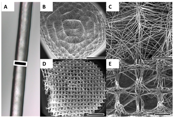
Figure 1
Melt electrospinning of PCL was used to produce 3D scaffolds. ( A ) Optical microscopy image of a representative melt electrospun PCL fibre; ( B , C ) Due to structured and curved metallic collector s...
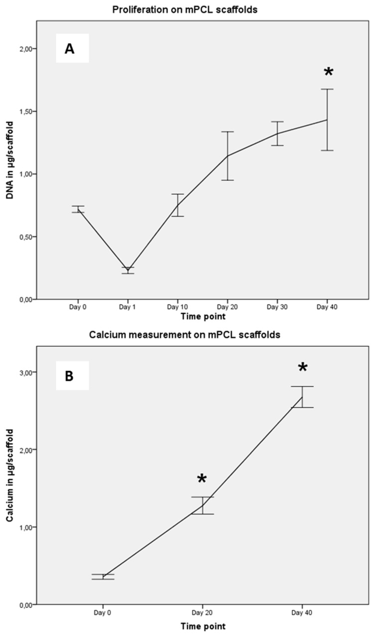
Figure 2
( A ) Proliferation potential of osteoblasts (n = 6) was determined on Days 0 (seeding), 1, 10, 20, 30 and 40. Approximately one-third of seeded cells attached to the scaffold (Day 1). Osteoblasts pro...
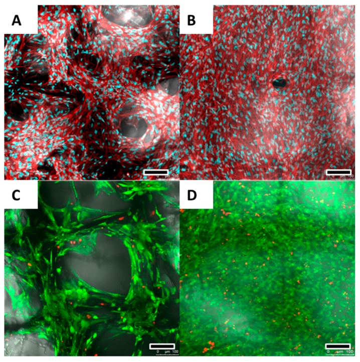
Figure 3
After 20 ( A ) and 40 ( B ) days, Actin/nuclei (Phalloidin/PicoGreen u00ae ) staining revealed an elongated and spindle-shaped morphology for the osteoblasts. The nuclei appear blue while actin cytosk...
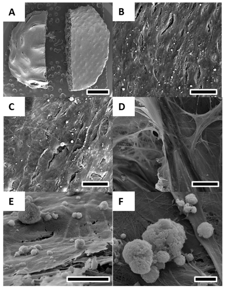
Figure 4
SEM of osteoblasts cultured for 40 days in osteogenic differentiation media on melt electrospun scaffolds. ( A ) Overview revealed a solid layer of osteoblasts covering the entire scaffold on both the...
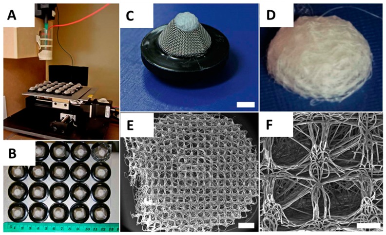
Figure 5
( A ) Melt Electrospinning apparatus spinning molten PCL onto an array of 20 structured metallic substrates with raised architecture. X-y-stage moves substrates into path of melt electrospinning jet. ...
Figure images are served from the NIH/NLM PubMed Central Open Access Subset or Europe PMC; copyright remains with the publishers and authors.
💬 Discussion
0 commentsNo comments yet. Be the first to start a discussion!
Leave a Comment