Abstract
Deubiquitinating enzymes are now emerging as potential therapeutic targets that control many cellular processes, but few have been demonstrated to control cell motility. Here, we show that ubiquitin-specific protease 17 (USP17) is rapidly and transiently induced in response to chemokines SDF-1/CXCL12 and IL-8/CXCL8 in both primary cells and cell lines, and that its depletion completely blocks chemokine-induced cell migration and cytoskeletal rearrangements. Using live cell imaging, we demonstrate that USP17 is required for both elongated and amoeboid motility, in addition to chemotaxis. USP17 has previously been reported to disrupt Ras localization and we now find that USP17 depletion blocks chemokine-induced subcellular relocalization of GTPases Cdc42, Rac and RhoA, which are GTPases essential for cell motility. Collectively, these results demonstrate that USP17 has a critical role in cell migration and may be a useful drug target for both inflammatory and metastatic disease.
🔬 Techniques
🔭 Microscopes
💻 Software
✨ Fluorophores
🧪 Sample Preparation
🔬 Cell Lines
🏭 Microscope Brands
🧪 Reagent Suppliers
💻 Software Details
🏛️ Research Organizations (ROR)
Affiliated research institutions:
📋 Methods
Plasmids pSUPER-scrambled shRNA and pSUPER-USP17 shRNA1 (target sequence 5′-GCAGGAAGATGCCCATGAA-3′) were kind gifts from Dr Rene Medema (University Medical Center, Utrecht, the Netherlands). pRS-USP17 shRNA2 (target sequence 5′-AAGAGCCACTCAGGAAAGCACCTTAGACC-3′) and pRS-USP17 shRNA3 (target sequence 5′-GATGATTTGGCTCCTGTGGCAAGACAGCT-3′) were purchased from Origene Technologies. pGFP-RS-USP17 shRNA (target sequence 5′-GCAGGAAGATGCCCATGAA-3′) was purchased from Origene technologies. pCAGGS-eGFP-Cdc42, pCAGGS-eGFP-Rac1 and pCAGGS-eGFP-RhoA were kind gifts from Dr Michiyuki Matsuda (Osaka University, Osaka, Japan). pDQ-EV (His), pDQ-USP17 (His) and pDQ-USP17CS (His) were kind gifts from Dr Derek Quinn (Queen's University, Belfast).
Cell culture and transfection
Hut78 and U937 cells were grown in RPMI 1640 (PAA) and HeLa and MDA-MB-231 cells (American Type Culture Collection, Manassas, USA) in DMEM, supplemented with 10% FCS, 1% penicillin (10,000 U ml −1 )/streptomycin (10,000 μg ml −1 ) and 1% L -glutamine (200 mM), grown at 37 °C in a 5% CO 2 humidified incubator. Hut78 cells were transfected using the Amaxa Nucleofector System (Lonza). U937, MDA-MB-231 and Hela cells were transfected with FuGENE6 (Roche) according to the manufacturer's instructions. For those experiments with CXCL12 or CXCL8 stimulation, cells were rested for 12 h in DMEM without serum, then stimulated with the chemokine for the indicated times and concentrations indicated.
Primary cell isolation
Whole peripheral blood packs were donated by Northern Ireland Blood Service (Belfast, UK) or peripheral blood was drawn from healthy donors using 0.5 ml of 0.5 M EDTA (pH 8.0) as an anticoagulant. PBMCs and polymononuclear neutrophils (PMNs) from healthy volunteers were isolated from 50 ml of whole blood by density gradient centrifugation at 650 g for 30 min over Ficoll (Amersham). PMNs were isolated by centrifugation and hypotonic lysis of erythrocytes. The cells were then washed twice at 290 g for 10 min with RPMI medium before counting. RT–PCR mRNA was extracted from cells using STAT60 reagent (Biogenesis), and RNA isolation was performed according to the manufacturer's instructions. RT–PCR analysis of 1 μg total RNA was performed using the OneStep RT–PCR kit (Qiagen) according to the manufacturer's instructions, and separated on 1% agarose gels 11 . The primer sets used were USP17 forward 5′-CAGTGAATTCGTGGGAATGGAGGACGACTCACTCTAC-3′ and USP17 reverse 5-AGTCATCGATCTGGCACACAAGCATAGCCCTC-3′, and β-actin forward 5′-GGACTTCGAGCAAGAGATGG-3′ and β-actin reverse 5′-AGCACTGTGTTGGCGTACAG-3′.
Show full methods section
Plasmids pSUPER-scrambled shRNA and pSUPER-USP17 shRNA1 (target sequence 5′-GCAGGAAGATGCCCATGAA-3′) were kind gifts from Dr Rene Medema (University Medical Center, Utrecht, the Netherlands). pRS-USP17 shRNA2 (target sequence 5′-AAGAGCCACTCAGGAAAGCACCTTAGACC-3′) and pRS-USP17 shRNA3 (target sequence 5′-GATGATTTGGCTCCTGTGGCAAGACAGCT-3′) were purchased from Origene Technologies. pGFP-RS-USP17 shRNA (target sequence 5′-GCAGGAAGATGCCCATGAA-3′) was purchased from Origene technologies. pCAGGS-eGFP-Cdc42, pCAGGS-eGFP-Rac1 and pCAGGS-eGFP-RhoA were kind gifts from Dr Michiyuki Matsuda (Osaka University, Osaka, Japan). pDQ-EV (His), pDQ-USP17 (His) and pDQ-USP17CS (His) were kind gifts from Dr Derek Quinn (Queen's University, Belfast).
Cell culture and transfection
Hut78 and U937 cells were grown in RPMI 1640 (PAA) and HeLa and MDA-MB-231 cells (American Type Culture Collection, Manassas, USA) in DMEM, supplemented with 10% FCS, 1% penicillin (10,000 U ml −1 )/streptomycin (10,000 μg ml −1 ) and 1% L -glutamine (200 mM), grown at 37 °C in a 5% CO 2 humidified incubator. Hut78 cells were transfected using the Amaxa Nucleofector System (Lonza). U937, MDA-MB-231 and Hela cells were transfected with FuGENE6 (Roche) according to the manufacturer's instructions. For those experiments with CXCL12 or CXCL8 stimulation, cells were rested for 12 h in DMEM without serum, then stimulated with the chemokine for the indicated times and concentrations indicated.
Primary cell isolation
Whole peripheral blood packs were donated by Northern Ireland Blood Service (Belfast, UK) or peripheral blood was drawn from healthy donors using 0.5 ml of 0.5 M EDTA (pH 8.0) as an anticoagulant. PBMCs and polymononuclear neutrophils (PMNs) from healthy volunteers were isolated from 50 ml of whole blood by density gradient centrifugation at 650 g for 30 min over Ficoll (Amersham). PMNs were isolated by centrifugation and hypotonic lysis of erythrocytes. The cells were then washed twice at 290 g for 10 min with RPMI medium before counting. RT–PCR mRNA was extracted from cells using STAT60 reagent (Biogenesis), and RNA isolation was performed according to the manufacturer's instructions. RT–PCR analysis of 1 μg total RNA was performed using the OneStep RT–PCR kit (Qiagen) according to the manufacturer's instructions, and separated on 1% agarose gels 11 . The primer sets used were USP17 forward 5′-CAGTGAATTCGTGGGAATGGAGGACGACTCACTCTAC-3′ and USP17 reverse 5-AGTCATCGATCTGGCACACAAGCATAGCCCTC-3′, and β-actin forward 5′-GGACTTCGAGCAAGAGATGG-3′ and β-actin reverse 5′-AGCACTGTGTTGGCGTACAG-3′.
Immunoprecipitation and immunoblotting
Cells were washed once with ice-cold PBS and resuspended in 1% Triton lysis buffer containing protease inhibitors (1 mM Na 3 V0 4, 10 μg ml −1 leupeptin, 10 μg ml −1 aprotinin and 1 mM phenylmethylsulphonyl fluoride). Where indicated, cells were immunoprecipitated using the monoclonal USP17 antibody (Fusion Antibodies).
Immunoprecipitations or whole-cell lysates
(IB) were separated by SDS polyacrylamide gel electrophoresis, transferred to polyvinylidene difluoride membrane and probed with the appropriate antibodies. Antibodies used are listed in Supplementary Table S1 . Proteins were detected using the enhanced chemiluminescence detection method (Amersham), exposed using the ChemiDoc XRS imaging system (Bio-Rad) and analysed with The Discovery Series software from Quantity One version 4.5.0 (Bio-Rad). GTPase activation pulldowns Cells were washed once with ice-cold PBS and lysed in 200 μl lysis buffer (50 mM Tris-HCl, pH 7.5, 1% Triton X-100, 100 mM NaCl, 10 mM MgCl 2 , 5% glycerol, 1 mM Na 3 VO 4 ) containing protease inhibitors (1 mM Na 3 V0 4 , 10 μg ml −1 leupeptin, 10 μg ml −1 aprotinin and 1 mM phenylmethylsulphonyl fluoride). Cell lysates were incubated at 4 °C for 1 h with GST-fusion protein (GST-PAK for CDC42 and Rac1 of GST-Rhotekin for RhoA) coupled to glutathione-sepharose beads (Amersham Biosciences) as described previously 11 . Protein was run on SDS polyacrylamide gel electrophoresis gels and analysed as indicated in the previous section. Active GTPases and whole-cell lysates were detected by immunoblotting with the appropriate antibodies.
Boyden chamber cell migration assay
MDA-MB-231 and HeLa cells were plated in 12 mm-diameter Transwells (Corning Costar), transfected with the indicated plasmids and stimulated with 20 ng ml −1 CXCL12 (PeproTech). Plates were then incubated at 37 °C in 5% CO 2 for 20 h. Migrated cells at the bottom of the top chamber were fixed for 10 min in methanol, stained with crystal violet solution (0.05% crystal violet and 25% methanol), washed and air-dried for 1 h. The filters were then destained with cell migration destaining solution (1:1 ethanol: 0.2 M sodium citrate) and measured using a microplate reader read at A 570 .
Fixed cell confocal imaging
Cells were seeded and transfected on LabTek II, CC2-treated chamber slides as described, fixed in 4% paraformaldehyde for 20 min, permeabilized with 0.5% Triton for 5 min and stained with the indicated antibodies and/or costain, which are listed in Supplementary Table S2 . Slides were viewed using a Leica TCS SP5 DMI6000 CS Confocal Microscope (Leica) using the following lasers: ultraviolet (355 nm), Argon (488 nm) and HeNe (543 nm, 633 nm). Fluorescent images were captured with a 40× lens zoomed in 1–3× with a 1,024×1,024 frame and 400 Hz scanning speed. Images were analysed using Leica LAS AF software (Leica) and ImageJ (NIH).
Live cell migration assay
Hut78 migration assays were carried out in nunclon 96-well plates. On the evening before the migration, a goat anti-mouse capture antibody (Sigma) was added to the wells and left overnight at 4 °C. Wells were washed and incubated with a mouse anti-CD11a antibody (Monosan Clone SPV-L7) for 3 h at 37 °C. Wells were washed three times, and 10,000 cells per 100 μl were added to the wells and left for 15 min before beginning live cell image capture using the IN Cell Imager 1000 (GE Healthcare). Brightfield images were captured every 60 s for 90 min and images at 480×535 nm were taken at 0 and 90 min and cell migration was measured using ImageJ software (NIH). Alternatively, HeLa cells were plated onto glass-bottomed 35 mM μ-dishes (Ibidi) and transfected as previously described. At 48 h post infection, images were captured every 15 s for 10 min using a Leica TCS SP5 Confocal Microscope and analysed using ImageJ software (NIH).
TIRF microscopy
HeLa cells were plated onto 35 mM MatTek glass-bottomed dishes (MatTek), and transfected with GFP-tagged RhoA along with scrambled shRNA, USP17 shRNA1 or USP17 shRNA2. At 48 h post transfection, cells were serum starved for 12 h. Brightfield and TIRF images were captured using the Nikon Eclipse Ti both before and after stimulation with CXCL12. TIRF settings were kept at 200 ms exposure and gain 10 and brightfield at 100 ms exposure and gain 10 at 60× magnification. Analysis for Green TIRF fluorescence was performed using NIS Elements AF 3.1 Software.
Supplementary Material Supplementary Figures and Tables Supplementary Figures S1-S7 and Supplementary Tables S1-S2. Supplementary Movie 1 Hut-78 T-cells transfected with scrambled shRNA on LFA-1 Supplementary Movie 2 Hut-78 T-cells transfected with USP17 shRNA1 on LFA-1 Supplementary Movie 3 Unstimulated HeLa cells transfected with scrambled shRNA. Supplementary Movie 4 HeLa cells transfected with scrambled shRNA and stimulated with CXCL12. Supplementary Movie 5 Unstimulated HeLa cells transfected with USP17 shRNA1. Supplementary Movie 6 HeLa cells transfected with USP17 shRNA1 and stimulated with CXCL12.
📊 Figures
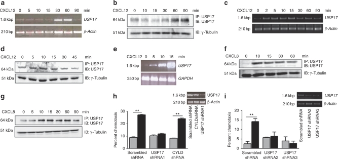
Figure 1
USP17 expression is required for chemotaxis.
Human PBMCs ( a , b ) or HeLa cells ( c , d ) were stimulated with CXCL12 (100 ng ml u22121 ) for the indicated times. USP17 mRNA expression was assessed by RTu2013PCR ( a , c ) and USP17 protein was ...
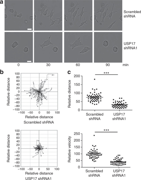
Figure 2
USP17 depletion inhibits chemokinesis.
Hut78 T cells were cotransfected with either scrambled shRNA or USP17 shRNA1 together with pMAX-GFP to determine transfected cells. Cells were allowed to adhere to culture plates coated with a CD11a/l...
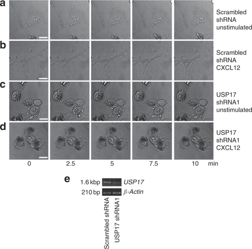
Figure 3
USP17 shRNA alters cell morphology.
HeLa cells transfected with scrambled shRNA were left unstimulated ( a ) or stimulated with CXCL12 (100 ng ml u22121 ) ( b ). USP17 shRNA1 cells were unstimulated ( c ) or stimulated with CXCL12 (100 ...
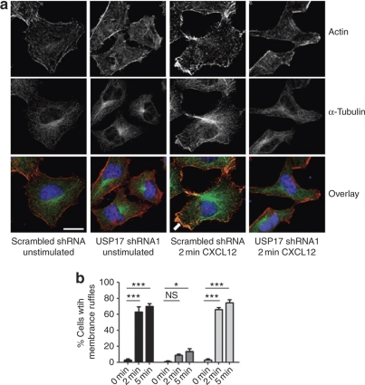
Figure 4
USP17 depletion inhibits cytoskeleton rearrangements.
HeLa cells were transfected with scrambled shRNA or USP17 shRNA1, and stimulated with CXCL12 (100 ng ml u22121 ) for 0, 2 or 5 min. ( a ) Cells were costained for actin-phalloidin (red), u03b1-tubulin...
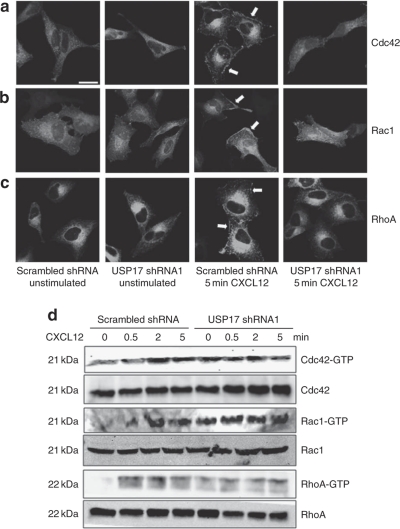
Figure 5
Knockdown of USP17 inhibits GTPase localization and activation.
HeLa cells were transfected with scrambled shRNA or USP17 shRNA1 along with GFP-tagged Cdc42 ( a ), Rac1 ( b ) and RhoA ( c ). Cells were either left unstimulated or stimulated with CXCL12 (100 ng ml ...
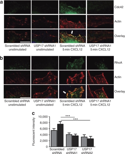
Figure 6
GTPase mislocalization at the leading edge by USP17 depletion.
HeLa cells were transfected with GFP-tagged Cdc42 ( a ) or RhoA ( b ) and either scrambled shRNA or USP17 shRNA1 as indicated. Cells were serum starved and either left unstimulated or stimulated with ...
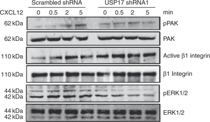
Figure 7
USP17 depletion results in blunted downstream signalling.
HeLa cells were transfected with either scrambled shRNA or USP17 shRNA1 and stimulated with CXCL12 (100 ng ml u22121 ) for 0, 0.5, 2 or 5 min. Protein was collected and whole-cell lysates were immunob...
Figure images are served from the NIH/NLM PubMed Central Open Access Subset or Europe PMC; copyright remains with the publishers and authors.
💬 Discussion
0 commentsNo comments yet. Be the first to start a discussion!
Leave a Comment