Abstract
Abstract Central serotonin (5-HT) orchestrates myriad cognitive processes and lies at the core of many stress-related psychiatric illnesses. However, the basic relationship between its brain-wide axonal projections and functional dynamics is not known. Here we combine optogenetics and fMRI to produce a brain-wide 5-HT evoked functional map. We find that DRN photostimulation leads to an increase in the hemodynamic response in the DRN itself, while projection areas predominately exhibit a reduction of cerebral blood volume mirrored by suppression of cortical delta oscillations. We find that the regional distribution of post-synaptically expressed 5-HT receptors better correlates with DRN 5-HT functional connectivity than anatomical projections. Our work suggests that neuroarchitecture is not the primary determinant of function for the DRN 5-HT. With respect to two 5-HT elevating stimuli, we find that acute stress leads to circuit-wide blunting of the DRN output, while the SSRI fluoxetine noticeably enhances DRN functional connectivity. These data provide fundamental insight into the brain-wide functional dynamics of the 5-HT projection system.
🔬 Techniques
🔭 Microscopes
💻 Software
✨ Fluorophores
🧪 Sample Preparation
🏭 Microscope Brands
🧪 Reagent Suppliers
💻 Software Details
💻 Code & Software
💾 Data Repositories
🏛️ Research Organizations (ROR)
Affiliated research institutions:
📋 Methods
Experimental subjects
All experiments and manipulations conformed to the guidelines set by the Animal Care Commission of Switzerland and were covered under the authority of animal permit ZH263/14 belonging to B.J.S. and in accordance with the UK Animals (Scientific Procedures) Act 1986. All possible measures were taken to ensure minimal pain and discomfort. B6.Cg-Tg(Fev-cre)1Esd/J (ePet-cre mice; RRID:IMSR_JAX:012712) males and females, 8–16 weeks of age, were used in this study. ePet-cre Genotyping was complete using forward primer AAAATTTGCCTGCATTACCG, reverse primer ATTCTCCCACCGTCACG and an annealing temperature of 57 °C.
Histology
Fluorescent imaging: Animals were anesthetized with a lethal cocktail of ketamine (120–150 mg kg −1 ) and medetomidine (0.5–1.0 mg kg −1 ) administered i.p. and then perfused with 10–15 ml ice-cold PBS followed by 10–15 ml ice-cold 4% paraformaldehyde (PFA) in phosphate buffered saline (PBS). The whole brain was then removed and stored in 4% PFA in PBS at 4 °C for at least 24 h. Brains were sectioned on a vibratome (Leica, Germany), permeabilized with 0.1% Triton-X in PBS for 10 min, processed according to target of interest (Supplementary Table 1 ) and mounted in VectaShield medium (Vector Laboratories, CA, USA) according to manufacturer instructions. Fluorescence was captured using a Leica DFC365FX camera mounted on a Leica M165F6 wide-field fluorescent stereoscope (Leica, Germany), or confocal microscope (Leica SP8, or Olympus Fluoview 1000).
Surgical procedures
Virus delivery: The surgery area and equipment were sterilized with 70% ethanol, a bead sterilizer and/or autoclaving where possible. Subjects were anesthetized with 3% isoflurane in an anaesthetic 12 cm 3 chamber. Once fully anesthetized, subjects were weighed and transferred to a stereotaxic apparatus by gently fixing the head with ear bars and softly clamping the open snout on a nose piece that provided continuous isoflurane as anaesthetic blended with oxygen and air to a minimum of 30% oxygen. Throughout surgery, subjects overlaid a feedback-controlled heating pad receiving information from a lubricated rectal probe to assess core temperature (maintained between 35 and 37 °C). Subject breathing was continually monitored and anaesthesia regulated accordingly. The subject’s eyes were protected with Vaseline or vitamin A tear gel. Betadine ointment as aseptic and lidocaine/prilocaine as a topical analgesic (EMLA cream) were applied topically to the precise incision area on the scalp. An s.c. injection of Meloxicam (Metacam) analgesic was given in sterile saline (0.5 mg ml −1 ; 5 µl g −1 ) using a 30G needle. After testing for analgesia by gentle tail and/or hind paw pinch, a sharp scalpel was used to expose the skull. All membranes were pushed aside and the skull surface cleaned with mild hydrogen peroxide (not exceeding 10%) to remove remaining membranes and bleach the connective tissue, enabling clear visualization of cranial reference points. A remote, pedal-driven drill affixed to the stereotaxic manipulator was next used to create a 400 µm diameter craniotomy at coordinates −0.6 mm from Lamda, 1.0 mm from midline. A stainless steel 33G infusion cannula (Plastics One, WV, USA) affixed to the manipulator and connected to a 50 µl gas-tight syringe (Hamilton, Switzerland) via infusion tubing (Plastics One, WV, USA) and loaded with AAV packaged with EF1a.DIO.hChR2(H134R)-eYFP.WPRE.hGH (AV-1-20298P; Penn Vector Core, PA, USA) was then lowered at 20° off the normal axis, 3.6 mm beyond the brain dura. Infusion of 1.0 µl AAV ensued over the course of 10 min (0.1 µl min −1 ). An additional 5 min were then allowed for diffusion before the infusion cannula was gradually removed. Finally, the skin was pulled over the skull and sutured with a sterile curved needed and non-absorbable sutures. Betadine and EMLA cream were again applied to the now closed surgery area and the subject was removed from the apparatus, weighed and placed in a clean and heated recovery chamber with close monitoring until behaving normally. The subjects were then returned to their home cage (group housed) and monitored at least once a day for 3 days. If any measure from the postoperative monitoring sheet received a score greater than 1, an additional dose of general analgesic (Metacam in sterile saline; 0.5 mg ml −1 ; 5 µl g −1 ; s.c.) was administered. Non-absorbable sutures typically grew out from the skin within 2 weeks, and if not, were removed during subsequent optical cannula implant. Optical implants: Implantation of fMRI-friendly optical fibre cannulae occurred 1–2 weeks post-viral infusion and at least 1 week prior to ofMRI. Preparation of the surgery area and initial surgery steps proceeded identically to viral infusions (see ‘virus delivery’ above). Once the skull was exposed and membranes cleared, a 600 µm hole was drilled into the position for fibre implantation, directly on midline at Lamda −0.6 mm. A 400 µm optic fibre extending 3.3 mm beyond the fibre casing obtained from Doric Lenses (Quebec, Canada) was gently lowered until the casing became flush with the skull. Any bleeding was cleaned away with the finely-twisted end of a sterile cotton swab. The skull was then re-hydrated by applying PBS with a cotton swap. After providing about 30 s for the skull to hydrate, a layer of etching reagent (iBond Total Etch, Heraeus Kulzer, Germany) was applied to the skull using the manufacturer’s accompanying applicator. After 30 s, another layer of etching reagent was applied and then fixed with 15 s illumination with a 6 mW blue LED (Elipar S10, 3M, Switzerland). Optic cannulae were then cemented in place using light-curing dental cement (Tetric EvoFlow, Ivoclar Vivadent, NY, USA), providing a minimum of 3 mm of unobstructed cannula above the cement layer to enable coupling to the optic fibre. Finally, the cannula holder was raised away from the fibre and the skin fixed to the base of the cement with veterinary tissue glue (Surgibond, Eisenhut-Vet, Switzerland). Finally, animals were allowed to recover in an identical manner reported above (see ‘virus delivery’). Craniotomies for acute electrophysiological recordings in vivo: Subjects containing optic implants targeting the DRN were anesthetized using 3% isoflurane in a 30%-minimum oxygen/air blend, and transferred to a mouse stereotaxic frame providing continuous circulating isoflurane at roughly 2% in the same gas vehicle mixture as required according to the animal’s breathing. To provide access of a recording electrode to 5-HT neurons of the DRN, dental cement was progressively removed using a foot-powered drill on the right side of the optical cannula until the skull was exposed. For subjects in which dual recordings were to be made, the cement overlaying the projection ROI was also removed in an identical manner. Craniotomies were performed by removing the skull overlaying the cortex lateral to the DRN using the drill at low speed to gently grind off successive layers of a circle encasing the desired region. Once the skull at the circle’s edge was completely removed, fine tip forceps were used to lift away the remaining plate of cortex, and the dura was punctured and removed with the same tool. Finally, anti-coagulate sponge fully-hydrated with room temperature PBS was added over the exposed brain. Once all craniotomies were complete, a bolus of medetomidine (0.1 mg kg −1 ) was delivered s.c., and after 5 min, the concentration of isoflurane was reduced to 0.5% and medetomidine was continuously delivered at (0.2 mg kg −1 per hour) s.c., in a manner identical to the ofMRI experiments.
Show full methods section
Experimental subjects
All experiments and manipulations conformed to the guidelines set by the Animal Care Commission of Switzerland and were covered under the authority of animal permit ZH263/14 belonging to B.J.S. and in accordance with the UK Animals (Scientific Procedures) Act 1986. All possible measures were taken to ensure minimal pain and discomfort. B6.Cg-Tg(Fev-cre)1Esd/J (ePet-cre mice; RRID:IMSR_JAX:012712) males and females, 8–16 weeks of age, were used in this study. ePet-cre Genotyping was complete using forward primer AAAATTTGCCTGCATTACCG, reverse primer ATTCTCCCACCGTCACG and an annealing temperature of 57 °C.
Histology
Fluorescent imaging: Animals were anesthetized with a lethal cocktail of ketamine (120–150 mg kg −1 ) and medetomidine (0.5–1.0 mg kg −1 ) administered i.p. and then perfused with 10–15 ml ice-cold PBS followed by 10–15 ml ice-cold 4% paraformaldehyde (PFA) in phosphate buffered saline (PBS). The whole brain was then removed and stored in 4% PFA in PBS at 4 °C for at least 24 h. Brains were sectioned on a vibratome (Leica, Germany), permeabilized with 0.1% Triton-X in PBS for 10 min, processed according to target of interest (Supplementary Table 1 ) and mounted in VectaShield medium (Vector Laboratories, CA, USA) according to manufacturer instructions. Fluorescence was captured using a Leica DFC365FX camera mounted on a Leica M165F6 wide-field fluorescent stereoscope (Leica, Germany), or confocal microscope (Leica SP8, or Olympus Fluoview 1000).
Surgical procedures
Virus delivery: The surgery area and equipment were sterilized with 70% ethanol, a bead sterilizer and/or autoclaving where possible. Subjects were anesthetized with 3% isoflurane in an anaesthetic 12 cm 3 chamber. Once fully anesthetized, subjects were weighed and transferred to a stereotaxic apparatus by gently fixing the head with ear bars and softly clamping the open snout on a nose piece that provided continuous isoflurane as anaesthetic blended with oxygen and air to a minimum of 30% oxygen. Throughout surgery, subjects overlaid a feedback-controlled heating pad receiving information from a lubricated rectal probe to assess core temperature (maintained between 35 and 37 °C). Subject breathing was continually monitored and anaesthesia regulated accordingly. The subject’s eyes were protected with Vaseline or vitamin A tear gel. Betadine ointment as aseptic and lidocaine/prilocaine as a topical analgesic (EMLA cream) were applied topically to the precise incision area on the scalp. An s.c. injection of Meloxicam (Metacam) analgesic was given in sterile saline (0.5 mg ml −1 ; 5 µl g −1 ) using a 30G needle. After testing for analgesia by gentle tail and/or hind paw pinch, a sharp scalpel was used to expose the skull. All membranes were pushed aside and the skull surface cleaned with mild hydrogen peroxide (not exceeding 10%) to remove remaining membranes and bleach the connective tissue, enabling clear visualization of cranial reference points. A remote, pedal-driven drill affixed to the stereotaxic manipulator was next used to create a 400 µm diameter craniotomy at coordinates −0.6 mm from Lamda, 1.0 mm from midline. A stainless steel 33G infusion cannula (Plastics One, WV, USA) affixed to the manipulator and connected to a 50 µl gas-tight syringe (Hamilton, Switzerland) via infusion tubing (Plastics One, WV, USA) and loaded with AAV packaged with EF1a.DIO.hChR2(H134R)-eYFP.WPRE.hGH (AV-1-20298P; Penn Vector Core, PA, USA) was then lowered at 20° off the normal axis, 3.6 mm beyond the brain dura. Infusion of 1.0 µl AAV ensued over the course of 10 min (0.1 µl min −1 ). An additional 5 min were then allowed for diffusion before the infusion cannula was gradually removed. Finally, the skin was pulled over the skull and sutured with a sterile curved needed and non-absorbable sutures. Betadine and EMLA cream were again applied to the now closed surgery area and the subject was removed from the apparatus, weighed and placed in a clean and heated recovery chamber with close monitoring until behaving normally. The subjects were then returned to their home cage (group housed) and monitored at least once a day for 3 days. If any measure from the postoperative monitoring sheet received a score greater than 1, an additional dose of general analgesic (Metacam in sterile saline; 0.5 mg ml −1 ; 5 µl g −1 ; s.c.) was administered. Non-absorbable sutures typically grew out from the skin within 2 weeks, and if not, were removed during subsequent optical cannula implant. Optical implants: Implantation of fMRI-friendly optical fibre cannulae occurred 1–2 weeks post-viral infusion and at least 1 week prior to ofMRI. Preparation of the surgery area and initial surgery steps proceeded identically to viral infusions (see ‘virus delivery’ above). Once the skull was exposed and membranes cleared, a 600 µm hole was drilled into the position for fibre implantation, directly on midline at Lamda −0.6 mm. A 400 µm optic fibre extending 3.3 mm beyond the fibre casing obtained from Doric Lenses (Quebec, Canada) was gently lowered until the casing became flush with the skull. Any bleeding was cleaned away with the finely-twisted end of a sterile cotton swab. The skull was then re-hydrated by applying PBS with a cotton swap. After providing about 30 s for the skull to hydrate, a layer of etching reagent (iBond Total Etch, Heraeus Kulzer, Germany) was applied to the skull using the manufacturer’s accompanying applicator. After 30 s, another layer of etching reagent was applied and then fixed with 15 s illumination with a 6 mW blue LED (Elipar S10, 3M, Switzerland). Optic cannulae were then cemented in place using light-curing dental cement (Tetric EvoFlow, Ivoclar Vivadent, NY, USA), providing a minimum of 3 mm of unobstructed cannula above the cement layer to enable coupling to the optic fibre. Finally, the cannula holder was raised away from the fibre and the skin fixed to the base of the cement with veterinary tissue glue (Surgibond, Eisenhut-Vet, Switzerland). Finally, animals were allowed to recover in an identical manner reported above (see ‘virus delivery’). Craniotomies for acute electrophysiological recordings in vivo: Subjects containing optic implants targeting the DRN were anesthetized using 3% isoflurane in a 30%-minimum oxygen/air blend, and transferred to a mouse stereotaxic frame providing continuous circulating isoflurane at roughly 2% in the same gas vehicle mixture as required according to the animal’s breathing. To provide access of a recording electrode to 5-HT neurons of the DRN, dental cement was progressively removed using a foot-powered drill on the right side of the optical cannula until the skull was exposed. For subjects in which dual recordings were to be made, the cement overlaying the projection ROI was also removed in an identical manner. Craniotomies were performed by removing the skull overlaying the cortex lateral to the DRN using the drill at low speed to gently grind off successive layers of a circle encasing the desired region. Once the skull at the circle’s edge was completely removed, fine tip forceps were used to lift away the remaining plate of cortex, and the dura was punctured and removed with the same tool. Finally, anti-coagulate sponge fully-hydrated with room temperature PBS was added over the exposed brain. Once all craniotomies were complete, a bolus of medetomidine (0.1 mg kg −1 ) was delivered s.c., and after 5 min, the concentration of isoflurane was reduced to 0.5% and medetomidine was continuously delivered at (0.2 mg kg −1 per hour) s.c., in a manner identical to the ofMRI experiments.
Electrophysiology
Patch-clamp recordings in vitro: Mice were decapitated under isoflurane anaesthesia, and the brains removed in ice-cold oxygenated cutting solution, containing (in mM): N-methyl d-glucamine (135), KCl (1), CaCl 2 (0.5), MgCl 2 (1.5), KH 2 PO 4 (1.2), choline bicarbonate (20), D-glucose (10), with pH adjusted to 7.4 with HCl (resulting in a final [Cl-] of ~145 mM). Coronal slices (350 μm) were prepared using a Vibratome VT1200S (Leica, Germany), transferred to an interface recovery chamber filled with artificial cerebrospinal fluid (aCSF) containing (in mM): 126 NaCl, 3 KCl, 1.25 NaH 2 PO 4 , 1.2 MgSO 4 , 1 CaCl 2 , 26 NaHCO 3 and 10 glucose, with pH 7.2–7.4 when bubbled with carbogen gas (95% O 2 and 5% CO 2 ). The slices were maintained at 32–34 °C for at least 30 min, before being allowed to cool to room temperature. For recordings, slices were transferred to a submerged chamber, and superfused with carbogenated aCSF heated to 32–34 °C at 2–4 ml min −1 . Neurons were visualized under infrared oblique illumination (Olympus, BX51WI, 40× water-immersion objective). Whole-cell current-clamp recordings were performed with glass pipettes (5–8 MΩ), pulled from standard borosilicate glass, and filled with a pipette solution containing (in mM): 110 potassium-gluconate, 40 HEPES, 2 ATP-Mg, 0.3 GTP, 4 NaCl and 4% biocytin (wt/vol) (pH 7.2–7.3; osmolarity 280–290 mosmol l −1 ). Recordings were acquired using a Multiclamp 700B amplifier (Molecular Devices), and digitised using an ITC-18 A/D board (Instrutech). Blue light was delivered via a galvanometer-based movable spot illumination system coupled to the epifluorecscence port of the microscope using a single mode fibre (473 nm, 5–25 ms, UGA-40, Rapp OptoElectronic). Stimulation and recordings were controlled via custom-written procedures in Igor Pro (Wavemetrics). Isoflurane was dissolved in an air-tight container of aCSF using conditions previously shown to induce a final concentration comparable to 1 MAC for C57Bl/6 mice. Multielectrode recordings and optogenetic activation in vivo: Recordings were performed using single-shank 16 site silicon probes, with electrode spacings of 25 µm, 100 µm (Neuronexus Technologies Inc., MI, USA) or 200 µm (Cambridge NeuroTech, UK). Recordings from the dorsal raphe were performed with 25/100 µm spaced electrodes, with recordings from target structures performed using 100/200 µm spaced electrodes. Each shank was gently lowered progressively to the desired coordinates (Supplementary Table 2 ). Recordings from multielectrode arrays were performed using Brainware (Tucker Davis Technologies, Alachua, FL, USA), with traces for detecting multiunit activity band-pass filtered between 0.3 and 3 kHz and digitised at 25 kHz, and traces for LFP recordings low-pass filtered at 1.9 kHz, digitised at 25 kHz, and down-sampled by a factor of 8 for file storage. The source of blue light was a 473 nm laser (Thorlabs, Germany; selected for ease of transport) used to deliver 4–40 mW of power (Fig. 1I ). Laser power was controlled with the bench-top unit, and verified with a light meter (PM 160, Thorlabs, Germany). Pulse duration, inter-stimulus and inter-train intervals were controlled with in-house software designed in LabView (National Instruments, Switzerland). At the end of the recording session, the animal was overdosed with sodium pentobarbitone and perfused with 10–15 ml ice-cold PBS followed by 10–15 ml ice-cold 4% PFA in PBS. Analysis of electrophysiological data: Data were analysed using custom-written procedures in Igor Pro (Wavemetrics). Extracellular spikes were detected as signals exceeding 5 standard deviations of the noise. For recordings from the dorsal raphe using 25 µm spaced linear probes, an adapted spike sorting procedure 32 was used to explore whether neurons displaying specific spike waveforms were selectively recruited by optogenetic stimulation. Briefly, spike metrics were converted into z scores, over-clustered using an in-built k means algorithm, and progressively aggregated if the intercluster distance was 50 spikes were included for subsequent analysis. Spike metrics from the average waveform for each cluster were used to identify different waveform types via a k means algorithm. This clustering procedure is likely to be conservative, and underestimate the firing rate of individual neurons, but was deemed sufficiently robust to detect any bias in optogenetic recruitment. Significant differences in spiking behaviour were examined with a Kruskal–Wallis test followed by Dunn’s post-hoc comparison test. Statistics are reported for combined analysis of stereo and single channel clusters, but the same pattern of statistical significance was also observed for stereo clusters alone.
Functional magnetic resonance imaging
Animal preparation: Animals were anesthetized with isoflurane (induction 3%, preparation 2%) in a 20/80% O 2 /air mixture. Animals were positioned on a MRI-compatible cradle equipped with a face mask, rectal thermometer and adjustable warm water flowing within the support. Animal temperature was kept at 36.5 ± 0.5 °C throughout the experiment. A cannulae was placed in the tail vein to administer agents. A s.c. line was placed on the animal flank to administer complementary anaesthetic (Dormitor, medetomidine hydrochloride; Pfizer Pharmaceuticals, UK). After animal positioning, a bolus of medetomidine was injected s.c. at 0.1 mg kg −1 . After 5 min post-bolus, isoflurane was reduced to 0.5% at the initiation of continuous infusion of medetomidine was initiated (0.2 mg kg −1 per hour) to maintain the sedation for the remainder of the scanning session. For CBV fMRI experiments, the paramagnetic iron oxide nanoparticle-based intravascular contrast agent Endorem® (Laboratoire Guerbet SA, France) was injected at a dose of 30 mg kg −1 Fe, and given 10 min to reach steady-state prior to imaging. MRI: Functional MRI was performed on a 7 T Pharmascan scanner (Bruker BioSpin MRI, Ettlingen, Germany), operating at 300 MHz. A custom-built transmit-receive surface coil was positioned on the head of the animal. A light fibre connected to a laser (LuxX® 488-60, Omicron, Germany) was positioned through the coil and attached to the zirconia fibre insert on the mouse head with a zirconia sleeve. Images acquisition was performed with Paravision 6 software. High-resolution anatomical images were acquired using a gradient echo FLASH sequence to serve as references with repetition time (TR) 1500 ms, echo time (TE) 1.97 ms, flip angle (FA) 50°, matrix size (MS) 120 × 120, field of view 20 × 17.5 mm, slice thickness 0.5 mm, slice gap 0.15 mm, 14 slices. CBV fMRI was acquired with multi-shot gradient echo EPI using the same geometry as the anatomical image, 2 segments, TR 1000 ms. TE 5.6 ms, FA 90°, MS 64 × 64, bandwidth 250,000 Hz, 360 or 720 repetitions for a total duration of 12 and 24 min, corresponding to the short-block (Fig. 2a ) and long-block protocol, respectively. The echo time was changed to 15 ms for BOLD fMRI. Correction for magnetic field inhomogeneity was performed with Mapshim using an ellipsoid ROI covering the whole brain. A trigger device was used to control laser onset with respect to the fMRI scan. Laser power was controlled via the accompanying Omicron software. Laser stimulation was performed with 6 blocks of 20 s ON and 40 s OFF for 12 min scans (short-block protocol), and 20 s ON and 160 s OFF for 24 min scans (long-block protocol), and controlled via an in-house LabView program (National Instruments, Switzerland). Conditions: Short CBV scans (12 min) were performed in a series of 3 per session: (a) 3 scans with laser power set at 100%, used as a control group for the subsequent analysis, (b) 3 scans with laser power set at 100%, 66% and 33% in varying order, (c) 1 baseline scan, i.v. administration of Fluoxetine (4.5 mg kg −1 29 ), 2 scans post Fluoxetine, (d) 60 min pre-scan animal restrain, 3 scans post restrain. Data processing: Data processing was performed with FSL (5.0.8, https://fsl.fmrib.ox.ac.uk/ ) and AFNI (2011_12_21_1014, https://afni.nimh.nih.gov/ ) and BROCCOLI (2015-09-11, https://github.com/wanderine/BROCCOLI ) 33 . Anatomical images from each scan session were linearly aligned with one another, flipped and merged to generate a symmetrical reference template. Linear and non-linear transformations were estimated between the anatomical images and the reference template using FLIRT and FNIRT. Functional images were temporally realigned (3dVolreg), the linear and non-linear transformations from the anatomical images were then applied to the functional images. The temporal signal for each region was extracted from a set of ROIs based on the reference anatomical images. The time series were linearly detrended to account for the iron nanoparticle clearance, normalised as percent change to baseline, and the sign inverted. For voxel-wise analysis, the functional images were smoothed with a 0.45 mm 2 kernel (3dBlurtoFWHM). A GLM first-level analysis was applied to each scan individually using BROCCOLI. The parameters of the response to the stimulation blocks were modelled into separate regressors using the default hemodynamic response function convolution together with temporal derivatives for the activity regressors and polynomial detrending regressors to account for linear and non-linear drifts. A contrast was designed to obtain COPE at every voxel. Visual inspection of the residuals from the analysis for each scan suggested the model accounted fully for the response in the time series for each condition. Statistical analysis: Second-level between-group voxel-wise statistics was carried using non-parametric permutation testing implemented in BROCCOLI. A design matrix modelling scan order for each condition (control, fluoxetine and restraint) and gender as a covariate was used for the analysis. Fluoxetine versus control comparisons were tested by using a within-session correction to account for the inter-animal variability. Contrasts were designed so that the two scans post-drug injection were averaged, and subtracted with the pre-drug scan and compared against the control scans processed similarly. For the restrain versus control comparison, within-session correction could not be applied; all 3 scans in the sessions were averaged and compared against the control scans. The null distributions for each comparison were estimated using 5000 permutations, and the estimated p -values were corrected using cluster extend correction. Corrected t -statistic maps are shown as overlay on the AMBMC template (Australian Mouse Brain Mapping Consortium, https://www.imaging.org.au/AMBMC ).
Statistical analysis across
ROIs were corrected using false discovery rate (FDR). Descriptive statistics are given as mean ± 1 standard deviation. Reporting summary Further information on experimental design is available in the Nature Research Reporting Summary linked to this article.
Experimental subjects
All experiments and manipulations conformed to the guidelines set by the Animal Care Commission of Switzerland and were covered under the authority of animal permit ZH263/14 belonging to B.J.S. and in accordance with the UK Animals (Scientific Procedures) Act 1986. All possible measures were taken to ensure minimal pain and discomfort. B6.Cg-Tg(Fev-cre)1Esd/J (ePet-cre mice; RRID:IMSR_JAX:012712) males and females, 8–16 weeks of age, were used in this study. ePet-cre Genotyping was complete using forward primer AAAATTTGCCTGCATTACCG, reverse primer ATTCTCCCACCGTCACG and an annealing temperature of 57 °C.
Surgical procedures
Virus delivery: The surgery area and equipment were sterilized with 70% ethanol, a bead sterilizer and/or autoclaving where possible. Subjects were anesthetized with 3% isoflurane in an anaesthetic 12 cm 3 chamber. Once fully anesthetized, subjects were weighed and transferred to a stereotaxic apparatus by gently fixing the head with ear bars and softly clamping the open snout on a nose piece that provided continuous isoflurane as anaesthetic blended with oxygen and air to a minimum of 30% oxygen. Throughout surgery, subjects overlaid a feedback-controlled heating pad receiving information from a lubricated rectal probe to assess core temperature (maintained between 35 and 37 °C). Subject breathing was continually monitored and anaesthesia regulated accordingly. The subject’s eyes were protected with Vaseline or vitamin A tear gel. Betadine ointment as aseptic and lidocaine/prilocaine as a topical analgesic (EMLA cream) were applied topically to the precise incision area on the scalp. An s.c. injection of Meloxicam (Metacam) analgesic was given in sterile saline (0.5 mg ml −1 ; 5 µl g −1 ) using a 30G needle. After testing for analgesia by gentle tail and/or hind paw pinch, a sharp scalpel was used to expose the skull. All membranes were pushed aside and the skull surface cleaned with mild hydrogen peroxide (not exceeding 10%) to remove remaining membranes and bleach the connective tissue, enabling clear visualization of cranial reference points. A remote, pedal-driven drill affixed to the stereotaxic manipulator was next used to create a 400 µm diameter craniotomy at coordinates −0.6 mm from Lamda, 1.0 mm from midline. A stainless steel 33G infusion cannula (Plastics One, WV, USA) affixed to the manipulator and connected to a 50 µl gas-tight syringe (Hamilton, Switzerland) via infusion tubing (Plastics One, WV, USA) and loaded with AAV packaged with EF1a.DIO.hChR2(H134R)-eYFP.WPRE.hGH (AV-1-20298P; Penn Vector Core, PA, USA) was then lowered at 20° off the normal axis, 3.6 mm beyond the brain dura. Infusion of 1.0 µl AAV ensued over the course of 10 min (0.1 µl min −1 ). An additional 5 min were then allowed for diffusion before the infusion cannula was gradually removed. Finally, the skin was pulled over the skull and sutured with a sterile curved needed and non-absorbable sutures. Betadine and EMLA cream were again applied to the now closed surgery area and the subject was removed from the apparatus, weighed and placed in a clean and heated recovery chamber with close monitoring until behaving normally. The subjects were then returned to their home cage (group housed) and monitored at least once a day for 3 days. If any measure from the postoperative monitoring sheet received a score greater than 1, an additional dose of general analgesic (Metacam in sterile saline; 0.5 mg ml −1 ; 5 µl g −1 ; s.c.) was administered. Non-absorbable sutures typically grew out from the skin within 2 weeks, and if not, were removed during subsequent optical cannula implant. Optical implants: Implantation of fMRI-friendly optical fibre cannulae occurred 1–2 weeks post-viral infusion and at least 1 week prior to ofMRI. Preparation of the surgery area and initial surgery steps proceeded identically to viral infusions (see ‘virus delivery’ above). Once the skull was exposed and membranes cleared, a 600 µm hole was drilled into the position for fibre implantation, directly on midline at Lamda −0.6 mm. A 400 µm optic fibre extending 3.3 mm beyond the fibre casing obtained from Doric Lenses (Quebec, Canada) was gently lowered until the casing became flush with the skull. Any bleeding was cleaned away with the finely-twisted end of a sterile cotton swab. The skull was then re-hydrated by applying PBS with a cotton swap. After providing about 30 s for the skull to hydrate, a layer of etching reagent (iBond Total Etch, Heraeus Kulzer, Germany) was applied to the skull using the manufacturer’s accompanying applicator. After 30 s, another layer of etching reagent was applied and then fixed with 15 s illumination with a 6 mW blue LED (Elipar S10, 3M, Switzerland). Optic cannulae were then cemented in place using light-curing dental cement (Tetric EvoFlow, Ivoclar Vivadent, NY, USA), providing a minimum of 3 mm of unobstructed cannula above the cement layer to enable coupling to the optic fibre. Finally, the cannula holder was raised away from the fibre and the skin fixed to the base of the cement with veterinary tissue glue (Surgibond, Eisenhut-Vet, Switzerland). Finally, animals were allowed to recover in an identical manner reported above (see ‘virus delivery’). Craniotomies for acute electrophysiological recordings in vivo: Subjects containing optic implants targeting the DRN were anesthetized using 3% isoflurane in a 30%-minimum oxygen/air blend, and transferred to a mouse stereotaxic frame providing continuous circulating isoflurane at roughly 2% in the same gas vehicle mixture as required according to the animal’s breathing. To provide access of a recording electrode to 5-HT neurons of the DRN, dental cement was progressively removed using a foot-powered drill on the right side of the optical cannula until the skull was exposed. For subjects in which dual recordings were to be made, the cement overlaying the projection ROI was also removed in an identical manner. Craniotomies were performed by removing the skull overlaying the cortex lateral to the DRN using the drill at low speed to gently grind off successive layers of a circle encasing the desired region. Once the skull at the circle’s edge was completely removed, fine tip forceps were used to lift away the remaining plate of cortex, and the dura was punctured and removed with the same tool. Finally, anti-coagulate sponge fully-hydrated with room temperature PBS was added over the exposed brain. Once all craniotomies were complete, a bolus of medetomidine (0.1 mg kg −1 ) was delivered s.c., and after 5 min, the concentration of isoflurane was reduced to 0.5% and medetomidine was continuously delivered at (0.2 mg kg −1 per hour) s.c., in a manner identical to the ofMRI experiments.
Supplementary information Supplementary Information Reporting Summary
📊 Figures
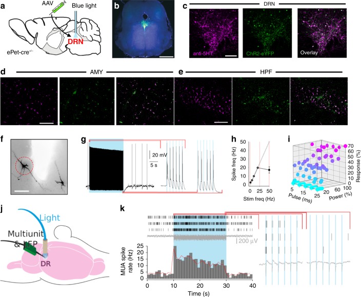
Fig. 1
Optogenetic targeting of DRN 5-HT neurons. a Infusion of AAV harbouring cre-dependent ChR2-eYFP into the DRN of ePet-cre +/u2212 mice, followed by implantation of a MRI-friendly optic fibre used to de...
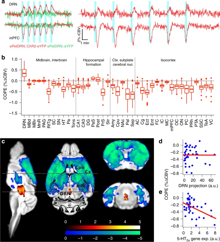
Fig. 2
A whole-brain functional map of the DRN 5-HT circuit. a CBV traces from the DRN and medial prefrontal cortex (mPFC) during block stimulation with blue light (20u2009Hz, 5u2009ms pulse width, 6u2009u00...
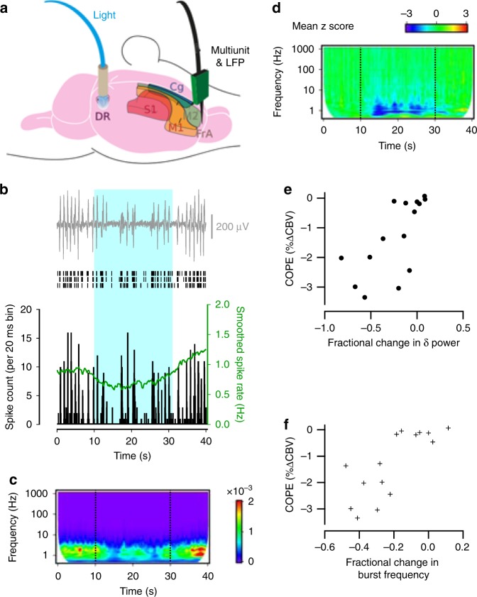
Fig. 3
Photoactivation of DRN 5-HT neurons dampens MUA and LFP power in cortical projection areas. a Schematic of the experimental set-up for paired MUA and LFP recordings in the DRN and projection areas dur...
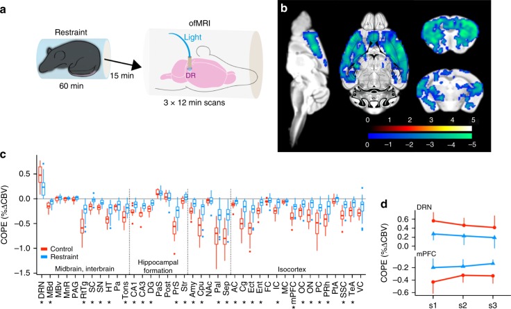
Fig. 4
Acute stress occludes photoactivation of the DRN 5-HT circuit. a Experiment schematic. b Second level analysis comparing restraint ( n =u20097) to control ( n =u20094) condition indicate a decrease in...
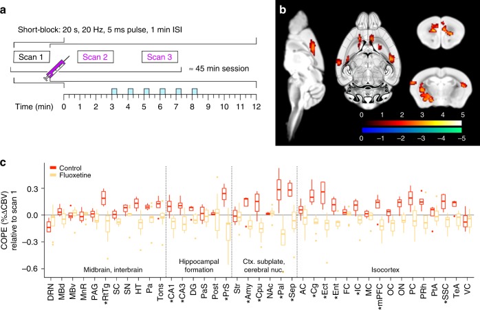
Fig. 5
Circuit regulation of DRN 5-HT functional connectivity by fluoxetine. a Experiment schematic. b Second level analysis comparing fluoxetine ( n =u20099) to control ( n =u20094) condition indicate an in...
Figure images are served from the NIH/NLM PubMed Central Open Access Subset or Europe PMC; copyright remains with the publishers and authors.
💬 Discussion
0 commentsNo comments yet. Be the first to start a discussion!
Leave a Comment