Abstract
Connexin43 (CX43), a protein that forms gap junction channels and hemichannels in astrocytes, is downregulated in high-grade gliomas. Its relevance for glioma therapy has been thoroughly explored; however, its positive effects on proliferation are counterbalanced by its effects on migration and invasion. Here, we show that a cell-penetrating peptide based on CX43 (TAT-Cx43266-283) inhibited c-Src and focal adhesion kinase (FAK) and upregulated phosphatase and tensin homolog in glioma stem cells (GSCs) derived from patients. Consequently, TAT-Cx43266-283 reduced GSC motility, as analyzed by time-lapse microscopy, and strongly reduced their invasive ability. Interestingly, we investigated the effects of TAT-Cx43266-283 on freshly removed surgical specimens as undissociated glioblastoma blocks, which revealed a dramatic reduction in the growth, migration, and survival of these cells. In conclusion, a region of CX43 (amino acids 266-283) exerts an important anti-tumor effect in patient-derived glioblastoma models that includes impairment of GSC migration and invasion.
🔬 Techniques
🔭 Microscopes
💻 Software
✨ Fluorophores
🧪 Sample Preparation
🔬 Cell Lines
🏭 Microscope Brands
🧪 Reagent Suppliers
💻 Software Details
🏛️ Research Organizations (ROR)
Affiliated research institutions:
📋 Methods
Ethics Statement
Patients provided written informed consent to participate in the study, and tumor samples and cell culture brain tumor samples were obtained following local ethical board approval at the Service of Neurosurgery in the Hospital Universitario de Salamanca (Spain). The study was approved by the bioethics committee of the University of Salamanca and Junta de Castilla y León (Spain). GSC Cultures G166 GSCs were obtained from BioRep (Milan, Italy) ( Pollard et al., 2009 ). Primary GSCs were obtained and cultured as described previously ( Thirant et al., 2011 ). In brief, immediately after surgery, the tumor samples (G9, G12, G13, G15, and G16, diagnosed as classic glioblastomas; N6, a neuroblastoma; and O17, an oligodendroglioma) were washed and deprived of vessels in PBS. After mechanical dissociation, the samples were subjected to enzymatic dissociation with Accutase (Sigma-Aldrich Química, Madrid, Spain) for 15–20 min at 37°C. These solutions were then filtered and centrifuged at 1,000 × g for 5 min. The G166 and primary GSCs were cultured in RHB-A medium (Takara Bio, Condalab, Madrid, Spain) supplemented with 2% B27 (Life Technologies, Thermo Fisher Scientific, Waltham, USA), 1% N2 supplement (Life Technologies), 20 ng/mL epidermal growth factor (EGF), and 20 ng/mL basic fibroblast growth factor (b-FGF) (PeproTech, London, UK) under adherent conditions as described by Pollard et al. (2009) . Culture plates were coated with 10 μg/mL laminin (Life Technologies) for 2 hr before use. The cells were maintained at 37°C in an atmosphere of 95% air/5% CO 2 and with 90%–95% humidity. The G166 and primary GSCs were grown to confluency, dissociated using Accutase, and then split. We routinely used cultures expanded for no more than 15 passages.
Show full methods section
Ethics Statement
Patients provided written informed consent to participate in the study, and tumor samples and cell culture brain tumor samples were obtained following local ethical board approval at the Service of Neurosurgery in the Hospital Universitario de Salamanca (Spain). The study was approved by the bioethics committee of the University of Salamanca and Junta de Castilla y León (Spain). GSC Cultures G166 GSCs were obtained from BioRep (Milan, Italy) ( Pollard et al., 2009 ). Primary GSCs were obtained and cultured as described previously ( Thirant et al., 2011 ). In brief, immediately after surgery, the tumor samples (G9, G12, G13, G15, and G16, diagnosed as classic glioblastomas; N6, a neuroblastoma; and O17, an oligodendroglioma) were washed and deprived of vessels in PBS. After mechanical dissociation, the samples were subjected to enzymatic dissociation with Accutase (Sigma-Aldrich Química, Madrid, Spain) for 15–20 min at 37°C. These solutions were then filtered and centrifuged at 1,000 × g for 5 min. The G166 and primary GSCs were cultured in RHB-A medium (Takara Bio, Condalab, Madrid, Spain) supplemented with 2% B27 (Life Technologies, Thermo Fisher Scientific, Waltham, USA), 1% N2 supplement (Life Technologies), 20 ng/mL epidermal growth factor (EGF), and 20 ng/mL basic fibroblast growth factor (b-FGF) (PeproTech, London, UK) under adherent conditions as described by Pollard et al. (2009) . Culture plates were coated with 10 μg/mL laminin (Life Technologies) for 2 hr before use. The cells were maintained at 37°C in an atmosphere of 95% air/5% CO 2 and with 90%–95% humidity. The G166 and primary GSCs were grown to confluency, dissociated using Accutase, and then split. We routinely used cultures expanded for no more than 15 passages.
Cell Treatments
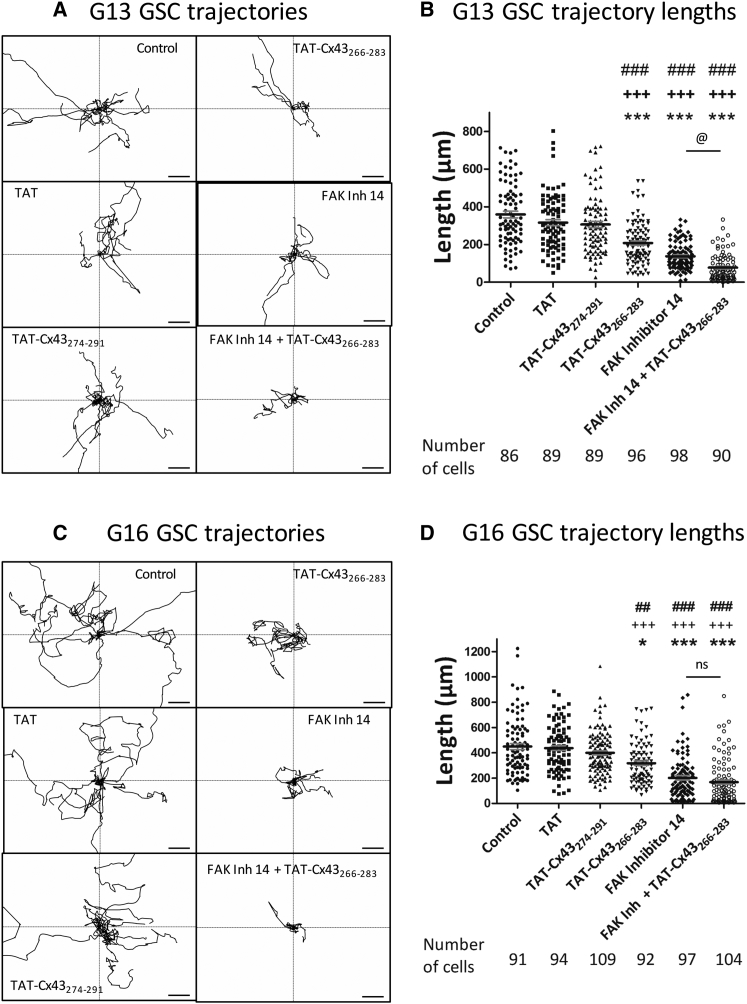
The synthetic peptides (>85% pure) were obtained from GenScript (Piscataway, NJ, USA). YGRKKRRQRRR was used as the TAT sequence, which is responsible for the cell penetration of the peptides ( Gangoso et al., 2014 ). The TAT-Cx43 266-283 sequence was TAT-AYFNGCSSPTAPLSPMSP and the TAT-Cx43 274-291 sequence was TAT-PTAPLSPMSPGYKLVTG. The peptides were used at different concentrations (25, 50, or 100 μM) in culture medium at 37°C for the indicated time. FAK inhibitor 14 (SML0837) was obtained from Sigma and used at 5 μM.
Immunocytochemistry
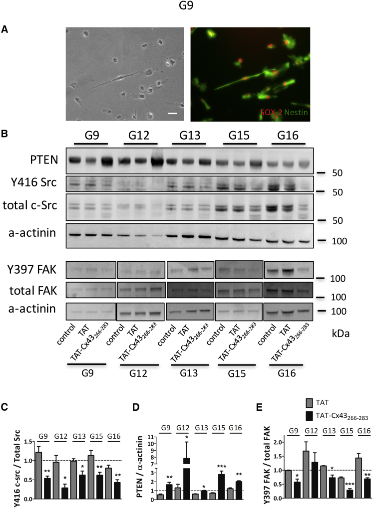
Cells were fixed in 4% paraformaldehyde for 20 min. A mouse monoclonal antibody against human Nestin (1:200; Abcam, Cambridge, UK; Ref. ab18102) was applied overnight at 4°C, followed by incubation with an Alexa Fluor 488-conjugated anti-mouse immunoglobulin G (IgG) antibody (1:1,000; Life Technologies; Ref. A-11029) for 2 hr. A rabbit polyclonal antibody against SOX-2 (1:200; Abcam; Ref. ab97959) was applied overnight at 4°C, followed by incubation with an Alexa Fluor 594-conjugated anti-rabbit IgG antibody (1:1,000; Life Technologies; Ref. A-11012) for 2 hr. The cells were then mounted using a SlowFade Light Antifade Kit (Life Technologies), and they were analyzed on a Leica inverted fluorescence microscope connected to a digital video camera (Leica DC 100; Leica Microsystems, Wetzlar, Germany).
Migration Assays
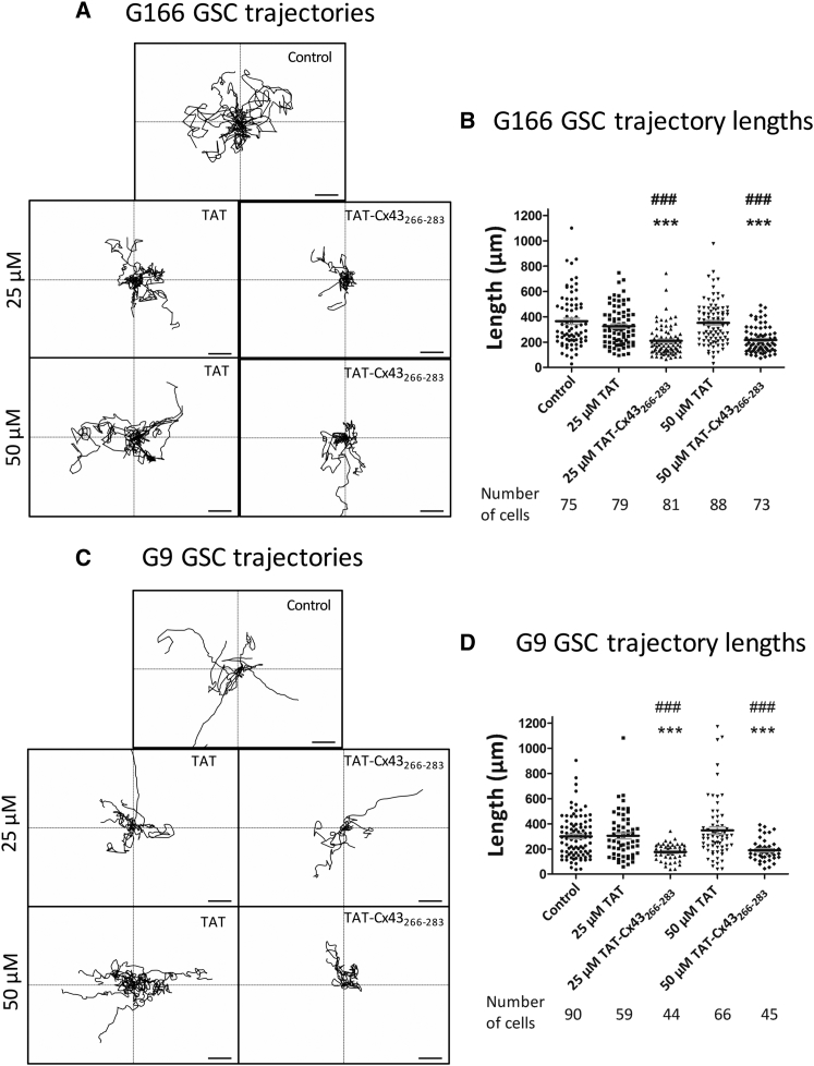
G166 or primary GSCs were plated at a low density (5,000 cells/cm 2 ) in 24-well plates. Once the cells were attached, TAT or TAT-Cx43 266-283 was added at 25 or 50 μM, and the cells were allowed to equilibrate for 1–3 hr in the microscope incubator before imaging. Random cell movement was recorded by time-lapse live-cell imaging for 12–14 hr. The total duration of the treatment was always 15 hr. Every 10 min, phase-contrast photographs of each experimental condition were taken with an inverted Zeiss Axio Observer Z1 microscope for live-cell imaging (Carl Zeiss Microscopy, LLC, USA) coupled to an AxioCam MRm camera. The system included an automated XY stage controller and a humidified incubator set at 37°C and 5% CO 2 . Image stacks were processed, and cell movement was manually tracked and further analyzed using Zen imaging software (Carl Zeiss Microscopy). Average cell length was calculated in at least six independent movies from three independent experiments.
Glioblastoma Explant Cultures
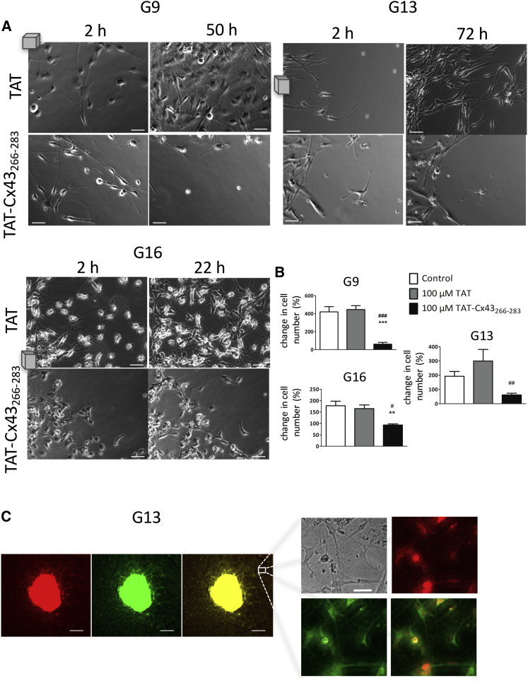
The tumor samples, immediately after surgery, were washed, deprived of vessels in PBS and finely minced into approximately 1-mm 3 pieces. The explants were plated individually in 24-well plates and cultured in RHB-A medium (Takara Bio, Condalab, Madrid, Spain) supplemented with 2% B27 (Life Technologies, Thermo Fisher Scientific, Waltham, USA), 1% N2 supplement (Life Technologies), 20 ng/mL EGF, and 20 ng/mL b-FGF (PeproTech, London, UK), as described previously ( Bayin et al., 2016 ). Culture plates were coated with 10 μg/mL laminin (Life Technologies) for 2 hr before use. The tumor blocks were maintained at 37°C in an atmosphere of 95% air/5% CO 2 and with 90%–95% humidity. Once the explants were attached (after 24–72 hr), TAT or TAT-Cx43 266-283 was added at 100 μM, and the cells were allowed to equilibrate for 1–3 hr in the microscope incubator before imaging. The cells were recorded by time-lapse live-cell imaging for the indicated times. Every 10 min, phase-contrast photographs of each experimental condition were taken with an inverted Zeiss Axio Observer Z1 microscope for live-cell imaging (Carl Zeiss Microscopy, LLC, USA) coupled to an AxioCam MRm camera. The system included an automated XY stage controller and a humidified incubator set at 37°C and 5% CO 2 . Image stacks were processed using Zen imaging software (Carl Zeiss Microscopy).
Invasion Assays
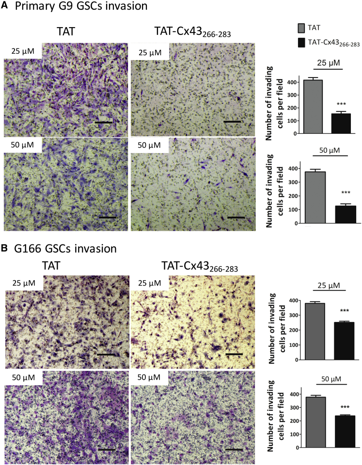
Cell invasion was measured in Matrigel (Corning, Amsterdam, The Netherlands)-coated transwell inserts (Merck Millipore, Madrid, Spain) containing polyethylene terephthalate filters with 8-μm pores. The inserts were coated with 100 μL of 1 mg/mL Matrigel matrix according to the manufacturer's recommendations. Next, 7.5 × 10 4 cells in 200 μL of serum-free medium were plated in the upper chamber, whereas 500 μL of medium supplemented with 10% fetal bovine serum (Gibco, Life Technologies) was added to the lower well. The indicated treatments were added, and the cells were allowed to invade for 15 hr. Non-invading cells were carefully removed with wet cotton swabs from the top of the membranes. The invading cells of the lower surface were fixed with 4% paraformaldehyde for 10 min, washed with PBS, and stained with Giemsa for 10 min. The inserts were washed with PBS and allowed to dry. The invading cells were counted in at least five random fields per insert from three independent experiments. Images were taken using a Leica microscope connected to a digital camera (Leica DFC500).
Western Blot Analysis
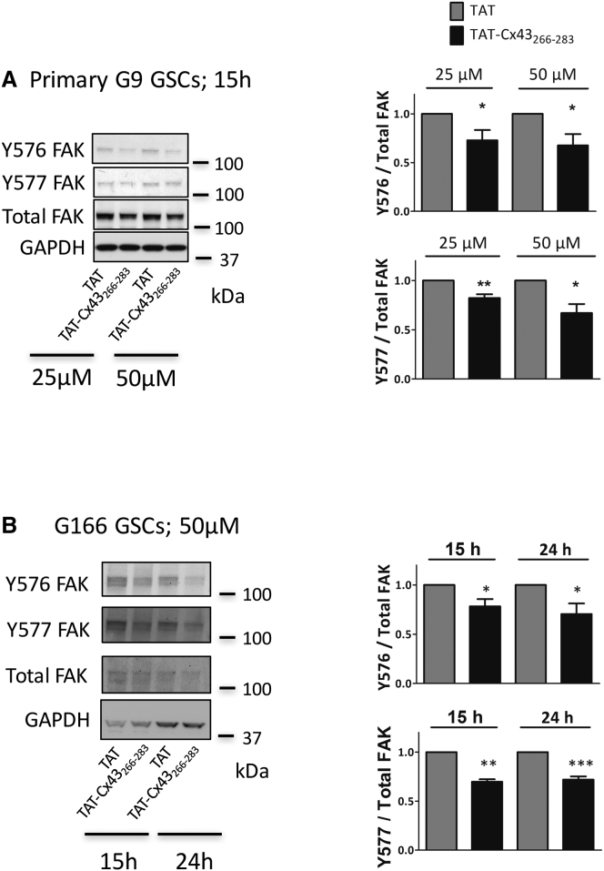
Western blotting was performed as described previously ( Herrero-Gonzalez et al., 2010 ). In brief, equivalent amounts of proteins (20 μg per lane) were separated on NuPAGE Novex Bis-Tris (4%–12%) midi gels (Life Technologies). The proteins were transblotted using an iBlot dry blotting system (Life Technologies). After blocking, the membranes were incubated overnight at 4°C with the primary antibodies against Y416 Src (1:200; Cell Signaling, Danvers, MA, USA; Ref. 2101), total Src (1:500; Cell Signaling; Ref. 2108), PTEN (1:500; Cell Signaling; Ref. 9556S), Y397 FAK (1:1,000; Life Technologies; Ref. 44-624G), Y576 FAK (1:500; Life Technologies; Ref. 44652G), Y577 FAK (1:500; Life Technologies; Ref. 44-614G), and total FAK (1:500; Life Technologies; Ref. AHO0502). The antibodies against glyceraldehyde phosphate dehydrogenase (GAPDH, 1:15,000; Ambion, Thermo Fisher Scientific; Ref. AM4300) or alpha-actinin (1:1,000; Chemicon International, Merck Millipore; Ref. MAB1682) were used as a loading control. After extensive washing, the membranes were incubated with peroxidase-conjugated anti-rabbit IgG or the anti-mouse IgG antibody (Santa Cruz Biotechnology, Dallas, TX, USA; Refs. sc-2030 and sc-2005) in TTBS and developed with a chemiluminescent substrate (Western Blotting Luminol Reagent; Santa Cruz Biotechnology). X-ray films were obtained from Fujifilm (Madrid, Spain).
MTT Assay
Cells cultured at 37°C in 24-well plates were incubated in the dark for 75 min with 300 μL of RHB-A medium containing 0.5 mg/mL MTT (Sigma). The medium was then removed, and the cells were incubated for 10 min in the dark with DMSO (500 μL per well) with mild shaking. Finally, the absorbance was measured at a wavelength of 570 nm using a microplate reader (Appliskan 2001; Thermo Electron Corporation, Thermo Scientific, Madrid, Spain).
Statistical Analysis
The results are expressed as the mean ± SEM of at least three independent experiments. Statistical analyses were carried out using Student's t test when two groups were compared. For the comparison of more than two groups, an ANOVA (one-way ANOVA) was used, followed by the appropriate post-test (Tukey). Values were considered significant when p < 0.05.
Supplemental Information Document S1. Figures S1–S4 Movie S1. Time-Lapse Movie of Control G9 GSCs, Related to Figure 3 Movie S2.
Time-Lapse Movie of G9 GSCs
Treated with 50 μM TAT, Related to Figure 3 Movie S3.
Time-Lapse Movie of G9 GSCs
Treated with 50 μM TAT-Cx43266-283, Related to Figure 3 Movie S4.
Time-Lapse Movie of G9 Explant
Treated with 100 μM TAT, Related to Figure 6 Movie S5.
Time-Lapse Movie of G9 Explant
Treated with 100 μM TAT-Cx43266-283, Related to Figure 6 Movie S6.
Time-Lapse Movie of G13 Explant
Treated with 100 μM TAT, Related to Figure 6 Movie S7.
Time-Lapse Movie of G13 Explant
Treated with 100 μM TAT-Cx43266-283, Related to Figure 6 Movie S8.
Time-Lapse Movie of G16 Explant
Treated with 100 μM TAT, Related to Figure 6 Movie S9.
Time-Lapse Movie of G16 Explant
Treated with 100 μM TAT-Cx43266-283, Related to Figure 6 Document S2. Article plus Supplemental Information
📊 Figures
Figureu00a01
TAT-Cx43 266-283 Targets c-Src, PTEN, and FAK in Primary GSCs (A) Phase-contrast images (left) and SOX-2 (red) and Nestin (green) immunostaining (right) of the same field showing that primary G9 GSCs ...
Figureu00a02
TAT-Cx43 266-283 Reduces Y576 and Y577 FAK Phosphorylation in GSCs Primary G9 (A) or G166 (B) GSCs were incubated with TAT or TAT-Cx43 266-283 . Western blot analysis for FAK, Y576 FAK, and Y577 FAK. ...
Figureu00a03
TAT-Cx43 266-283 Reduces GSC Motility G166 (A and B) or primary G9 GSCs (C and D) were incubated with 25 or 50u00a0u03bcM TAT or TAT-Cx43 266-283 , and their random movements were recorded by time-lap...
Figureu00a04
Specificity of the Effect of TAT-Cx43 266-283 on FAK-Dependent GSC Motility Primary G13 (A and B) or G16 (C and D) GSCs were incubated with 50u00a0u03bcM TAT, TAT-Cx43 274-291 , TAT-Cx43 266-283 , 5u0...
Figureu00a05
TAT-Cx43 266-283 Reduces GSC Invasion Primary (A) or G166 (B) GSC invasion was analyzed using a transwell Matrigel invasion assay. Cells were incubated with TAT or TAT-Cx43 266-283 and were allowed to...
Figureu00a06
TAT-Cx43 266-283 Reduces Growth, Migration, and Survival in Patient-Derived Glioblastoma Explants G9, G13, and G16 glioma explants were cultured in GSC medium and incubated in the absence (control) or...
Figureu00a07
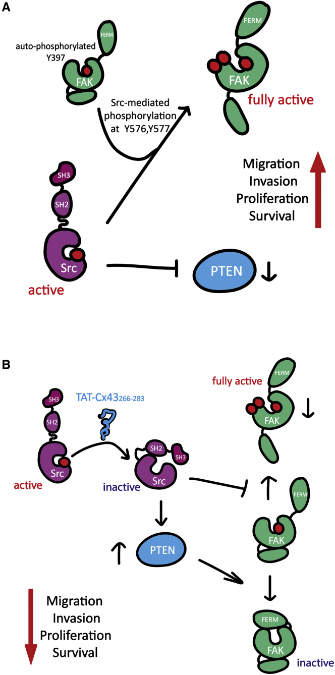
TAT-Cx43 266-283 Targets Src, PTEN, and FAK in GSCs (A) In GSCs, active c-Src phosphorylates FAK at Y576 and Y577, thus fully activating FAK. Active c-Src also promotes the degradation of PTEN, an imp...
Figure images are served from the NIH/NLM PubMed Central Open Access Subset or Europe PMC; copyright remains with the publishers and authors.
💬 Discussion
0 commentsNo comments yet. Be the first to start a discussion!
Leave a Comment