Abstract
As one of the most powerful tools in the biological investigation of cellular structures and dynamic processes, fluorescence microscopy has undergone extraordinary developments in the past decades. The advent of super-resolution techniques has enabled fluorescence microscopy - or rather nanoscopy - to achieve nanoscale resolution in living specimens and unravelled the interior of cells with unprecedented detail. The methods employed in this expanding field of microscopy, however, are especially prone to the detrimental effects of optical aberrations. In this review, we discuss how super-resolution microscopy techniques based upon single-molecule switching, stimulated emission depletion and structured illumination each suffer from aberrations in different ways that are dependent upon intrinsic technical aspects. We discuss the use of adaptive optics as an effective means to overcome this problem.
🔬 Techniques
🧬 Organisms
✨ Fluorophores
🧪 Sample Preparation
🏭 Microscope Brands
🏛️ Research Organizations (ROR)
Affiliated research institutions:
📊 Figures
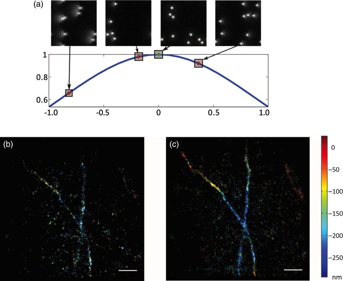
Fig. 1.
Aberration correction in a SMS microscope. (a) Principle of image-based optimization. For each mode used in the aberration correction procedure, different amplitudes are applied and their corresponden...
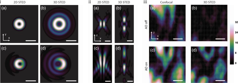
Fig. 2.
Effect of aberrations on STED microscopy. (i) Calculations of the effect of coma in the focal plane of STED depletion profiles. (a and b) Ideal focal plane intensity distributions of STED beams. (c an...
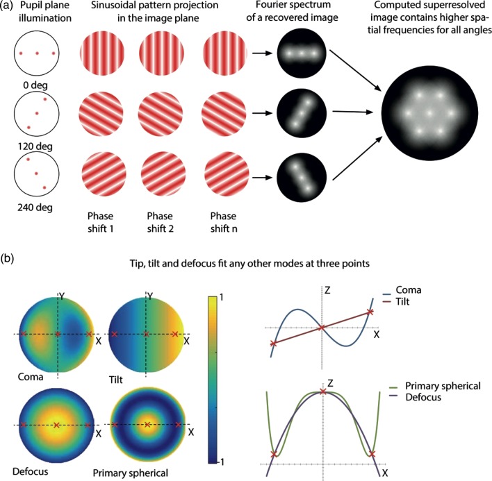
Fig. 3.
Effect of aberrations on SIM. (a) Schematic of the principles of SIM. (b) Schematic showing how aberrations in the illumination pattern are equivalent to an offset in the spatial coordinates of the in...
Figure images are served from the NIH/NLM PubMed Central Open Access Subset or Europe PMC; copyright remains with the publishers and authors.
💬 Discussion
0 commentsNo comments yet. Be the first to start a discussion!
Leave a Comment