Abstract
Impaired immune responses in the elderly lead to reduced vaccine efficacy and increased susceptibility to viral infections. Although several groups have documented age-dependent defects in adaptive immune priming, the deficits that occur prior to antigen encounter remain largely unexplored. Herein, we identify novel mechanisms for compromised adaptive immunity that occurs with aging in the context of infection with West Nile virus (WNV), an encephalitic flavivirus that preferentially causes disease in the elderly. An impaired IgM and IgG response and enhanced vulnerability to WNV infection during aging was linked to delayed germinal center formation in the draining lymph node (DLN). Adoptive transfer studies and two-photon intravital microscopy revealed a decreased trafficking capacity of donor naïve CD4+ T cells from old mice, which manifested as impaired T cell diapedesis at high endothelial venules and reduced cell motility within DLN prior to antigen encounter. Furthermore, leukocyte accumulation in the DLN within the first few days of WNV infection or antigen-adjuvant administration was diminished more generally in old mice and associated with a second aging-related defect in local cytokine and chemokine production. Thus, age-dependent cell-intrinsic and environmental defects in the DLN result in delayed immune cell recruitment and antigen recognition. These deficits compromise priming of early adaptive immune responses and likely contribute to the susceptibility of old animals to acute WNV infection.
🔬 Techniques
🧬 Organisms
✨ Fluorophores
🧪 Sample Preparation
🔬 Cell Lines
🏭 Microscope Brands
🧪 Reagent Suppliers
💻 Software Details
💾 Data Repositories
🏛️ Research Organizations (ROR)
Affiliated research institutions:
📋 Methods
Ethics statement
This study was carried out in strict accordance with the recommendations in the Guide for the Care and Use of Laboratory Animals of the National Institutes of Health. The protocols were approved by the Institutional Animal Care and Use Committee at the Washington University School of Medicine (Assurance Number: A3381-01). Dissections and footpad injections were performed under anesthesia that was induced and maintained with ketamine hydrochloride and xylazine, and all efforts were made to minimize suffering. Reagents and antibodies Cell Trackers: Cell Tracker Orange CMTMR (5-(and-6)-(((4-Chloromethyl) Benzoyl) Amino) Tetramethylrhodamine), CellTrace CFSE (carboxyfluorescein diacetate, succinimidyl ester) and CellTrace Violet were purchased from Life Technologies. The following fluorochrome or biotin-conjugated antibodies were purchased from BD Biosciences, Biolegend, and eBioscience (clone name in parenthesis): CD3ε (145-2C11), CD4 (RM4-5), CD8α (53–6.7) CD11b (M1/70), CD11c (HL3), CD19 (1D3), CD28 (E18), CD44 (IM7), CD45R (RA3-6B2), CD62L (MEL-14), CD69 (H1.2F3), OX40/CD134 (OX-86), Ki67 (SolA15), PD1 (29F.1A12), FAS (Jo2), GL7, F4/80 (BM8), TCRγ (eBioGL3), NK1.1 (PK136), VLA4 (R1-2), LFA-1 (2D7), PSGL-1 (2PH1), CCR7 (4B12), CXCR4 (L276F12), and CXCR5 (2G8). Viruses and cells The WNV strain (3000.0259) was isolated in New York in 2000 and passaged once in C6/36 Aedes albopictus cells. The WNV-KUN strain (CH16532) was a gift of R. Tesh (World Reference Center of Emerging Viruses and Arboviruses, Galveston, TX) and amplified as described previously [ 48 ]. Viral titer was determined by plaque assay using Vero cells, as described previously [ 49 ]. Mouse experiments C57BL/6 mice were purchased from The Jackson Laboratory (4 month-old) (Bar Harbor, ME) or acquired from the National Institute of Aging dedicated breeding colony at Charles River Laboratories (18 month-old) (Wilmington, MA). TCR β/δ -/- mice were purchased from The Jackson Laboratory and bred in the animal facilities at the Washington University School of Medicine. All mice were housed in a pathogen-free mouse facility at the Washington University. Mice were inoculated subcutaneously via footpad injection with 10 2 plaque-forming units (PFU) of WNV-NY or 10 4 PFU of WNV-KUN diluted in 50 μl of Hanks balanced salt solution (HBSS) supplemented with 0.1% heat-inactivated fetal bovine serum (FBS). For intravital imaging of the draining cervical lymph node, mice were infected subcutaneously in the nape of the neck.
Show full methods section
Ethics statement
This study was carried out in strict accordance with the recommendations in the Guide for the Care and Use of Laboratory Animals of the National Institutes of Health. The protocols were approved by the Institutional Animal Care and Use Committee at the Washington University School of Medicine (Assurance Number: A3381-01). Dissections and footpad injections were performed under anesthesia that was induced and maintained with ketamine hydrochloride and xylazine, and all efforts were made to minimize suffering. Reagents and antibodies Cell Trackers: Cell Tracker Orange CMTMR (5-(and-6)-(((4-Chloromethyl) Benzoyl) Amino) Tetramethylrhodamine), CellTrace CFSE (carboxyfluorescein diacetate, succinimidyl ester) and CellTrace Violet were purchased from Life Technologies. The following fluorochrome or biotin-conjugated antibodies were purchased from BD Biosciences, Biolegend, and eBioscience (clone name in parenthesis): CD3ε (145-2C11), CD4 (RM4-5), CD8α (53–6.7) CD11b (M1/70), CD11c (HL3), CD19 (1D3), CD28 (E18), CD44 (IM7), CD45R (RA3-6B2), CD62L (MEL-14), CD69 (H1.2F3), OX40/CD134 (OX-86), Ki67 (SolA15), PD1 (29F.1A12), FAS (Jo2), GL7, F4/80 (BM8), TCRγ (eBioGL3), NK1.1 (PK136), VLA4 (R1-2), LFA-1 (2D7), PSGL-1 (2PH1), CCR7 (4B12), CXCR4 (L276F12), and CXCR5 (2G8). Viruses and cells The WNV strain (3000.0259) was isolated in New York in 2000 and passaged once in C6/36 Aedes albopictus cells. The WNV-KUN strain (CH16532) was a gift of R. Tesh (World Reference Center of Emerging Viruses and Arboviruses, Galveston, TX) and amplified as described previously [ 48 ]. Viral titer was determined by plaque assay using Vero cells, as described previously [ 49 ]. Mouse experiments C57BL/6 mice were purchased from The Jackson Laboratory (4 month-old) (Bar Harbor, ME) or acquired from the National Institute of Aging dedicated breeding colony at Charles River Laboratories (18 month-old) (Wilmington, MA). TCR β/δ -/- mice were purchased from The Jackson Laboratory and bred in the animal facilities at the Washington University School of Medicine. All mice were housed in a pathogen-free mouse facility at the Washington University. Mice were inoculated subcutaneously via footpad injection with 10 2 plaque-forming units (PFU) of WNV-NY or 10 4 PFU of WNV-KUN diluted in 50 μl of Hanks balanced salt solution (HBSS) supplemented with 0.1% heat-inactivated fetal bovine serum (FBS). For intravital imaging of the draining cervical lymph node, mice were infected subcutaneously in the nape of the neck.
Measurement of viral burden
At specified time points after WNV infection, serum was obtained by intracardiac heart puncture, followed by intracardiac perfusion (20 ml of PBS), and organ recovery. Organs were weighed, homogenized using a bead-beater apparatus, and titrated by plaque assay on BHK21-15 cells as described previously [ 49 ]. Bone marrow chimera and transfers Bone marrow cells were collected from adult or old C57BL/6 (CD45.2) mice and adult B-cell deficient μMT mice. Adult or old bone marrow cells were mixed with an equal number of μMT bone marrow cells and 1 x 10 7 cells were transferred adoptively by retroorbital injection into 8 to 12-wk-old 800 cGy–irradiated B6.SJL (CD45.1) mice (National Cancer Institute). Twelve weeks later, reconstitution was validated by flow cytometry and mice were infected with WNV. Ovalbumin immunizations Mice were injected subcutaneously in the footpad with an emulsion containing CFA and 10 nmoles of OVA 323–337 peptide, and DLN were analyzed at indicated time points. The ELISPOT assay has been described previously [ 50 , 51 ]. To evaluate MHC class II specific T cell responses in adult and old mice, 5 x 10 5 cells from the DLN were harvested at day 7 post-immunization and plated on 96-well plates with Immobilon-P membrane coated with anti-mouse IL-2 antibody. After overnight incubation, plates were washed, incubated with a secondary antibody and developed as previously described [ 50 , 51 ]. Spots were analyzed on dedicated equipment and software (Cellular Technology Ltd along with Immunospot software Version 5.0.9) to calculate the total number of antigen-specific CD4 + T cells in DLN.
Antibody responses The levels of WNV-specific
IgM and IgG were determined using an ELISA against purified WNV E protein [ 52 ]. Focus reduction neutralization assays were performed on Vero cells after mixing serial dilutions of serum with a fixed amount (100 FFU) of WNV [ 49 ].
ELISPOT assay
WNV antigen-specific antibody secreting cells were quantified via an enzyme-linked immunospot assay [ 28 ]. Briefly, mixed cellulose esters filter plates (Millipore) were coated with 20 μg/ml of purified WNV E protein. Bone marrow cells were harvested from infected mice 90 days post infection, and 200,000 cells were seeded per well with 8 wells per mouse. Antibody spots were developed with chromogen substrate (3-amino-9-ethyl-carbazole) and wells were imaged and enumerated with an ImmunoSpot plate reader from Cellular Technology Ltd.
Cell purification
To isolate naïve CD4 + T cells, splenocytes were isolated from mice and erythrocytes depleted with ACK lysis buffer (Invitrogen). Cells were then stained in buffer (PBS, 1% FBS, and 2 mM EDTA) with biotinylated antibodies against CD8, CD45R (B220), NK1.1, and MHC class II as well as fluorescently conjugated antibodies against CD4, CD44, and CD62L. Cells were washed with 10 mL of staining buffer and stained with anti-biotin magnetic beads (Miltenyi Biotec). Magnetically labeled cells were then depleted by passing through a LS magnetic column (Miltenyi Biotec) leaving roughly 90% pure CD4 + T cells. CD4 + CD44 - CD62L + were then isolated on an Aria-II (BD Biosciences) fluorescence-activated cell sorter (see S2 Fig ). B cells were isolated using a B Cell Isolation Kit (Miltenyi Biotech). Briefly, cells were stained with a biotinylated-antibody cocktail followed by anti-biotin labeled magnetic beads. Cells were loaded onto a magnetic LS column and the flow-through was collected. Adoptive cell transfers Naïve CD4 + T cells or B cells from uninfected adult or old mice were sorted as detailed above. For cell tracking, isolated cells from adult and old donors were labeled differentially with vital dyes (CellTracker Orange CMTMR, CellTrace CFSE, or CellTrace Violet) according to the manufacturer recommendations. Importantly, dyes were switched between old and adult cells in some experiments and dye-dependent effects on cell trafficking were not observed under our staining conditions (data not shown). Isolated cells were incubated at 37°C and 5% CO 2 in pre-warmed CO 2 -Independent Medium (Invitrogen) with 2 μM of CellTrace CFSE, 5 μM CellTracker Orange, or 10 μM of CellTrace Violet (Invitrogen) for 15 minutes. Subsequently, cells were centrifuged (300 x g, 5 minutes) and the pellet was resuspended in pre-warmed medium for another 30 minutes. The incubation was stopped after two washes with cold PBS. Finally, cells were counted and adjusted to equal number for homing or microscopy imaging experiments. Cells were transferred adoptively via an intravenous route into recipient mice that had been infected subcutaneously with WNV-KUNV 48 hours earlier. For transfer into TCR β/δ -/- mice, 3 x 10 6 sorted naïve CD4 + T cells were injected intravenously into each recipient mouse that had been infected subcutaneously with WNV-NY 48 hours earlier. T cell homing experiments For analysis of T cell homing to the LN and spleen, naïve CD4 + T cells or B cells were sorted from adult and old mice and labeled with different dyes. Cells were suspended in PBS at a ratio 1:1 (adult to old) and injected intravenously (2 x 10 6 from each donor). One hour post transfer, LN and spleens were harvested and processed into single cell suspensions. Cells were analyzed by flow cytometry on an LSRII (BD Biosciences).
Intravital two-photon microscopy
We analyzed the trafficking behavior of lymphocytes in lymphoid organs using published methods for ex vivo (LN explants) and with minor modifications for in vivo imaging of cervical LNs [ 53 ]. Recipient mice (adult or old) infected with WNV-KUN 48 hours earlier were given 5 x 10 6 cells CellTrace CFSE or Violet labeled naïve T cells (from adult and old donors). For ex vivo analysis, LN were harvested 6 to 8 hours later, glued to plastic cover slips with VetBond (3M), placed under the flow of warm (35°C to 37°C) oxygenated medium (DMEM, without phenol red) [ 54 ]. Intravital imaging of T cell extravasation was performed as described [ 55 ] with the following modifications. Mice were anesthetized with avertin (250 mg/kg) and connected to a MiniVet Mouse Ventilator (Harvard Apparatus) to reduce movement artifacts and improve mouse viability. The skin of the neck was opened and the cervical DLN exposed on a skin flap and the inner surface was attached to a plastic cover slip using VetBond with the LN protruding through a hole in the center. Imaging was performed on A custom-build two-photon microscope equipped with an Olympus 1.0 NA 20x water-immersion objective, a Vision II Coherent Ti:Sapphire laser and running ImageWarp (A&B Software) for hardware and acquisition control [ 56 ]. Dyes were excited at 820 nm and detected using 480 nm and 570 nm dichroic filters. T cell rolling in vessels was imaged at 25 f/sec and 3D images collected ~ 2 per minute with image dimensions of ~250 x 225 x 75 μm (X,Y,Z; 2.5 μm Z steps). Blood vessels in the LN were labeled with QTracker 655 (10 μl in 50 μl of PBS given intravenously, Life Technologies).
Image analysis
Multidimensional data rendering and cell tracking were performed using Imaris software (version 7.3, Bitplane). For quantitative and statistical analysis of T cell motility we used either, T Cell Analyzer Software (version 1.7.0, Dr. J. Dempster, University of Strathclyde, UK) and an open-source software suite developed by Dr. J Textor (University of Ultrecht, The Netherlands). For quantitative and statistical analysis of T and B cell motility, we used the X and Y dimensions of the cell tracks to avoid bias arising from the intrinsically poorer Z resolution [ 57 ]. Displacement factors were defined as the distance between the endpoints of a track divided by the square root of its duration. The unpaired Mann-Whitney U test was used to compare track motility parameters. To control for variation between experiments, data were normalized prior to comparison by equalizing the medians across independent experiments. Hotelling's test for directionality was performed as described previously [ 58 ]. Briefly, cell tracks were broken down into steps (each step being a pair of consecutive cell positions), and the mean speed vector was computed per population of interest. Under the null hypothesis, the cells perform a random walk and the mean speed vector is zero in each dimension. Ellipses in Figs 4K , 5E , 7F and 7J show 95% confidence regions for the mean speed vector. If an ellipse does not contain the origin (0,0), then the null hypothesis is rejected for that population, and there is some directionality in the cell motion. For all cell populations studied in this paper, directionality was of a small magnitude ( 0.8 are commonly associated with chemotaxis, whereas values of < 0.5 are consistent with random cell migration. The slope indicates how fast the mean square displacement increases with time, and thus is a measure of maintenance of motility. F-G . Time-lapse image sequences of transferred adult and old naïve CD4 + T cells in explanted LN in recipient mice 48 h after WNV-KUN infection. Differentially labeled (blue = old, green = adult) naïve CD4+ T cells were adoptively transferred into WNV-KUNV infected adult recipient mice (48 h after infection) and DLN were harvested 6 to 8 h later followed by ex vivo imaging. Panel F presents all tracked adult and old cells during the first 12 minutes. Panel G and others presents time-lapse image sequences of adult and old cell tracking during first 12 minutes. Yellow and red arrows indicate which old and adult cells were analyzed and their starting points, respectively. Scale bar is indicated in white. (TIF) Click here for additional data file. S5 Fig Surface staining of naïve CD4 + T cells from adult and old mice for adhesion molecules and chemokine receptors. Splenocytes from adult or old C57BL/6 mice were stained on their surface for markers for naïve CD4 + T cells (CD4 + , CD44 - , CD62L + ) and for levels of adhesion molecules ( A ) (VLA-4 (CD49d), ( B ) L-selectin (CD62L), ( C ) LFA1 (CD11a), and ( D ) PSGL1 (CD162) or chemokine receptors (( E ) CCR7 (CD197) or ( F ) CXCR4). The geometric mean fluorescent intensity was measured in individual experiments and normalized to the levels seen with cells from adult mice. The data is the average of at least two independent experiments comprising a total of 4 to 6 mice per group. None of the differences were statistically significant. Also shown are representative histograms of each surface marker on naïve CD4 + T cells from an adult and old mouse along with the fluorescence minus one (FMO) control. Because CD44- CD62L + cells were gated for the flow cytometric analysis, an FMO control is not included in the L-selectin (CD62L) histogram. (TIF) Click here for additional data file. S6 Fig Cytokine-induced cytoskeletal rearrangement and ICAM-1 binding of naïve CD4 + T cells from adult and old mice. A . CD4 + T cells were isolated from adult and old mice and stimulated with 100 ng/mL of both CCL19 and CCL21. Cells were fixed at the indicated time post stimulation and stained for naïve CD4 + T cell markers (CD4 + CD44 - CD62L + ) and phalloidin-A647 (which binds to filamentous actin). Cells were analyzed by flow cytometry and the geometric mean fluorescent intensity was measured. Shown is the mean + SEM from three independent experiments. B-C . Naïve CD4 + T cells were isolated from adult or old mice. ICAM-1 Fc (0, 4, or 40 μg/ml) was coated on ( B ) polystyrene Petri dishes or ( C ) 5 μm transwell membranes. B. Cells were added to the ICAM-1 coated Petri dishes in the presence of 200 ng/ml of CCL19 and CCL21. After a one-hour incubation, adherent cells were counted. Plotted is the number of adherent cells (mean ± SEM) minus the background (to 0 μg/ml of ICAM-1 Fc). Data is pooled from three independent experiments. C. CCL19 and CCL21 (0.1, 1, or 10 ng/ml) were plated in the bottom compartment of a Boyden chamber and cells were added to the top chamber above the transwell membrane insert. The number of cells migrating through the membrane was quantified and normalized to the number observed at 40 μg/mL ICAM-1 Fc in the presence of 1 ng/mL of CCL19 and CCL21. Plotted is the mean number of migrating cells (± SEM) from three independent experiments. (TIF) Click here for additional data file. S1 Movie Intravital microscopy showing differential movement of adoptively transferred naïve CD4 + T cells from adult and old mice across the HEV. Naïve CD4 + T cells from adult and old mice were sorted, differentially labeled with fluorescent dyes (adult = green; old = blue) and adoptively transferred into adult mice that had been infected subcutaneously with WNV-KUN 48 hours earlier. One hour later, the DLN was evaluated by intravital microscopy and vascular structures were visualized with labeled quantum dots. One representative experiment of three is shown. The time of analysis and scale bar are indicated. (MOV) Click here for additional data file. S2 Movie Higher-magnification images showing differential movement of adoptively transferred naïve CD4 + T cells from adult and old mice across the HEV. These images was obtained from S1 Movie at higher magnification to highlight differential trafficking capacity of naïve CD4 + T cells from adult and old mice. The time of analysis and scale bar are indicated. (MOV) Click here for additional data file. S3 Movie Intravital microscopy showing differential movement of adoptively transferred naïve CD4 + T cells from adult and old mice in explanted LN. Naïve CD4 + T cells from adult and old mice were sorted, differentially labeled with fluorescent dyes (adult = green; old = blue) and adoptively transferred into adult mice that had been infected subcutaneously with WNV-KUN 48 hours earlier. Six hours later, the DLN was explanted and evaluated by intravital microscopy. Measurements of velocity and distance movement by individual naïve CD4 + T cells (see Fig 4 and S4 Fig ) were obtained from these videos. One representative experiment of several independent trials is shown. The time of analysis and scale bar are indicated. (MOV) Click here for additional data file. S4 Movie Intravital microscopy showing similar movement patterns of adoptively transferred CD19 + B cells from adult and old mice in explanted LN. CD19 + B cells from adult and old mice were sorted, differentially labeled with fluorescent dyes (adult = green; old = blue) and adoptively transferred into adult mice that had been infected subcutaneously with WNV-KUN 48 hours earlier. Six hours later, the DLN was explanted and evaluated by intravital microscopy. Measurements of velocity and distance movement by individual CD19 + B cells (see Fig 7 ) were obtained from these videos. One representative experiment of several independent trials is shown. The time of analysis and scale bar are indicated. (MOV) Click here for additional data file.
📊 Figures
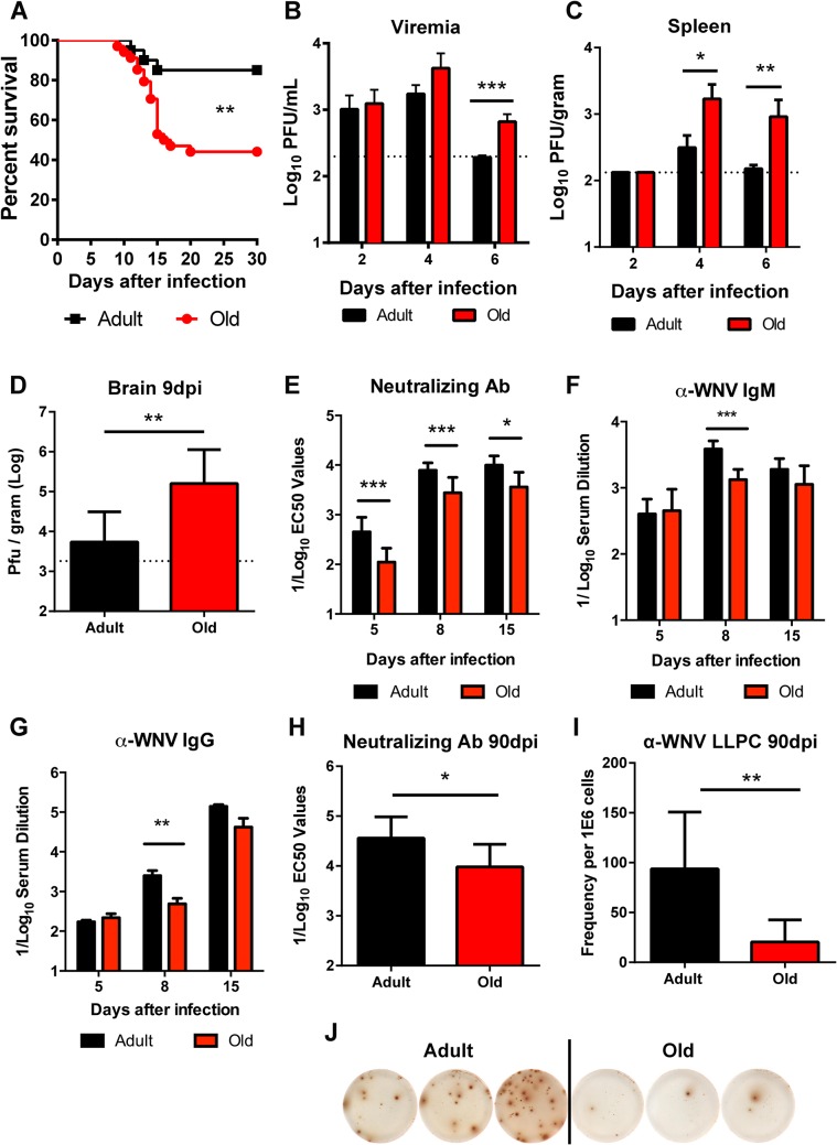
Fig 1
Survival rate, viral burden, and B cell responses after WNV infection.
A . Adult or old mice were infected with 10 2 PFU of WNV (New York strain) via subcutaneous injection in the footpad. Survival was monitored for 30 days: n = 20 adult (16 weeks) and n = 34 old (18 mon...
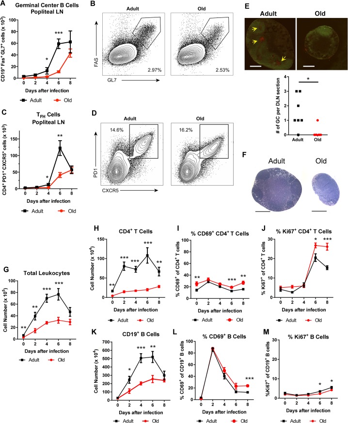
Fig 2
Delayed germinal center formation and decreased accumulation of CD4 + T cells and CD19 + B cells in DLN after WNV infection of old mice.
Mice were infected with 10 2 PFU of WNV (New York strain) in the footpad. A-D . Draining popliteal LN cells were analyzed by flow cytometry at the indicated days after infection. Numbers of GC B cells...
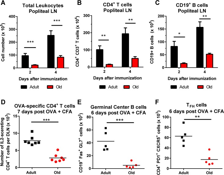
Fig 3
Decreased accumulation of CD4 + T cells and CD19 + B cells in DLN of old mice after immunization with ovalbumin.
A-C . Adult and old mice were immunized with ovalbumin (OVA) complexed with complete Freundu2019s adjuvant in the footpad. At the indicated days post infection, the draining popliteal LN was harvested...
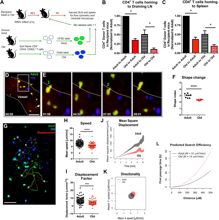
Fig 4
Defects in migration of nau00efve CD4 + T cells from old mice into inflamed LN.
A. Scheme of adoptive transfer studies. 2 x 10 6 FACS-sorted nau00efve (CD44 - CD62L + ) CD4 + T cells from adult or old mice were differentially labeled with fluorescent dyes, mixed in a 1:1 ratio, a...
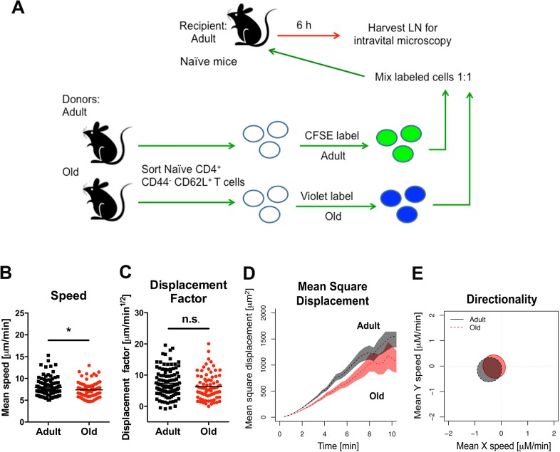
Fig 5
Migration of nau00efve CD4 + T cells from old mice into naive LN.
A. Scheme of adoptive transfer studies of nau00efve CD4 + T cells. 2 x 10 6 FACS-sorted nau00efve (CD44 - CD62L + ) CD4 + T cells from adult or old mice were differentially labeled with fluorescent dy...
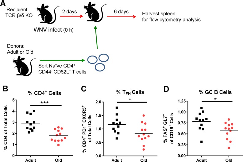
Fig 6
Transfer of nau00efve CD4 + T cells from old mice results in reduced GC responses.
A. TCR u03b2/u03b4 -/- recipient mice were infected with WNV (New York strain). Two days after infection, 3 x 10 6 nau00efve sorted CD4 + T cells from adult or old mice were transferred adoptively int...
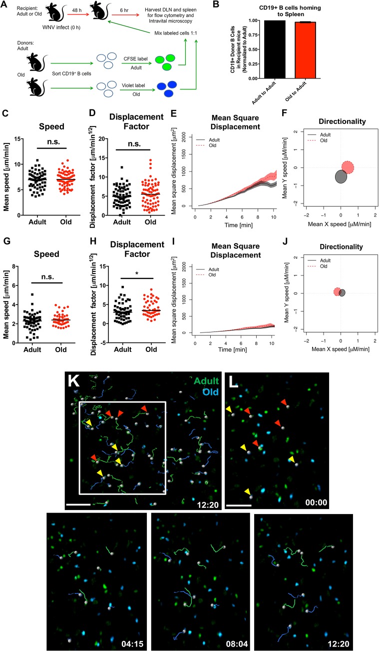
Fig 7
Normal migration of CD19 + B cells from old mice.
A. Scheme of adoptive transfer studies of CD19 + B cells. 5 x 10 6 isolated B cells from adult or old mice were differentially labeled with fluorescent dyes, mixed in a 1:1 ratio, and transferred to r...

Fig 8
Humoral immune response of donor bone marrow into irradiated recipient mice.
A. Scheme of reconstitution studies with bone marrow from adult or old mice. 10 7 bone marrow cells from adult or old mice were mixed with 10 7 bone marrow cells from u03bcMT (B cell deficient) mice a...
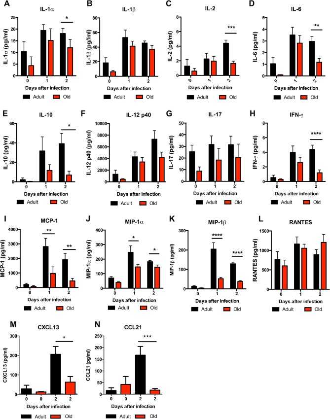
Fig 9
Decreased levels of cytokines and chemokines in the DLN of old mice.
Adult and old mice were infected bilaterally with 10 2 PFU of WNV (New York strain) in the footpad and DLN were harvested at days 0, 1, and 2 after infection. A-L . Levels of cytokines (IL-1u03b1 ( A ...

Fig 10
Leukocyte infiltration in the DLN of WNV-infected adult and old mice.
Adult and old mice were infected with 10 2 PFU of WNV (New York strain) in the footpad. One or two days later, the draining popliteal LN was harvested, cells were stained with antibodies to detect spe...
Figure images are served from the NIH/NLM PubMed Central Open Access Subset or Europe PMC; copyright remains with the publishers and authors.
💬 Discussion
0 commentsNo comments yet. Be the first to start a discussion!
Leave a Comment