Abstract
Many antibiotics stop bacterial growth by inhibiting different steps of protein synthesis. However, no specific inhibitors of translation termination are known. Proline-rich antimicrobial peptides, a component of the antibacterial defense system of multicellular organisms, interfere with bacterial growth by inhibiting translation. Here we show that Api137, a derivative of the insect-produced antimicrobial peptide apidaecin, arrests terminating ribosomes using a unique mechanism of action. Api137 binds to the Escherichia coli ribosome and traps release factor (RF) RF1 or RF2 subsequent to the release of the nascent polypeptide chain. A high-resolution cryo-EM structure of the ribosome complexed with RF1 and Api137 reveals the molecular interactions that lead to RF trapping. Api137-mediated depletion of the cellular pool of free release factors causes the majority of ribosomes to stall at stop codons before polypeptide release, thereby resulting in a global shutdown of translation termination.
🔬 Techniques
🔭 Microscopes
🧬 Organisms
💻 Software
🧪 Sample Preparation
🔬 Cell Lines
🏭 Microscope Brands
🧪 Reagent Suppliers
📷 Detectors
💻 Software Details
💾 Data Repositories
🏛️ Research Organizations (ROR)
Affiliated research institutions:
📋 Methods
On-line Methods
Peptides and oligonucleotides Api137 was synthesized by NovoPro Biosciences Inc. Onc112 was synthesized by GenScript. The ‘start-stop’ mRNA ( Supplementary Table 2 ) was purchased from IBA GmbH. The 2XermCL_S10_UAG construct was synthesized by Eurofins. DNA oligonucleotides were synthesized by Integrated DNA Technologies. Generation of templates for in vitro translation and toeprinting The DNA templates for toeprinting ( Supplementary Table 2 ) were generated by PCR using AccuPrime DNA Polymerase (Thermo Fisher Scientific) and primers listed in Supplementary Table 3 . The synthetic template yrbA-fs15 was prepared using 3 overlapping primers (T7-IR-AUG, IR-yrbA-fs15-RF1 and posT-NV1) in a single PCR reaction. The ermCL template was created by PCR amplification of the gene from the plasmid pERMCT7-M 32 using primers T7 and ermCL-UAG. The complete sequences of the templates are shown in Supplementary Table 2 . Toeprinting reactions were carried out in 5 μl of PURExpress transcription-translation system (New England Biolabs) as previously described 32 , 33 . The reverse transcription on the ermCL template was carried out using primer ermCL-TP-term. The final concentrations of Api137 and Onc112 in the reactions were 50 μM; the PrAMPs were added as stock solutions in water.
Selection of Api137-resistant mutants
The first round of selection of Api137 resistant mutants was performed with the E. coli strain SQ110, derived from the K12 strain ( Supplementary Table 4 ). An overnight culture grown in Luria-Bertani (LB) medium was diluted 100-fold into fresh medium containing subinhibitory concentration of Api137 (10 μM). After 24 h growth at 37°C, the culture was diluted 100-fold into 1 ml fresh LB medium containing 50 μM Api137. The culture was passaged one more time at 100 μM Api137 (8-fold MIC). The dilutions of cell culture were plated on LB agar. After overnight incubation, the sbmA gene was PCR amplified from 20 individual colonies using primers SbmA-seq-fwd and SbmA-seq-rev and sequenced. All but one clone had mutations in the sbmA gene. The Api137-resistant clone with the wt sbmA sequence (clone SQ110 ApiR21 in Supplementary Table 4 ) was grown in liquid culture; genomic DNA was isolated and prepared for sequencing using a Nextera XT kit (Illumina). Sequencing was performed on an Illumina NextSeq500 instrument (paired-end, 2×150 base reads) at the DNA Services facility at UIC. After mapping the reads to the genome of the strain SQ110 34 , the single mutation A722G in the prfA gene was identified. The presence of the mutation was verified by PCR-amplification of the prfA gene using primers PrfA-seq-fwd and PrfA-seq-rev from the parent and mutant strains and sequencing. E. coli strain BL21(DE3) ( Supplementary Table 4 ) was used in the second selection experiment. In order to avoid selection of sbmA mutants, prior to selection cells were transformed with the multicopy plasmid pZα-SbmA encoding the functional SbmA transporter. The pZα-SbmA plasmid was prepared by amplifying the E. coli sbmA gene using primers SbmA-seq-fwd and SbmA-EcoRI-rev, cutting the PCR product with restriction enzymes Nde I and Eco RI, and ligating the resulting DNA fragment into the pZα plasmid 35 cut with the same enzymes. For selection of Api137-resistant mutants, the overnight culture of BL21(DE3)/pZα-SbmA cells was diluted 1:100 in LB medium containing ampicillin (100 μg/ml) and 0.1 μM isopropyl-b-D-1-thiogalactopyranoside (IPTG) and grown at 37°C until reaching A 600 of 0.5. Two ml (approximately 10 9 cells) were plated on LB agar supplemented with 100 μg/ml ampicillin, 0.1 μM IPTG and 12 μM (4-fold MIC) Api137. After overnight incubation at 37°C, 10 colonies appeared. The prfA , prfB and prfC genes were PCR amplified using pairs of primers PrfA-seq-fwd with PrfA-seq-rev, PrfB-seq-fwd and PrfB-seq-rev, or PrfC-seq-fwd with PrfC-seq-rev, respectively, and sequenced. Five clones had mutations in the prfB gene: three of these had the C784T and two clones had the A839T mutation. The genome of one of the remaining five clones was sequenced and revealed the presence of the G241A mutation in the rplP gene encoding ribosomal protein uL16. The presence of this mutation in this and four remaining clones was verified by PCR-amplification of the rplP gene using primers RplP-seq-fwd and RplP-seq-rev and sequencing. The minimal inhibitory concentrations (MICs) of Api137 for the parental strains and selected resistant mutants were determined by microbroth dilution technique in 96-well plates. Specifically, exponentially growing cells were diluted to the final density A 600 = 0.002, 100 μl of the culture were placed in the wells and after addition of Api137 plates were incubated overnight at 37°C. The minimal Api137 concentration preventing appearance of the visible cell density was recoded as MIC.
Show full methods section
On-line Methods
Peptides and oligonucleotides Api137 was synthesized by NovoPro Biosciences Inc. Onc112 was synthesized by GenScript. The ‘start-stop’ mRNA ( Supplementary Table 2 ) was purchased from IBA GmbH. The 2XermCL_S10_UAG construct was synthesized by Eurofins. DNA oligonucleotides were synthesized by Integrated DNA Technologies. Generation of templates for in vitro translation and toeprinting The DNA templates for toeprinting ( Supplementary Table 2 ) were generated by PCR using AccuPrime DNA Polymerase (Thermo Fisher Scientific) and primers listed in Supplementary Table 3 . The synthetic template yrbA-fs15 was prepared using 3 overlapping primers (T7-IR-AUG, IR-yrbA-fs15-RF1 and posT-NV1) in a single PCR reaction. The ermCL template was created by PCR amplification of the gene from the plasmid pERMCT7-M 32 using primers T7 and ermCL-UAG. The complete sequences of the templates are shown in Supplementary Table 2 . Toeprinting reactions were carried out in 5 μl of PURExpress transcription-translation system (New England Biolabs) as previously described 32 , 33 . The reverse transcription on the ermCL template was carried out using primer ermCL-TP-term. The final concentrations of Api137 and Onc112 in the reactions were 50 μM; the PrAMPs were added as stock solutions in water.
Selection of Api137-resistant mutants
The first round of selection of Api137 resistant mutants was performed with the E. coli strain SQ110, derived from the K12 strain ( Supplementary Table 4 ). An overnight culture grown in Luria-Bertani (LB) medium was diluted 100-fold into fresh medium containing subinhibitory concentration of Api137 (10 μM). After 24 h growth at 37°C, the culture was diluted 100-fold into 1 ml fresh LB medium containing 50 μM Api137. The culture was passaged one more time at 100 μM Api137 (8-fold MIC). The dilutions of cell culture were plated on LB agar. After overnight incubation, the sbmA gene was PCR amplified from 20 individual colonies using primers SbmA-seq-fwd and SbmA-seq-rev and sequenced. All but one clone had mutations in the sbmA gene. The Api137-resistant clone with the wt sbmA sequence (clone SQ110 ApiR21 in Supplementary Table 4 ) was grown in liquid culture; genomic DNA was isolated and prepared for sequencing using a Nextera XT kit (Illumina). Sequencing was performed on an Illumina NextSeq500 instrument (paired-end, 2×150 base reads) at the DNA Services facility at UIC. After mapping the reads to the genome of the strain SQ110 34 , the single mutation A722G in the prfA gene was identified. The presence of the mutation was verified by PCR-amplification of the prfA gene using primers PrfA-seq-fwd and PrfA-seq-rev from the parent and mutant strains and sequencing. E. coli strain BL21(DE3) ( Supplementary Table 4 ) was used in the second selection experiment. In order to avoid selection of sbmA mutants, prior to selection cells were transformed with the multicopy plasmid pZα-SbmA encoding the functional SbmA transporter. The pZα-SbmA plasmid was prepared by amplifying the E. coli sbmA gene using primers SbmA-seq-fwd and SbmA-EcoRI-rev, cutting the PCR product with restriction enzymes Nde I and Eco RI, and ligating the resulting DNA fragment into the pZα plasmid 35 cut with the same enzymes. For selection of Api137-resistant mutants, the overnight culture of BL21(DE3)/pZα-SbmA cells was diluted 1:100 in LB medium containing ampicillin (100 μg/ml) and 0.1 μM isopropyl-b-D-1-thiogalactopyranoside (IPTG) and grown at 37°C until reaching A 600 of 0.5. Two ml (approximately 10 9 cells) were plated on LB agar supplemented with 100 μg/ml ampicillin, 0.1 μM IPTG and 12 μM (4-fold MIC) Api137. After overnight incubation at 37°C, 10 colonies appeared. The prfA , prfB and prfC genes were PCR amplified using pairs of primers PrfA-seq-fwd with PrfA-seq-rev, PrfB-seq-fwd and PrfB-seq-rev, or PrfC-seq-fwd with PrfC-seq-rev, respectively, and sequenced. Five clones had mutations in the prfB gene: three of these had the C784T and two clones had the A839T mutation. The genome of one of the remaining five clones was sequenced and revealed the presence of the G241A mutation in the rplP gene encoding ribosomal protein uL16. The presence of this mutation in this and four remaining clones was verified by PCR-amplification of the rplP gene using primers RplP-seq-fwd and RplP-seq-rev and sequencing. The minimal inhibitory concentrations (MICs) of Api137 for the parental strains and selected resistant mutants were determined by microbroth dilution technique in 96-well plates. Specifically, exponentially growing cells were diluted to the final density A 600 = 0.002, 100 μl of the culture were placed in the wells and after addition of Api137 plates were incubated overnight at 37°C. The minimal Api137 concentration preventing appearance of the visible cell density was recoded as MIC.
Preparation of PreHC for fast kinetics experiments
All experiments were performed in buffer A (50 mM Tris-HCl, pH 7.5, 70 mM MgCl 2 , 30 mM KCl, 7 mM MgCl 2 ) at 37°C if not stated otherwise. Ribosomes from the E. coli strain MRE600, E. coli initiation factors IF1, IF2 and IF3, f[ 3 H]Met-tRNA fMet and its fluorescein-labeled version f[ 3 H]Met-tRNA fMet (Flu) were prepared as described 36 , 37 . PreHC was assembled on the synthetic ‘start-stop’ mRNA ( Supplementary Table 2 ) and purified through sucrose cushion as described 38 . The extent of f[ 3 H]Met-tRNA fMet binding was better than 95% as determined by nitrocellulose filter binding. The pellets of PreHC were resuspended in buffer A, flash-frozen in liquid nitrogen, and stored at −80°C. Single-cysteine mutants RF1(S167C), RF1(S167C/D241G) and the K12-type RF2(A246T) variant were generated by site-directed mutagenesis of the corresponding plasmids. C-terminally 6xHis-tagged RF1 and RF2 were purified and in vitro methylated by PrmC according to the published protocol 22 . RF3 was purified as described 38 . Peptide hydrolysis assay f[ 3 H]Met-tRNA fMet hydrolysis was monitored at single round conditions, by mixing [ 3 H]-PreHC (0.1 μM), preincubated with 0–100 μM Api137, with RF1 (1 μM) in a quench-flow apparatus at 37°C. Reactions were quenched with a 10% trichloroacetic acid (TCA) solution in 50% ethanol. The extent of hydrolysis was assessed by liquid scintillation counting of the supernatants after centrifugation for 30 min at 16000 x g at 4°C. For measuring peptide release under multi-turnover conditions, [ 3 H]-PreHC (0.1 μM) was preincubated with RF3 (0.1 μM), GTP (1 mM), pyruvate kinase (0.1 mg/ml), and phosphoenol pyruvate (3 mM) for 15 min at 37°C. The concentration of Api137, when present, was 1 μM. Time courses were started by addition of RF1 or RF2 (10 nM) and after quenching the reactions with a 10% TCA solution in 50% ethanol, the samples were processed as described above.
Preparation of quencher-labeled RF1 Qsy
Prior to labeling, RF1s containing a single cysteine was incubated for 30 min at room temperature with a 10-fold molar excess of Tris(2-carboxyethyl)phosphine (TCEP, Sigma). The quencher dye QSY9 (Thermo Fisher) was dissolved in dimethyl sulfoxide (DMSO) and added to the RF1 solution at a 10-fold molar excess. Labeling reaction was incubated for 1 h at room temperature with vigorous shaking and stopped by addition of 2 mM dithiothreitol (DTT). The excess dye was removed by gel filtration on a PD10 column (GE Healthcare) and protein purity was checked by SDS-PAGE. The extent of RF1 labeling (as analyzed by absorbance) was greater than 80%. Measuring kinetics of RF1 binding and dissociation Rapid kinetics measurements were performed on an SX-20MV stopped-flow apparatus (Applied Photophysics, Leatherhead, UK). Experiments were performed by rapidly mixing equal volumes (60 μl) of f[ 3 H]Met-tRNA fMet (Flu)-carrying PreHC (0.05 μM), preincubated with Api137 for 2 min at room temperature and RF1 Qsy (0.15 μM) at 37°C. Fluorescein was excited at 470 nm and fluorescence emission was monitored after passing a KV500 filter (Schott). Time courses were evaluated by fitting using exponential functions by GraphPad Prism software. Dissociation rates (k off ) were determined by chase experiments: PreHC flu (0.05 μM) was preincubated with 0.15 μM RF1 Qsy to generate PostHC flu in the absence or presence of 1 μM Api137. PreHC was then rapidly mixed with a 10-fold excess of unlabeled RF1 and RF3·GTP (1 mM); pyruvate kinase (0.1 mg/ml), and phosphoenol pyruvate (3 mM) were present in both syringes. The increase of fluorescence upon dissociation of RF1 Qsy was monitored as described above.
Chemical probing of Api137 interaction with the ribosome
PostHC was prepared by incubating 70S ribosomes (9 μM) with tRNA fMet (18 μM) and start-stop mRNA (18 μM) at 37°C for 30 min in buffer A containing 20 mM MgCl 2 . PostHC (0.2 μM) was incubated in 50 μl of reaction buffer B (250 mM K-Borate, 50 mM MgCl 2 , 500 mM NH 4 Cl) with RF1 (1 μM) and/or Api137 (50 μM) at 37°C for 10 min. Modification with dimethylsulfate (Sigma-Aldrich) and quenching were carried out at 37°C for 10 min as described 39 . rRNA was isolated by phenol extraction and the distribution of modifications was analyzed by primer extension using primers L2667 and L2180.
Cell-free translation and analysis of peptidyl-tRNA accumulation
To prepare the templates for translation in the E. coli S30 Extract System for Linear Templates (Promega), the tnaC gene was first amplified by PCR from genomic DNA of E. coli MG1655 using primer Ptrc-tnaC-2 in combination with either tnaC-UGA-rev or tnaC-UAG-rev. These PCR fragments were cloned into the SmaI site of pUC18 and the tnaC template was re-amplified with primers Ptrc-eCLi and rev-44. The transcription–translation reactions were carried out in a total volume of 5 μl. The reactions contained 0.5 pmol of the tnaC DNA template, 2 μCi [ 35 S]-L-methionine (specific activity 1,175 Ci/mmol, MP Biomedicals). When needed, the reactions were supplemented with 50 μM of Api137 or 5 mM tryptophan, or 3.7 μM of purified RF1. The reactions were incubated at 37°C for 30 min and then, when needed, split in two aliquots, one of which was treated for 5 min at 37°C with 0.5 μg RNase A (Sigma-Aldrich). The translation products were precipitated with four volumes of cold acetone and resolved in a 16.5% Tris-Tricine gels that preserve the integrity of peptidyl-tRNA 40 . Gels were dried, exposed to the phosphoimager screen and scanned on a Typhoon scanner (GE). In vivo suppression of premature stop codon The E. coli strain with a premature stop codon in the lacZ gene was generated by subjecting the SQ171-Δ tolC strain ( Supplementary Table 4 ) to chemical mutagenesis and selecting lacZ deficient mutants. For that, an overnight culture of SQ171-Δ tolC was diluted 1:200 into fresh LB medium supplemented with kanamycin (30 μg/ml), grown at 37°C until reaching A 600 of 0.1 and then exposed to 0.1% of ethyl methanesulfonate (EMS) for 1 hr. Cells were washed twice with LB medium and plated at high density on LB-agar supplemented with kanamycin (50 μg/ml), X-gal (40 μg/ml), and IPTG (0.3 mM). White colonies were selected and re-streaked on fresh kanamycin (50 μg/ml), X-gal (40 μg/ml), and IPTG (0.3 mM) LB-agar plates. The presence of mutations was detected by PCR amplification of the lacZ gene and sequencing. The clone designated SQ171-ΔtolC/W3 ( Supplementary Table 4 ) contained the C2035T mutation, which changed Gln679 of the encoded β-galactosidase to a UAG stop codon. For testing the stop codon suppressing activity of Api137, SQ171-ΔtolC/W3 cells were grown in LB medium supplemented with 50 μg/ml of kanamycin. Upon reaching A 600 of 1.0, 0.5 ml were mixed with 3.5 mL of LB agar (0.6%) kept at 50°C and poured on an LB-agar plate containing kanamycin (50 μg/ml), IPTG (0.2 mM) and X-gal (80 μg/ml). After solidification of the soft agar, 1 μl of 50 mg/ml solution of streptomycin (100 μg) or 1 μl of 2 mM solution of Api137 (4.6 μg) were spotted on top of the cell lawn. The plate was incubated overnight at 37°C. Stop-codon read-through activity was revealed by a blue halo around the spotted antibiotic.
Purification of RF1 for cryo-EM N-terminally 6xHis-tagged E. coli
RF1 was overexpressed in BL21 E. coli cells grown at 37°C from overnight culture in LB medium and in presence of 100 μg/mL Ampicillin. Protein expression was induced at A 600 of 0.4 by adding IPTG to a final concentration of 1 mM. RF1 was expressed from pET28-plasmid kindly provided by Rachel Green (John Hopkins University, Baltimore). After 1 h of expression, cells were lysed using a microfluidizer. The cell lysate was cleared by centrifugation in a SS34 rotor (Sorval) at 4°C and 44,100 x g for 30 min.
Purification of His-tagged
RF1 was done with Protino Ni-NTA agarose beads (Macherey-Nagel). The final eluate was applied onto a Superdex HiLoad S75 16/600 column (GE Healthcare) to yield the final concentrated protein in gel filtration buffer (50 mM HEPES pH 7.4, 50 mM KCl, 100 mM NaCl, 2% glycerol and 5 mM 2-mercaptoethanol). Sample preparation for cryo-electron microscopy ErmCL_S10_UAG-SRCs ( s talled r ibosome c omplexes) were generated following the same disome purification procedure as previously described 41 , 42 . The 2XermCL_S10_UAG template was based on the 2XermCL_disome construct described by 41 except that Ser10 codon was replaced by a UAG stop codon ( Supplementary Table 2 ). In vitro translation of the 2XermCL_S10_UAG template was performed using the Rapid Translation System RTS100 E. coli HY Kit (5PRIME) in the presence of 50 μM Api137. Disomes were isolated using sucrose density gradients (10–55% sucrose in buffer A, containing 50 mM HEPES-KOH, pH 7.4, 100 mM KOAc, 25 mM Mg(OAc) 2 , 6 mM 2-mercaptoethanol, 20 μM Api137 and one Complete EDTA-free Protease Inhibitor cocktail (Roche)) as previously described 41 , 42 . The final purified complex was re-incubated with a 2.5-fold excess of RF1 and 50 μM Api137 for 15 min at 37°C.
Cryo-electron microscopy and single particle reconstruction
A total of 5 A 260 /ml Api137-RF1 complex was applied to 2 nm pre-coated Quantifoil R3/3 holey carbon supported grids and vitrified using a Vitrobot Mark IV (FEI, Eindhoven). Data collection was performed using an FEI Titan Krios transmission electron microscope equipped with a Falcon II direct electron detector with a Falcon III chip (FEI, Eindhoven) at 300 kV using a pixel size of 1.084 Å and a defocus range of 0.7–2.5 μm. The data collection yielded a total number of 5132 micrographs. Each micrograph was recorded as a series of ten frames (2.5 e − /Å 2 dose per frame). All frames (accumulated dose of 28 e - /Å 2 ) were aligned using the Motion correction software 43 and power-spectra, defocus values, astigmatism and estimation of micrograph resolution were determined by CTFFIND4 44 . Micrographs showing Thon rings beyond 3.2 Å resolution were further manually inspected for good areas and power-spectra quality. Automatic particle picking was performed using SIGNATURE 45 and single particles were processed using the FREALIGN Software package 46 . Initial alignment was performed with 116,212 particles using E. coli 70S ribosome as a reference structure. Subsequently, particles were subjected to 3D-classification resulting in six classes with a maximum resolution extending to
📊 Figures
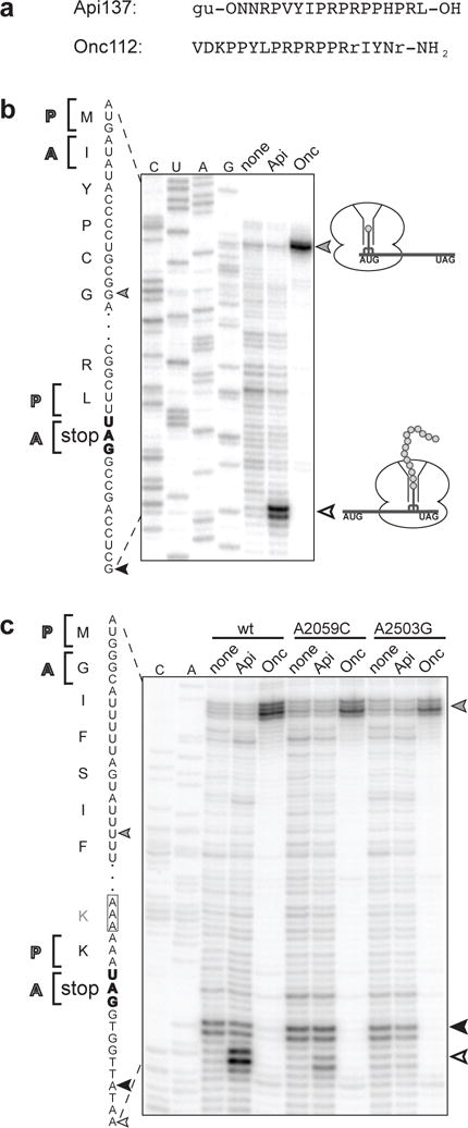
Figure 1
Api137 stalls ribosomes at the termination step of translation
a , Amino acid sequences of PrAMPs Api137 and Onc112. gu = N,N,Nu2032,Nu2032-tetramethylguanidino, O = L-ornithine, r = D-arginine. b, c , In vitro toeprinting analysis comparing the Onc112- or Api137...
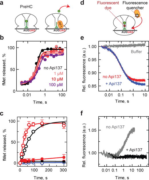
Figure. 2
Api137 allows peptide hydrolysis but inhibits turnover of RF1 and RF2
a , Schematics of the peptidyl-tRNA hydrolysis experiments. PreHC carrying f[ 3 H]Met-tRNA fMet is reacted with RF1 (shown) or RF2 and the release of f[ 35 S]Met is measured. b , Time courses of pepti...
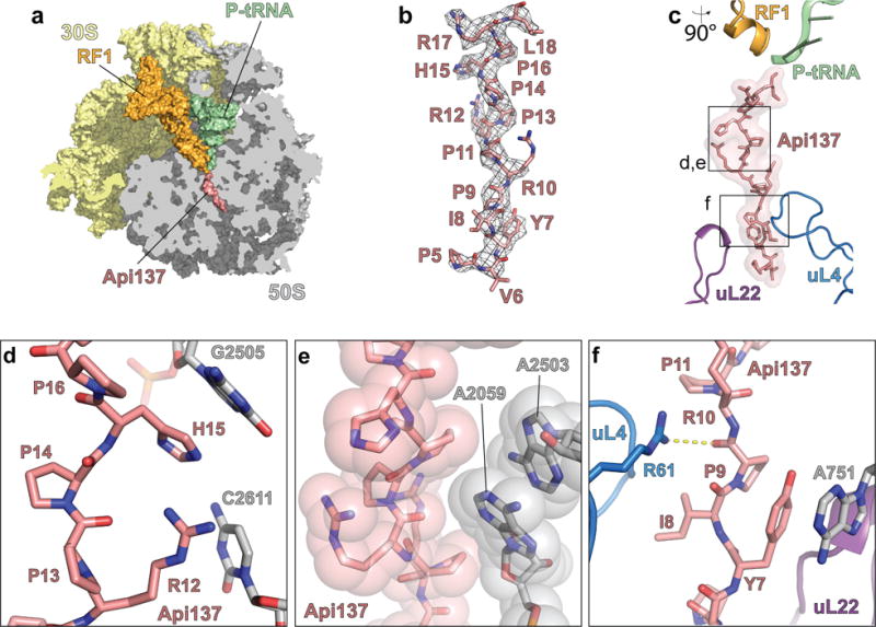
Figure. 3
Binding of Api137 to the terminating ribosome and its interactions with the exit tunnel
a , Transverse section of the 50S subunit (grey) showing the binding site of Api137 (salmon) on the 70S ribosome (30S subunit in yellow) within the polypeptide exit tunnel relative to RF1 (orange) and...
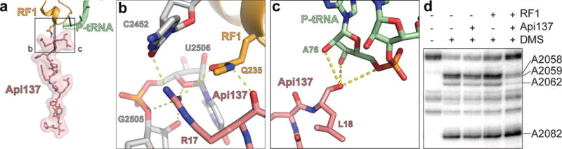
Figure 4
Inhibitory action of Api137 is mediated by its interactions with RF1 and P-site tRNA
a , Position of Api137 (salmon) relative to RF1 (orange) and P-site tRNA (green). The boxed regions are enlarged in panels (b,c). b , Interactions of Api137 with RF1. Arg17 of Api137 is coordinated by...
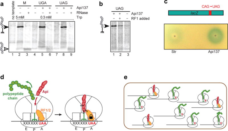
Figure. 5
Api137 induces accumulation of peptidyl-tRNA and stop codon read-through
a , Gel electrophoresis analysis of the [ 35 S]-labeled products of the in vitro translation of the tnaC gene with its original UGA stop codon (lanes 1u20136) or with the UAG stop codon (lanes 7u20139...
Figure images are served from the NIH/NLM PubMed Central Open Access Subset or Europe PMC; copyright remains with the publishers and authors.
💬 Discussion
0 commentsNo comments yet. Be the first to start a discussion!
Leave a Comment