Abstract
Transfer of T cells to freshly irradiated allogeneic recipients leads to their rapid recruitment to nonlymphoid tissues, where they induce graft-versus-host disease (GVHD). In contrast, when donor T cells are transferred to established mixed chimeras (MCs), GVHD is not induced despite a robust graft-versus-host (GVH) reaction that eliminates normal and malignant host hematopoietic cells. We demonstrate here that donor GVH-reactive T cells transferred to MCs or freshly irradiated mice undergo similar expansion and activation, with similar up-regulation of homing molecules required for entry to nonlymphoid tissues. Using dynamic two-photon in vivo microscopy, we show that these activated T cells do not enter GVHD target tissues in established MCs, contrary to the dogma that activated T cells inevitably traffic to nonlymphoid tissues. Instead, we show that the presence of inflammation within a nonlymphoid tissue is a prerequisite for the trafficking of activated T cells to that site. Our studies help to explain the paradox whereby GVH-reactive T cells can mediate graft-versus-leukemia responses without inducing GVHD in established MCs.
🔬 Techniques
✨ Fluorophores
🧪 Sample Preparation
🔬 Cell Lines
🏭 Microscope Brands
🧪 Reagent Suppliers
🏛️ Research Organizations (ROR)
Affiliated research institutions:
📋 Methods
Animals. Animals were used under a protocol approved by our institutional Subcommittee on Research Animal Care, and experiments were performed in accordance with National Institutes of Health guidelines. Female BALB/cJ, C57BL/6, and B6.SJL (CD45.1) mice were purchased from the Frederick Cancer Research Facility. Female recipient B6 × DBA/2 (BDF1) mice were purchased from Charles River Laboratories. Female donor C57BL/6-transgenic (UBC-GFP)30Scha/J mice, which express GFP under the control of the human ubiqutin C promoter, were purchased from The Jackson Laboratory.
2C T cell receptor transgenic mice
(H2 b on C57BL/6 background) were provided by D. Loh (Washington University, St. Louis, MO). Donors were aged 6–13 wk and recipients were aged 11–12 wk at the time of BMT. BMT, donor leukocyte infusion, and purification of T cells. MCs were established by reconstitution of lethally irradiated recipients with a mixture of TCD allogeneic (15 × 10 6 ) and host-type (5 × 10 6 ) BM cells or TCD allogeneic (10–15 × 10 6 ) BM cells alone. Recipient mice were lethally irradiated (B6 and B6DF1 10.25 Gy, BALB/c 8 Gy, 137 Cs source, 0.8 Gy/min), and TCD BM cells were injected i.v. 4–8 h later. TCD was performed using anti-CD4/anti-CD8 microbeads (Miltenyi Biotec). In additional experiments, B6 + BDF1→BDF1 MCs were generated using a nonmyeloablative protocol as described previously ( 56 ). MCs were used in experiments between 2–3 mo after initial BMT. Splenocyte suspensions were used for DLIs and were prepared as described previously ( 57 ). In experiments that involved transfer of activated donor T cells derived from freshly irradiated mice, recipients of BMT and donor splenocytes were killed on day 4 and splenocytes were harvested. Day 4 T cells were purified by passage through nylon wool to remove dead cells and then by negative selection to remove non–T cells (pan T isolation kit; Miltenyi Biotec). In experiments that involved transfer of activated T cells derived from established MCs, DLI recipients were killed on day 12 and splenocytes were harvested. Day 12 CD45.1 + T cells were purified by negative selection to remove non–T cells (pan T isolation kit; Miltenyi Biotec) and then incubated with biotinylated anti-CD45.1 antibody followed by positive selection with anti-biotin–conjugated microbeads (Miltenyi Biotec). Where required, naive donor 2C CD8 + T cells were isolated (purity >93%) by using anti-CD8 microbeads (Miltenyi Biotec) or after activation in vivo by negative selection of untouched T cells and positive selection using IgG1 1B2 clonotypic antibody and anti–mouse IgG1 microbeads (>95% purity). In experiments to examine proliferation of transferred donor T cells, polyclonal B6 splenocytes and 2C CD8 + T cells were labeled with 1 μM CFSE (Invitrogen) as described previously ( 24 ). In experiments designed to evaluate the effect of systemic TLR activation, 12.5 μg of the TLR7 agonist R-848 was administered by i.p. injection every 72 h for four doses from the day of splenocyte transfer to established MCs. In additional experiments, 1.25 mg imiquimod (3M Pharmaceuticals) was applied to the ear pinnae or flank of MCs on days 0 and 5 after splenocyte transfer to MCs. Assessment of GVHD. Clinical GVHD was assessed by scoring five parameters, including hunching, ruffled fur, diarrhea, periorbital edema, and activity. Histopathologic analysis of GVHD target organs was performed single blind by scoring changes in skin (dermal/epidermal lymphocyte infiltration, dyskeratotic epidermal keratinocytes, and epidermal thickening) and colon (crypt regeneration, apoptosis in crypt epithelial cells, crypt loss, surface colonocyte attenuation, inflammatory cell infiltration in lamina propria, mucosal ulceration, and thickening of mucosa). A severity scale from 0 to 4 was used: 0, normal; 0.5, focal and rare; 1, focal and mild; 2, diffuse and mild; 3, diffuse and moderate; 4, diffuse and severe. Isolation of lymphocytes from intestines. Mice were killed and perfused via the heart with 40 ml PBS containing 10 U/ml heparin. The gut, cut longitudinally, was rinsed in cold HBSS, sectioned into 5-mm pieces, and shaken at 120 rpm at 37°C for 30 min in RPMI 1640/5% FCS. The suspension was then passed serially through a steel mesh, nylon mesh, and nylon wool column before enrichment for lymphocytes by density centrifugation (400 g for 30 min) over a Ficoll-Histopaque gradient (Sigma-Aldrich). Flow cytometry. The following antibodies were used: anti–LPAM-1 (α4β7, DATK32), anti–H2-D d -biotin (34-2-12), anti–CD4-PE (RM4-5), anti–CD8β-PE (H35-17.2), anti–CD11b-PE (M1/70), anti–CD44-FITC (IM7), anti–CD45.1-biotin (A20), anti–CD45.2-FITC (104), anti–CD62L-FITC (MEL-14), anti–rat IgG2a-biotin (RG7/1.30), and all appropriate isotype controls (all purchased from BD Biosciences). Detection of biotinylated antibodies was performed using either PE or APC linked to streptavidin (BD Biosciences). CD8 + T cells bearing the 2C TCR were identified via the clonotype-specific mAb, 1B2, and anti–mouse IgG1-APC (X56; BD Biosciences). Enumeration of CD8 + 1B2 + T cells in peripheral blood was performed using TRUCOUNT (BD Biosciences) beads according to the manufacturer's instructions. Quantitative real-time PCR. Methods for RNA extraction, RT, primers/conditions for quantitative PCR, and analysis of quantity values for gene expression have been described previously ( 58 ). Calculated values for the gene of interest were normalized to the housekeeping gene GAPDH. Intravital two-photon microscopy and flow cytometry. Trafficking of GFP + T cells to the skin was imaged noninvasively using a custom-built two-photon fluorescence microscope designed specifically for in vivo imaging. In brief, output of a femtosecond titanium sapphire laser (890 nm; Coherent Mira) was injected into a video rate laser scanning platform consisting of a spinning polygon and a galvanometer that steer the laser beam in the fast (x) and slow (y) axes, respectively. The laser beam was focused onto the sample (mouse ear skin) using a 60×, 1.2NA water immersion objective lens (Olympus). Two-photon excited fluorescence was collected by the same objective lens, separated from the excitation beam by a dichroic filter, and detected with a photomultiplier tube (HC-124-2; Hamamamtsu) through a band pass filter (500–600 nm; Edmund Optics). Images were acquired at 30 frames per second and either stored to a digital video recorder or to a computer hard drive. In the case of static images, a 1-s frame average was performed to improve the signal/noise ratio. Circulating T cells were measured with an in vivo flow cytometer as described previously ( 59 ). Statistical analysis. Survival data were analyzed using the log-rank test. Otherwise, statistical analyses were performed using the Student's t test or the Mann-Whitney test for nonparametric data. A p-value of
Show full methods section
Animals. Animals were used under a protocol approved by our institutional Subcommittee on Research Animal Care, and experiments were performed in accordance with National Institutes of Health guidelines. Female BALB/cJ, C57BL/6, and B6.SJL (CD45.1) mice were purchased from the Frederick Cancer Research Facility. Female recipient B6 × DBA/2 (BDF1) mice were purchased from Charles River Laboratories. Female donor C57BL/6-transgenic (UBC-GFP)30Scha/J mice, which express GFP under the control of the human ubiqutin C promoter, were purchased from The Jackson Laboratory.
2C T cell receptor transgenic mice
(H2 b on C57BL/6 background) were provided by D. Loh (Washington University, St. Louis, MO). Donors were aged 6–13 wk and recipients were aged 11–12 wk at the time of BMT. BMT, donor leukocyte infusion, and purification of T cells. MCs were established by reconstitution of lethally irradiated recipients with a mixture of TCD allogeneic (15 × 10 6 ) and host-type (5 × 10 6 ) BM cells or TCD allogeneic (10–15 × 10 6 ) BM cells alone. Recipient mice were lethally irradiated (B6 and B6DF1 10.25 Gy, BALB/c 8 Gy, 137 Cs source, 0.8 Gy/min), and TCD BM cells were injected i.v. 4–8 h later. TCD was performed using anti-CD4/anti-CD8 microbeads (Miltenyi Biotec). In additional experiments, B6 + BDF1→BDF1 MCs were generated using a nonmyeloablative protocol as described previously ( 56 ). MCs were used in experiments between 2–3 mo after initial BMT. Splenocyte suspensions were used for DLIs and were prepared as described previously ( 57 ). In experiments that involved transfer of activated donor T cells derived from freshly irradiated mice, recipients of BMT and donor splenocytes were killed on day 4 and splenocytes were harvested. Day 4 T cells were purified by passage through nylon wool to remove dead cells and then by negative selection to remove non–T cells (pan T isolation kit; Miltenyi Biotec). In experiments that involved transfer of activated T cells derived from established MCs, DLI recipients were killed on day 12 and splenocytes were harvested. Day 12 CD45.1 + T cells were purified by negative selection to remove non–T cells (pan T isolation kit; Miltenyi Biotec) and then incubated with biotinylated anti-CD45.1 antibody followed by positive selection with anti-biotin–conjugated microbeads (Miltenyi Biotec). Where required, naive donor 2C CD8 + T cells were isolated (purity >93%) by using anti-CD8 microbeads (Miltenyi Biotec) or after activation in vivo by negative selection of untouched T cells and positive selection using IgG1 1B2 clonotypic antibody and anti–mouse IgG1 microbeads (>95% purity). In experiments to examine proliferation of transferred donor T cells, polyclonal B6 splenocytes and 2C CD8 + T cells were labeled with 1 μM CFSE (Invitrogen) as described previously ( 24 ). In experiments designed to evaluate the effect of systemic TLR activation, 12.5 μg of the TLR7 agonist R-848 was administered by i.p. injection every 72 h for four doses from the day of splenocyte transfer to established MCs. In additional experiments, 1.25 mg imiquimod (3M Pharmaceuticals) was applied to the ear pinnae or flank of MCs on days 0 and 5 after splenocyte transfer to MCs. Assessment of GVHD. Clinical GVHD was assessed by scoring five parameters, including hunching, ruffled fur, diarrhea, periorbital edema, and activity. Histopathologic analysis of GVHD target organs was performed single blind by scoring changes in skin (dermal/epidermal lymphocyte infiltration, dyskeratotic epidermal keratinocytes, and epidermal thickening) and colon (crypt regeneration, apoptosis in crypt epithelial cells, crypt loss, surface colonocyte attenuation, inflammatory cell infiltration in lamina propria, mucosal ulceration, and thickening of mucosa). A severity scale from 0 to 4 was used: 0, normal; 0.5, focal and rare; 1, focal and mild; 2, diffuse and mild; 3, diffuse and moderate; 4, diffuse and severe. Isolation of lymphocytes from intestines. Mice were killed and perfused via the heart with 40 ml PBS containing 10 U/ml heparin. The gut, cut longitudinally, was rinsed in cold HBSS, sectioned into 5-mm pieces, and shaken at 120 rpm at 37°C for 30 min in RPMI 1640/5% FCS. The suspension was then passed serially through a steel mesh, nylon mesh, and nylon wool column before enrichment for lymphocytes by density centrifugation (400 g for 30 min) over a Ficoll-Histopaque gradient (Sigma-Aldrich). Flow cytometry. The following antibodies were used: anti–LPAM-1 (α4β7, DATK32), anti–H2-D d -biotin (34-2-12), anti–CD4-PE (RM4-5), anti–CD8β-PE (H35-17.2), anti–CD11b-PE (M1/70), anti–CD44-FITC (IM7), anti–CD45.1-biotin (A20), anti–CD45.2-FITC (104), anti–CD62L-FITC (MEL-14), anti–rat IgG2a-biotin (RG7/1.30), and all appropriate isotype controls (all purchased from BD Biosciences). Detection of biotinylated antibodies was performed using either PE or APC linked to streptavidin (BD Biosciences). CD8 + T cells bearing the 2C TCR were identified via the clonotype-specific mAb, 1B2, and anti–mouse IgG1-APC (X56; BD Biosciences). Enumeration of CD8 + 1B2 + T cells in peripheral blood was performed using TRUCOUNT (BD Biosciences) beads according to the manufacturer's instructions. Quantitative real-time PCR. Methods for RNA extraction, RT, primers/conditions for quantitative PCR, and analysis of quantity values for gene expression have been described previously ( 58 ). Calculated values for the gene of interest were normalized to the housekeeping gene GAPDH. Intravital two-photon microscopy and flow cytometry. Trafficking of GFP + T cells to the skin was imaged noninvasively using a custom-built two-photon fluorescence microscope designed specifically for in vivo imaging. In brief, output of a femtosecond titanium sapphire laser (890 nm; Coherent Mira) was injected into a video rate laser scanning platform consisting of a spinning polygon and a galvanometer that steer the laser beam in the fast (x) and slow (y) axes, respectively. The laser beam was focused onto the sample (mouse ear skin) using a 60×, 1.2NA water immersion objective lens (Olympus). Two-photon excited fluorescence was collected by the same objective lens, separated from the excitation beam by a dichroic filter, and detected with a photomultiplier tube (HC-124-2; Hamamamtsu) through a band pass filter (500–600 nm; Edmund Optics). Images were acquired at 30 frames per second and either stored to a digital video recorder or to a computer hard drive. In the case of static images, a 1-s frame average was performed to improve the signal/noise ratio. Circulating T cells were measured with an in vivo flow cytometer as described previously ( 59 ). Statistical analysis. Survival data were analyzed using the log-rank test. Otherwise, statistical analyses were performed using the Student's t test or the Mann-Whitney test for nonparametric data. A p-value of
📊 Figures
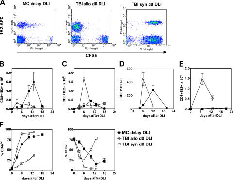
Figure 1.
Distribution of antihost CD8 + CTLs between lymphoid and nonlymphoid tissues differs according to host environment. (A) Proliferation of transferred 2C CD8 + T cells as evaluated by CFSE dilution. Dot...
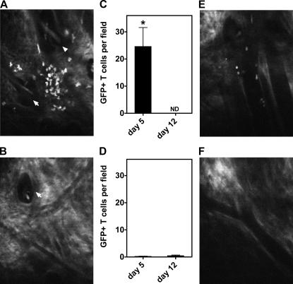
Figure 2.
Absence of donor T cells in skin after transfer to established MCs. Representative images of skin (A and B, day 5) and mean u00b1 SEM ( n = 3 per group) number of GFP + T cells per field (C and D) der...
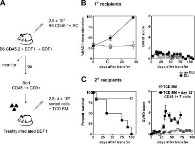
Figure 3.
T cells derived from MCs induce GVHD upon transfer to freshly irradiated recipients. (A) 2.5 u00d7 10 7 naive B6 CD45.1 splenocytes were transferred to established B6 + BDF1u2192BDF1 MCs. On day 12, C...
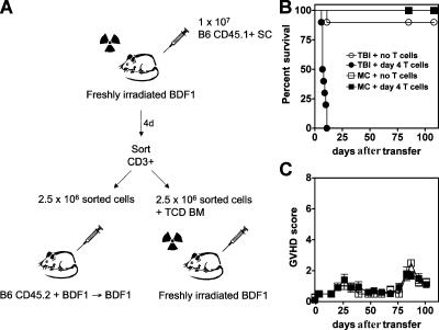
Figure 4.
T cells derived from mice developing GVHD are unable to induce GVHD upon transfer to MCs. (A) 10 7 naive B6 CD45.1 splenocytes plus TCD BM were transferred to freshly irradiated BDF1 recipients. On da...
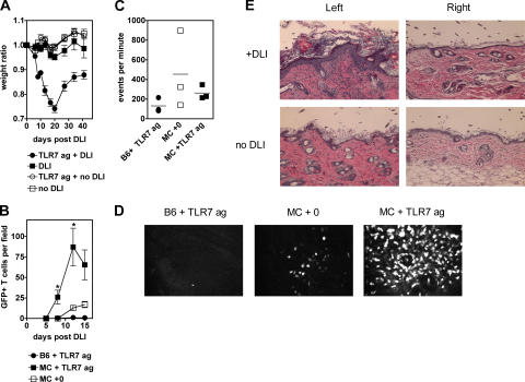
Figure 5.
Localized TLR-induced inflammation permits access to skin for activated T cells. (A) Mean u00b1 SEM weight ratio of untreated MCs (u25a1; n = 3) or at timed intervals in MCs after transfer of 3 u00d7 ...
Figure images are served from the NIH/NLM PubMed Central Open Access Subset or Europe PMC; copyright remains with the publishers and authors.
💬 Discussion
0 commentsNo comments yet. Be the first to start a discussion!
Leave a Comment